MRI Features of Ovarian Fibrothecoma
Received: 08-May-2021 / Accepted Date: 12-May-2021 / Published Date: 19-May-2021 DOI: 10.4172/2167-7964.1000327
Abstract
Fibrothecoma is considered as a benign tumor, usually in middle-aged women. Larger tumors can be associated with Meigs syndrome. Often an incidental finding, but a sudden onset of pelvic pain may indicate an ovarian torsion. We report a case of a post–menopausal woman with a history of vaginal bleeding. MRI revealed an endometrial hyperplasia with a left ovarian benign fibrothecoma, following surgery and histological findings confirmed the diagnosis.
Keywords: Ovary, Fibrothecoma, Magnetic Resonance Imaging
Text
Ovarian fibrothecomas are rare tumors of sex cord-stromal origin that represent <4% of all ovarian neoplasms [1] the majority of fibrothecomas are benign. Most occur in adult women, with 66% in postmenopausal women [2].
Generally, pelvic pain or distention and irregular vaginal bleeding are the main patient symptoms [3-5]. Although the ovarian mass by itself is supposedly benign in nature, estrogenic effects, such as endometrial hyperplasia, endometrial cancer and postmenopausal bleeding, commonly accompany fibrothecomas.
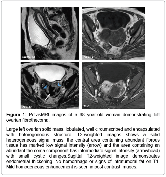
The parenchyma of fibrothecomas typically exhibits homogeneous low signal intensity on T1 and T2 when compared with myometrium; abundant collagen and fibrotic content of the tumor are the reasons that these features are observed on MRI. Scattered high signal areas on T2 may be present representing areas of cystic degeneration Figure 1. A prominent Diffusion restriction should not be misinterpreted as malignant lesion. Fibrothecoma shows a mild enhancement following contrast injection. The thickened endometrium observed in postmenopausal woman may also be a valuable imaging feature.
Figure 1: PelvisMRI images of a 68 year-old woman demonstrating left ovarian fibrothecoma:
Large left ovarian solid mass, lobulated, well circumscribed and encapsulated with heterogeneous structure. T2-
weighted images shows a solid heterogeneous signal mass; the central area containing abundant fibrous tissue has marked
low signal intensity (arrow) and the area containing an abundant the coma component has intermediate signal intensity
(arrowhead) with small cystic changes.Sagittal T2-weighted image demonstrates endometrial thickening. No hemorrhage or
signs of intratumoral fat on T1. Mild homogeneous enhancement is seen in post contrast images.
References
- Shinagare AB, Meylaerts LJ, Laury AR, Mortele KJ (2012)n MRI features of ovarian fibroma and fibrothecoma withnhistopathology correlation. AJR Am J Roentgenol 198:w296-w303.
- Yanxia Zhao, Jinghong Cao, Alexander Melamed (2019) Losartan treatment enhances chemotherapy efficacy andnreduces ascites in ovarian cancer models by normalizing the tumor stroma, Proc Natl Acad Sci USA 116(6 ):2210 -n2219
- Li X, Zhang W, Zhu G, Sun C, Liu Q, et al (2012) Imaging features and pathologic characteristics ofnovarian thecoma. J Comput Assist Tomogr 36:46-53.
- Yen P, Khong K, Lamba R, Corwin MT, Gerscovich EOn (2013) Ovarian fibromas and fibrothecomas:nSonographic correlation with computed tomography and magnetic resonance imaging: A 5-  A 5-year single-institutionnexperience. J Ultrasound Med 32:13-18.
- Wu B, Peng WJ, Gu YJ, Cheng YF, Mao J (2014) MRI diagnosis of ovarian fibrothecomas: Tumor appearancesnand estrogenic effect features. Br J Radiol 87:1038.
Citation: Lrhorfi N, Belkouchi L, El haddad S, Allali N, Chat L (2021) MRI Features of Ovarian Fibrothecoma. OMICS J Radiol 10: 327. DOI: 10.4172/2167-7964.1000327
Copyright: © 2021 Lrhorfi N, et al. This is an open-access article distributed under the terms of the Creative Commons Attribution License, which permits unrestricted use, distribution, and reproduction in any medium, provided the original author and source are credited.
Share This Article
Open Access Journals
Article Tools
Article Usage
- Total views: 2412
- [From(publication date): 0-2021 - Mar 29, 2025]
- Breakdown by view type
- HTML page views: 1643
- PDF downloads: 769

