MiR-124 could Inhibit CD151 Overexpression Associated with Non-Small Cell Lung Carcinoma Progression
Received: 21-Oct-2022 / Manuscript No. AOT-22-77978 / Editor assigned: 24-Oct-2022 / PreQC No. AOT-22-77978 (PQ) / Reviewed: 07-Nov-2022 / QC No. AOT-22-77978 / Revised: 06-Feb-2023 / Manuscript No. AOT-22-77978 (R) / Published Date: 13-Feb-2023
Abstract
Background: CD151 is a member of the transmembrane 4 super families and has been reported to be involved in cell adherence and migration. It has been reported that the overexpression of CD151 and the down regulation of miR-124 are all related to the progression of Non-Small Cell Lung Cancer (NSCLC). However, whether there is a regulatory relationship between them has not been examined. The purpose of this study is to further explore whether there is a correlation between the expression of CD151 and miR-124 in NSCLC.
Methods: The expression of CD151 and its functional partner α3β1 in NSCLC clinical tissue was detected by immunohistochemistry. The expression of miR-124 in lung tumor tissues and serums of NSCLC patients was detected by qPCR. The targeting of CD151 mRNA by miR-124 was confirmed by the dual luciferase reporter system. Cell migration and invasion were verified by scratch wound healing and transwell invasion assay.
Results: CD151 and α3β1 expression were up regulated, while miR-124 expression in the NSCLC group was decreased compared to that in the benign lesion group. Moreover, CD151 positive expression was associated with poor survival of NSCLC patients. MiR-124 targeted the 3’UTR of CD151 mRNA and regulated its expression. Knockdown of CD151 expression with CD151 shRNA or miR-124 mimics inhibited the migration and invasion of the A549 cell line.
Conclusion: The results indicated that up regulated miR-124 could inhibit CD151 overexpression, which is associated with NSCLC progression.
Keywords: CD151; α3β1; MiR-124; Non-small cell lung cancer; Cell migration
Introduction
Non-Small Cell Lung Cancer (NSCLC) accounts for approximately 85% of all lung cancers. It includes any type of epithelial lung cancer, except small cell lung carcinoma, such as squamous cell carcinoma, large cell carcinoma, adenocarcinoma, pleomorphic and other less frequent types, such as carcinoid tumour, salivary gland carcinoma and unclassified carcinoma [1]. These cancers are grouped together as NSCLC since the treatment approach and prognosis are often similar [2]. NSCLC is less sensitive to chemotherapy than small cell carcinoma. Definition of the biomarkers and critical pathways that affect NSCLC growth and progression is imperative for the treatment of the disease. Integrin adhesion receptors bind simultaneously to extracellular matrix ligands and cytoskeletal proteins. These proteins regulate cell migration; wound healing, tissue morphogenesis extracellular matrix assembly and remodelling [3]. CD151 is a tetraspanin that has transmembrane domains and conserved cysteine residues in a large extracellular loop [4]. CD151 is a pivotal regulator of signalling pathways associated with laminin binding integrin. It is widely expressed on the cell membrane and forms a stable complex with integrin α3β1, which is also ubiquitously expressed in many cell types as a laminin binding integrin [5]. The CD151-α3β1 complex promotes cellular invasion by regulating the actin assembly. Association of CD151 with α3β1 occurs dynamically in discrete subcellular compartmentsm and is involved in the local GTPase signaling for the promotion of tumor cell invasion [6]. CD151 is associated with cancer development and its roles in pro invasion and pro migration have been demonstrated in many in vitro and in vivo models [7-9]. CD151 is involved in signal transduction, which is crucial for the motility in many cancer cell types and acts on FAK, Src, Erk1/2, p38, Ick, Rac1, p130Cas, paxillin and JNK kinases in different cancer cell lines [10-15]. It also predicts the prognosis of patients with resected gastric cancer or endometrial cancer [16,17]. It is reported to have a basic expression in human lung especially in airway smooth muscle, while Tokuhara confirmed that it’s up regulation in lung tumor is associated with a poor prognosis, specially the most common subtype of NSCLC [18,19].
MicroRNA is a superfamily of small non-coding RNA molecules of 19 nucleotides-25 nucleotides in length that negatively regulate target gene expression by suppressing mRNA translation or promoting mRNA degradation [20]. Increasing studies have shown that the dysregulation of miRNAs is associated with cancer progression [21,22]. Human microRNA-124 (miR-124) is encoded by three loci: miR-124-1 (8p23.1), miR-124-2 (8q12.3) and miR-124-3 (20q13.33) [23]. Recently, numerous studies have focused on miR-124 due to its promising tumor suppressive effects in various cancers [24-28]. Reportedly, more and more miR-124 target genes have been identified and a lot of the target gene of miR-124 is consistent with malignancy and prognosis [29]. MiR-124 exerts its anti-tumor effects by regulating a wide range of target genes expression to regulate cell migration, invasion and proliferation. MiR-124 is down regulated in various cancer tissues and cell lines. However, the deeper molecular mechanism of their results is unclear.
In this study, we want to further verify the impact of the expression of CD151 and miR-124 on the prognosis of NSCLC patients and explore whether CD151 is one of the target genes of miR-124. We found that CD151 and α3β1 expression was up regulated in NSCLC tissues, while miR-124 expression was down regulated. The CD151 overexpression was associated with poor survival of NSCLC patients, which was consistent with previous studies. CD151 is a potential target gene of miR-124. The down regulation of CD151 with CD151 siRNA or miR-124 in the non-small cell lung cancer cell line A549 might attenuate cell migration.
Materials and Methods
Study design
This randomized, open label study, sponsor blind clinical prognostic follow up and histopathological analysis were conducted in Yunnan province, China, which has a high incidence of lung cancer. The study was done in accordance with the international conference on harmonization good clinical practice guidelines and applicable local regulations with approval from the ethics committee of Yunnan tumor hospital.
Patients and tissue samples
A total of 103 patients age from 27 years to 70 years old receiving treatment at Yunnan tumor hospital (Kunming, China) from January 2006 to December 2011 were enrolled in this study. Of these, 81 lung tumor tissue samples were collected from NSCLC patients (31 squamous carcinoma, 40 adenocarcinoma and 10 samples from other subtypes), 22 benign lung tissues were collected from 15 pulmonary tuberculosis patients and 7 inflammatory pseudo tumor patients. Serum samples were also collected from patients and healthy individuals. These samples were stored in liquid nitrogen until further use. All samples derived from human subjects were collected in accordance with the hospital regulations and were approved by the ethics committee of Yunnan tumor hospital. Written informed consent was obtained from each subject. Nine pathological features of eighty one NSCLCs are listed. All the NSCLC patients did not receive chemotherapy or radiotherapy before surgical excision of tumor tissues. A total of 56 patients out of 81 NSCLC subjects (69.1%) received 1 cycles-6 cycles of platinum based chemotherapy and several patients received local radiotherapy. Lung cancer was the only cause of death 5 years after surgery. All patients were followed up every 3 months by telephone, outpatient or inpatient visits [30]. The death, last follow up or lost follow up sessions were recorded up to the termination of the study. The follow up deadline was April 30, 2012. During the follow up period, 3 cases were lost to follow up.
Tumor specimens
81 lung tumor samples and control lung tissues were fixed for 24 h in 10% formaldehyde solution after thawing from liquid nitrogen. After fixation, the tissues were washed with water and dehydrated in alcohol. The alcohol was eliminated by incubation in xylene, embedded in paraffin at 60°C and allowed to harden overnight. The tissues were sliced into 8 μm-10 μm thick paraffin embedded sections.
Cell lines and Lentivirus
A549 cells and HEK293T cells were purchased from the Chinese Academy of Sciences Cell Bank (Shanghai, China). A549 cells were maintained in DMEM (Gibco, Thermo, US) containing 10% FBS (HyClone, US) at 37°C in a humidified incubator with 5% CO2. Plasmids that constitutively expressed CD151 shRNA or miR-124 were constructed by cloning CD151 shRNA (targeting CD151 open reading frame region at 5’-AAGTCTCAAGCTGGAGCACTAC-3’) or miR-124 sequence (5’-ATTCCGTGCGCCACTTACGG-3’) into the pLV-2 (pGLVU6/Puro) plasmid (Gene Pharma Inc., China). The control miR with sequence 5’-CTAGCTCCCTTCAATCCAAG-3 inserted into pLV-2 was used as the control. Lentiviruses were packaged into HEK293T cells by transfection with the pLV-2 plasmid and two other packaging plasmids that were supplied in the 2nd generation packaging mix (ABM Inc., US).
Immunohistochemical assays
The assays were carried out as described previously. Endogenous peroxidase activity was blocked with 3% H2O2 for 30 min. After deparaffinization and rehydration of the samples, the antigen was retrieved via microwave stimulation for 15 min in citrate buffer (pH=6.0). The sections were blocked with 10% BSA for 2 h at room temperature, followed by incubated with anti-CD151 monoclonal antibody (ab33315, Abcam Inc., US) or α3β1 (ab217145, Abcam Inc., US) antibody in a humidified chamber at 4°C. After three pieces of washing and diluted at a ratio of 1:200 with PBS, the sections were then incubated with an HRP conjugated secondary antibody, developed with DAB chromogen and counterstained with hematoxylin. Image J software was used to quantify IHC stain intensity. The stain percentage was evaluated under the same color threshold. The set of color threshold was based on the appropriate positive (visually distinguishable strong staining) and negative (no staining) staining of the typical images.
RT-PCR analysis
Total RNA, including miRNA was extracted from serum and cultured cells by using TRIzol universal reagent (Tiangen Inc., China). Small RNA from tumor tissues was isolated from 4 sections-5 sections of 20 uM of formalin fixed, paraffin embedded tissues using miRNeasy FFPE Kit (Qiagen Inc., GER) according to the manufacturer's instructions. Reverse transcription of the total RNA was performed using a FastKing gDNA dispelling RT SuperMix (Tiangen Inc., China). Reverse transcription of small RNA was performed by using the miRcute plus miRNA first strand cDNA kit (Tiangen Inc., CN). Real time PCR was performed by using the SuperReal PreMix plus (SYBR Green) (Tiangen Inc., China) or miRcute plus miRNA first strand cDNA kit (Tiangen Inc., China) on Qiagen Rotor-Gene Q-6000 system (Qiagen Inc., GER). U6 was used as the miRNA endogenous control. GAPDH was used as the mRNA endogenous control. Target gene expression was normalized to the endogenous control. Fold changes were determined by the 2^(-ddCt) method.
Luciferase reporter assay
Wild type and mutated 3’UTR of CD151 was synthesized and cloned into the pmiRGLO vector (Youbao lnc., China). HEK293 cells were maintained in a 6 well plate and transfected with 1 μg of the reporter plasmid vector, or the hsa-miR-124-3p.1 mimic (1 μM in each well) or both. Firefly and Renilla activity was determined at 24 h after transfection with the dual Luciferase reporter gene assay kit (Beyotime Inc., China).
Cell migration assay
Cell migration was evaluated by a cell scratch assay. Cells grown to 80%–90% confluence in a 6 well plate were gently scratched through the cell monolayer of the well with a new 20 μl pipette tip. After scratching, the wells were gently washed with PBS to remove detached cells. The wells were replenished with fresh medium. After 48 h of additional growth, the cells were washed with PBS and fixed with 4% paraformaldehyde for 10 min. The monolayer was photographed with a microscope.
Matrigel invasion assay
Cell invasion assays were performed according to Yang. The 24 well trans wells with an 8 mm pore size (Corning Inc. US) were pre coated with Matrigel (BD Biosciences Inc., BD). Approximately 10^4 cells were suspended in 100 μl DMEM containing 1% FBS and added to the upper chamber. Then, 600 μl DMEM containing 10% FBS was added to the lower chamber. After 48 h of incubation, cotton swabs were used to remove the remaining Matrigel and cells in the upper chamber. Cells on the lower surface of the membrane were fixed with 4% paraformaldehyde and stained with Giemsa.
Statistical analysis
All statistical analyses were performed with SPSS software (IBM, US). Counting data was expressed by frequency. The χ2 test was used to compare the rate values. The multivariate ANOVA test was used to compare the mean values of multiple groups. The t-test was used to compare the mean values of two groups. Pomit-biserial correlation test was used for correlation analysis. The Kaplan-Meier method and log rank test were used for survival analysis. Multivariate analysis of prognosis was performed with COX proportional risk model. P-value ≤ 0.05 was considered as statistically significant.
Results
Expression of CD151 and α3β1 in NSCLC lung tissues
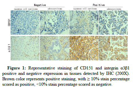
The expression of CD151 and α3β1 was detected by IHC (Figure 1). A total of 81 NSCLC patients were included in this study and 40 adenocarcinoma samples, 31 squamous cells (epidermoid) carcinoma samples and 10 samples of other NSCLC subtypes were obtained with surgical excision of NSCLC specimens that were confirmed with routine pathological examination. A total of 22 benign lesion samples, including 15 pulmonary tuberculosis and 7 inflammatory pseudotumor tissues obtained via surgery were used as control samples. The IHC results demonstrated that the percentages of CD151 positive and α3β1 positive samples in the total NSCLC group were significantly higher than those in the benign lesion group (Table 1). The paired χ2 test was used to analyze the statistical relationship between the expression of CD151 and integrin α3β1. The results are shown in Table 2. Their expression was statistically different (Pearson chi-square test, P<0.05). Their expression difference was not statistically significant (P-value of McNemar’s test>0.5). Although their expression consistency was statistically different, the consistency was poor (Kappa value<0.4). The statistical analysis examining the correlation of CD151 and α3β1 expression with nine clinical pathological factors indicated CD151 and α3β1 expression did not exhibit any association with gender, age, smoking, tumor T stages, tumor N stage, tumor TNM stages, tumor differential degree or patient Performance Status (PS). However, they exhibit an association with tumor subtypes (Table 3). CD151 positive expression is more frequent in adenocarcinoma than that in squamous carcinoma, while α3β1 positive expression in the two tumor subtypes is reverse, which may explain the poor agreement value (Kappa value<0.4) between the expression of CD151 and integrin α3β1 in NSCLC.
| CD151 | integrin α3β1 | ||||||||||
|---|---|---|---|---|---|---|---|---|---|---|---|
| Group | n | Negative | Positive | Positive rate (%) | χ2 | P | Negative | Positive | Positive rate (%) | χ2 | P |
| NSCLC | 81 | 36 | 45 | 55.6 | 9.7 | 0.002* | 41 | 40 | 49.4 | 4.8025 | 0.025* |
| Benign lesion | 22 | 18 | 4 | 18.2 | 17 | 5 | 22.7 | ||||
| Note: n=case; Chi-square test was performed to compare the rates. χ2, pearson Chi-square value; *P ≤ 0.05 | |||||||||||
Table 1: Expression of CD151 and integrin α3β1 in NSCLC and benign lesion.
| Groups | CD151 | α3β1 | |||||||
|---|---|---|---|---|---|---|---|---|---|
| n | Negative | Positive | χ2 | P | Negative | Positive | χ2 | P | |
| Gender | |||||||||
| Male | 56 | 25 | 31 | 0.003 | 0.957 | 27 | 29 | 0.419 | 0.517 |
| Female | 25 | 11 | 14 | 14 | 11 | ||||
| Age (Years) | |||||||||
| <60 | 44 | 19 | 25 | 0.062 | 0.803 | 23 | 22 | 1.049 | 0.306 |
| ≥ 60 | 37 | 17 | 20 | 19 | 18 | ||||
| Smoking | |||||||||
| No | 29 | 12 | 17 | 0.172 | 0.678 | 16 | 13 | 0.375 | 0.54 |
| Yes | 52 | 24 | 28 | 25 | 27 | ||||
| Pathological classification | |||||||||
| Adenocar-cinoma | 40 | 12 | 28 | 7.068 | 0.029* | 27 | 13 | 10.07 | 0.007* |
| Squamous carcinoma | 31 | 19 | 12 | 12 | 19 | ||||
| Others | 10 | 5 | 5 | 2 | 8 | ||||
| T stage | |||||||||
| T1-2 | 64 | 29 | 35 | 0.093 | 0.76 | 32 | 32 | 0.046 | 0.829 |
| T3-4 | 17 | 7 | 10 | 9 | 8 | ||||
| N stage | |||||||||
| N0-1 | 52 | 25 | 27 | 0.776 | 0.378 | 26 | 26 | 0.022 | 0.882 |
| N2-3 | 29 | 11 | 18 | 15 | 14 | ||||
| TNM stage | |||||||||
| I | 31 | 15 | 16 | 0.506 | 0.776 | 16 | 15 | 1.319 | 0.517 |
| II | 22 | 10 | 12 | 9 | 13 | ||||
| III-IV | 28 | 11 | 17 | 16 | 12 | ||||
| Tumor differentiation status | |||||||||
| Poorly | 26 | 13 | 13 | 0.479 | 0.489 | 13 | 13 | 0.006 | 0.939 |
| Medium/high | 55 | 23 | 32 | 28 | 27 | ||||
| Performance status score | |||||||||
| 0-1 | 50 | 22 | 28 | 0.01 | 0.919 | 27 | 23 | 0.598 | 0.439 |
| ≥ 2 | 31 | 14 | 17 | 14 | 17 | ||||
| Note: Chi-square test was performed to compare the rates. χ2, pearson Chi-square value; *P ≤ 0.05 | |||||||||
Table 2: Correlations between CD151 and α3β1 expression with clinical pathological factors.
| α3β1 | Total | ||||
|---|---|---|---|---|---|
| Negtive | Positive | ||||
| CD151 | Negtive | n | 9 | 27 | 36 |
| CD151 (%) | 25.00% | 75.00% | 100.00% | ||
| α3β1 (%) | 22.00% | 67.50% | 44.40% | ||
| Positive | n | 32 | 13 | 45 | |
| CD151 (%) | 71.10% | 28.90% | 100.00% | ||
| α3β1 (%) | 78.00% | 32.50% | 55.60% | ||
| Total | n | 41 | 40 | 81 | |
| CD151 (%) | 50.60% | 49.40% | 100.00% | ||
| 100.00% | 100.00% | 100.00% | |||
| Pearson chi-square test | χ2=17.0125 | P=0.000 | |||
| P value of McNemar’s Test | 0.603a | ||||
| Value Measure of Agreement Kappa | Value | P | |||
| -0.455 | 0 | ||||
Table 3: Correlation analysis between the protein expression of CD151 and α3β1 in NSCLC.
Expression of CD151 is associated with the prognosis of NSCLC
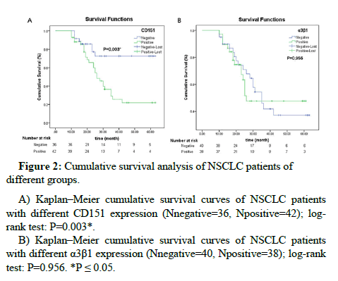
A total of 3 out of 81 cases were lost to follow up. Survival analysis and log rank tests were performed with 78 NSCLC patients by Kaplan-Meier. The results showed that there was a statistically significant difference in the survival between the positive and negative CD151 expression groups (P=0.003). The patients with positive CD151 expression exhibited a poor prognosis, as shown in Figure 2A. The difference between the survivals of patients that were α3β1 positive and α3β1 negative was not significant (Figure 2B). Cox regression model was used to analyze 11 factors including gender, age, smoking, pathological type, tumor T stage, tumor N stage, tumor TNM stage, tumor cell differentiation degree, PS score, CD151 protein expression and α3β1 protein expression. Based on the forward procedure used to screen the factors, we observed that CD151 expression might be an independent factor affecting the prognosis of NSCLC (P=0.001) (Table 4).
Figure 2: Cumulative survival analysis of NSCLC patients of different groups.
A) Kaplan–Meier cumulative survival curves of NSCLC patients with different CD151 expression (Nnegative=36, Npositive=42); log-rank test: P=0.003*.
B) Kaplan–Meier cumulative survival curves of NSCLC patients with different α3β1 expression (Nnegative=40, Npositive=38); log-rank test: P=0.956. *P ≤ 0.05.
| B | SE | Wald | df | Sig | Exp (B) | 95% CI for Exp (B) | ||
|---|---|---|---|---|---|---|---|---|
| Lower | Upper | |||||||
| Age | -0.017 | 0.019 | 0.806 | 1 | 0.369 | 0.983 | 0.948 | 1.020 |
| Gender | -0.088 | 0.429 | 0.042 | 1 | 0.838 | 0.916 | 0.395 | 2.125 |
| Pathological classification | 0.209 | 0.250 | 0.678 | 1 | 0.410 | 1.228 | 0.753 | 2.005 |
| T stage | 0.234 | 0.494 | 0.225 | 1 | 0.635 | 1.264 | 0.480 | 3.329 |
| N stage | 0.499 | 0.663 | 0.565 | 1 | 0.452 | 1.646 | 0.449 | 6.039 |
| TNM stage | -0.183 | 0.374 | 0.240 | 1 | 0.624 | 0.833 | 0.400 | 1.732 |
| Tumor differentiation status | 0.540 | 0.431 | 1.570 | 1 | 0.210 | 1.717 | 0.737 | 3.997 |
| Smoking | 0.378 | 0.397 | 0.906 | 1 | 0.341 | 1.459 | 0.670 | 3.175 |
| PS score | -0.142 | 0.379 | 0.141 | 1 | 0.707 | 0.867 | 0.413 | 1.822 |
| CD151 | 1.605 | 0.486 | 10.904 | 1 | 0.001 | 4.979 | 1.920 | 12.912 |
| α3β1 | 0.818 | 0.442 | 3.421 | 1 | 0.064 | 2.266 | 0.952 | 5.393 |
| Note: B=Constant value; SE=Standard Error; Wald=Chi-square value; df=degree of freedom; Sig, P value; Exp=Exponential; CI=Confidence Interval | ||||||||
Table 4: Multivariate regression analysis in predicting survival of 78 patients with NSCLC.
MiR-124 expression is inversely correlated with the expression of CD151
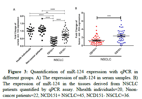
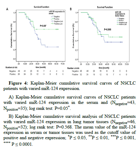
The expression of miR-124 is significantly down regulated in various tissues and cell lines of cancer and demonstrates tumor suppressive effect in various cancers. We examined miR-124 expression in the serum of 81 NSCLC patients, 20 non-cancer patients and 20 healthy individuals. The Q-PCR results indicated that the expression of miR-124 was down regulated in the serum of NSCLC patients, especially in CD151 positive patients (Figure 3A). The miR-124 expression in the serum of non-cancer patients and healthy individuals was similar. The expression pattern of miR-124 in CD151 positive and CD151 negative tumor tissues of NSCLC patients is consistent with that in the serum (Figure 3B). The expression of miR-124 in both serum and tumor tissue demonstrated a reverse correlation with CD151 protein expression in NSCLC tumor tissues (Table 5). This finding indicated that miR-124 and CD151 might have functional relationships. Multivariate ANOVA test of miR-124 expression with clinical parameters of NSCLC patients demonstrated that there was no significant relationship between miR-124 expression and gender, age, smoking, pathological type, tumor T stage, tumor N stage, tumor TNM stage, tumor cell differentiation degree and PS score in Serum or tumor tissues (Table 6). Kaplan-Meier analysis demonstrated a statistically significant difference in the survival between the groups with positive and negative miR-124 expression in serum samples (P=0.05). Patients with serum samples that were negative for miR-124 expression exhibited a poor prognosis (Figure 4A). However, miR-124 expression in lung tumor tissues did not predict the survival of NSCLC patients (Figure 4B).
Figure 3: Quantification of miR-124 expression with qPCR in different groups. A) The expression of miR-124 in serum samples. B) The expression of miR-124 in the tissues derived from NSCLC patients quantified by qPCR assay. Nhealth individuals=20, Nnon-cancer patients=22, NCD151+ NSCLC=45, NCD151- NSCLC=36.
| Serum miR-124 | Tumor miR-124 | |||
|---|---|---|---|---|
| n=81 | r | p | r | p |
| CD151 | -0.601 | 0.000*** | -0.417 | 0.000*** |
| Note: Pomit-biserial correlation test was performed to evaluate the correlation of miR-124 and CD151 expression. r=the Pomit-biserial correlation value; ***P ≤ 0.005 | ||||
Table 5: Pomit-biserial correlation analysis of the expression of CD151 and miR-124.
| Serum miR-124 | Tumor miR-124 | |||
|---|---|---|---|---|
| n=81 | F | P | F | P |
| Gender | 0.363 | 0.549 | 0.003 | 0.956 |
| Age | 0.013 | 0.909 | 1.106 | 0.297 |
| Smoking | 0.118 | 0.733 | 0.346 | 0.559 |
| Pathological classification | 2.477 | 0.092 | 0.677 | 0.511 |
| T | 0.296 | 0.588 | 0.645 | 0.425 |
| N | 0.111 | 0.74 | 1.197 | 0.279 |
| TNM | 0.195 | 0.823 | 0.778 | 0.464 |
| Tumor differentiation status | 0.036 | 0.85 | 0.29 | 0.593 |
| Performance status score | 1.237 | 0.297 | 0.893 | 0.415 |
| Note: Multivariate ANOVA test was performed to evaluate the correlation of miR-124 expression with the lung clinical parameters. P>0.05 | ||||
Table 6: Multivariate ANOVA test of miR-124 expression with the lung clinical parameters.
Figure 4: Kaplan-Meier cumulative survival curves of NSCLC patients with varied miR-124 expression.
A) Kaplan-Meier cumulative survival curves of NSCLC patients with varied miR-124 expression in the serum and (Nnegative=43, Npositive=35); log rank test: P=0.05*.
B) Kaplan-Meier cumulative survival analysis of NSCLC patients with varied miR-124 expression in lung tumor tissues (Nnegative=46, Npositive=32); log rank test: P=0.568. The mean value of the miR-124 expression in serum or tumor tissues was used as the cutoff value of positive and negative expression; *P ≤ 0.05, **P ≤ 0.01, ***P ≤ 0.001, ****P ≤ 0.0001.
MiR-124 regulates CD151 expression in the NSCLC lung cancer cell line A549
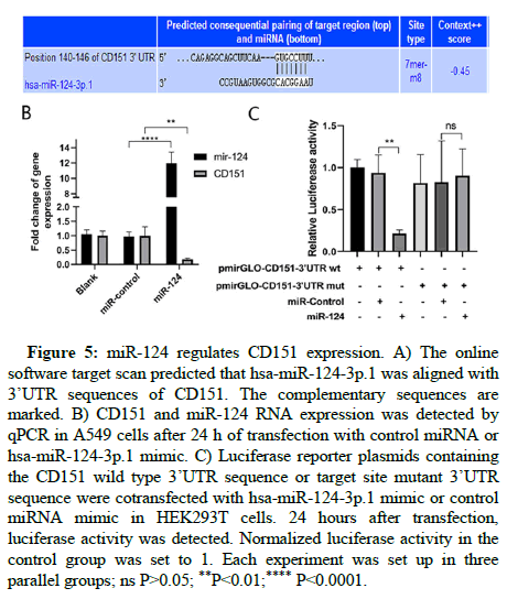
MiR-124 expression was down regulated in the serum and tissue samples derived from NSCLC patients. However, whether this down regulated expression was responsible for CD151 positive expression in NSCLC patient samples remained unclear. In this study, the 3’UTR region of CD151 mRNA (5’-GUGCCUU-3’) was predicted to be targeted by miR-124 sequence (5’-AAGGGCUC-3’) through the online software target scan. Thus we employed the A549 lung adenocarcinoma cell line to investigate the regulation of miR-124 on CD151 expression. A549 cells transfected with miR-124 were collected, and the expression of CD151 was analyzed by qPCR. The results showed that CD151 was sharply down regulated by miR-124 (Figure 5). We cloned the wild type 3’UTR and its target site mutated 3’UTR sequence into the luciferase reporter vector plasmid. The vectors were co-transfected with miR-124 mimics to analyze the luciferase activity. The miR-124 mimics significantly inhibited the luciferase activity of CD151 wild type 3’UTR linked reporter gene compared to that in cells transfected with the miR control [31]. The luciferase activity of the target site mutated 3’UTR linked luciferase reporter was not altered by miR-124. This result demonstrated that miR-124 targeted on the CD151 3’UTR region and regulated its expression (Figure 6).
Figure 5: miR-124 regulates CD151 expression. A) The online software target scan predicted that hsa-miR-124-3p.1 was aligned with 3’UTR sequences of CD151. The complementary sequences are marked. B) CD151 and miR-124 RNA expression was detected by qPCR in A549 cells after 24 h of transfection with control miRNA or hsa-miR-124-3p.1 mimic. C) Luciferase reporter plasmids containing the CD151 wild type 3’UTR sequence or target site mutant 3’UTR sequence were cotransfected with hsa-miR-124-3p.1 mimic or control miRNA mimic in HEK293T cells. 24 hours after transfection, luciferase activity was detected. Normalized luciferase activity in the control group was set to 1. Each experiment was set up in three parallel groups; ns *P>0.05; **P<0.01; ****P<0.0001.
Knockdown of CD151 expression with CD151 shRNA or miR-142 inhibits lung cancer cell migration and invasion
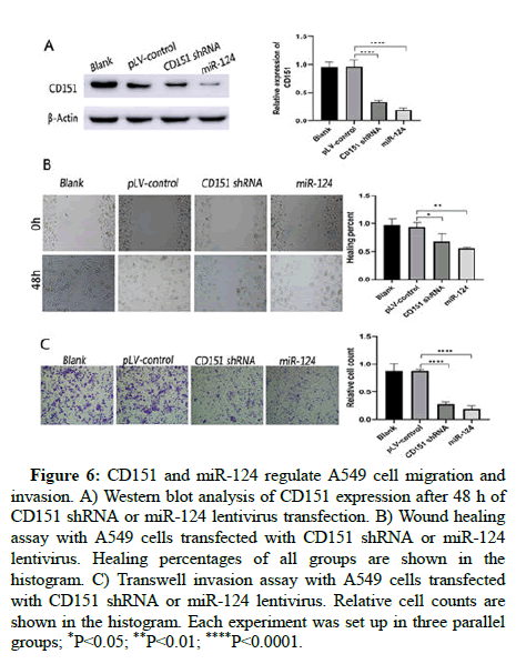
To further investigate the roles of CD151 in NSCLC progression, we knocked down its expression with CD151 shRNA or miR-124. The down regulation of CD151 was verified by western blot analysis. The wound healing assay was performed to detect the migration of A549 cells. The results showed that CD151 shRNA and miR-124 transfected A549 cells exhibited attenuated cell migration. We also detected the invasion ability of CD151 knockdown cells using transwell inserts coated with matrigel matrix. The results indicated that cells transfected with CD151 shRNA and miR-124 was less invasive. Hence, our results indicated that miR-124 and CD151 may influence cell migration and invasion in NSCLC. This result may account for the association of this protein with poor survival in CD151 positive patients (Figure 6) [32].
Figure 6: CD151 and miR-124 regulate A549 cell migration and invasion. A) Western blot analysis of CD151 expression after 48 h of CD151 shRNA or miR-124 lentivirus transfection. B) Wound healing assay with A549 cells transfected with CD151 shRNA or miR-124 lentivirus. Healing percentages of all groups are shown in the histogram. C) Transwell invasion assay with A549 cells transfected with CD151 shRNA or miR-124 lentivirus. Relative cell counts are shown in the histogram. Each experiment was set up in three parallel groups; *P<0.05; **P<0.01; ****P<0.0001.
Discussion
CD151 is expressed in a variety of tumors, including Hepatic Cellular Cancer (HCC), intrahepatic cholangio carcinoma, lung, colon, prostate and breast cancer [33-35]. CD151 overexpression is associated with poor prognosis in several cancer types [36,37]. Takahiro Tokuhara defined the significance of CD151 expression in non-small cell lung cancer in 2001. In this study, we analyzed its expression in several non-small cell lung cancer subtypes and firstly linked CD151 expression in NSCLC tumor tissues with mir-124 expression change in serum and tumor tissues.
The CD151-positive expression percentage in non-small cell lung carcinoma was 55.6%. This expression was significantly higher than that in the benign lesion group (18.2%). The percentage of CD151 positive expression in the adenocarcinoma group was 70%. This expression was higher than that in squamous carcinoma (38.7%) and other types of NSCLC (50.0%). This result is consistent with the previous study conducted by Takahiro Tokuhara. α3β1 is also up regulated in NSCLC lung tissues, but its expression in different subtypes of NSCLC is not consistent with CD151. In addition, there were significant differences in their expression (P<0.01), so the functional relationship between the two genes needs to be further studied.
The overexpression of CD151 and α3β1 indicates a high ability of migration and invasion. High CD151 expression in breast cancer tissues was associated with poor prognosis. In particular, higher CD151 expression demonstrated a significant correlation with larger tumor size, higher involvement of lymph nodes, and advanced stage of invasive breast cancer. The down regulation of CD151 inhibited tumor cell growth. Consistent with the poor prognosis associated with CD151 overexpression in breast cancer, the results obtained in this study indicated that CD151 expression was also associated with NSCLC progression and predicted a poor overall survival of NSCLC patients which is consistent with the former report that associated CD151 expression with NSCLC prognosis.
CD151 forms Tetraspanin Enriched Microdomains (TEMs) and may act as growth factor receptors and the laminin receptor via its extracellular domain. Tetraspanin proteins have been considered to be associated with signal transduction by modulating the organization and assembly of signaling complexes in membrane micro domains. The knock down of CD151 reduces in vitro cell migration and invasion may due to its downstream cell signal transduction is influenced. For example, CD151 interacts with EGFR to regulate cell cycle and migration, CD51 also regulates matrix metalloproteinase 9 and TGF- β1/Smad signaling pathway and involve in epithelial to mesenchymal transition [38-40]. CD151 functions through interacting with α3β1 and enhances its ability to bind to laminin. When CD151 was knocked down, the ligand binding activity of the integrin is decreased and the downstream cell signal transductions might be altered and lead to the change of cell motility.
MiR-124 exerts its anti-tumor effects by regulating a wide range of target genes to regulate cell migration, invasion and proliferation. MiR-124 expression was down regulated in various cancers, including breast, colon, prostate, stomach, liver, ovarian, gastric and brain cancers [41-47]. Yajun Zhang, et al., demonstrated that miR-124 expression was lower in NSCLC tissues than that in the adjacent non-tumor tissues. MiR-124 expression was an independent prognostic factor for both 5 year OS and 5 year DFS in NSCLC [48]. In this study, we demonstrated that the expression of miR-124 in the serum of NSCLC patients was down regulated. The miR-124 expression in the serum of NSCLC patients was associated with the 5 years OS. The patients with negative miR-124 expression in the serum samples exhibited a poor prognosis. The expression of miR-124 in the serum samples and lung tumor tissues derived from NSCLC patients was negatively correlated with CD151 expression (r=-0.601 and -0.412, p<0.01), which may reflect a feedback loop of the signal pathway and further demonstrated a functional association between them. The online software target scan was used to predict that CD151 was a target gene of miR-124, and then we verified that miR-124 targeted the 3’UTR of CD151. To further investigate the functional relationship of CD151 and miR-124 in NSCLC progression, we knocked down CD151 expression with shRNA and overexpressed miR-124 in A549 cells. When CD151 expression was knocked down, the migration and invasion of A549 cells were inhibited. MicroRNA expression changes in serum have been recognized as a reflection of some abnormal conditions in the body. For example, the up regulation of miR-421, miR-411 and miR-150 was associated with poor prognosis in non-small cell lung cancer [49,50]. The miR-146a-5p expression level in serum exosomes predicts the therapeutic effect of cisplatin in non-small cell lung cancer. MiR-124 expression in the serum of osteosarcoma patients was remarkably decreased compared to that in periostitis patients or healthy controls. However, whether miR-124 expression is associated with non-small cell lung cancer is unknown. In this study, we verified that miR-124 expression was decreased in the serum of NSCLC patients. This down regulation of miR-124 is particularly significant in CD151-positive patients. We demonstrated that miR-124 targeted the CD151 3’UTR directly via an in vitro experiment. This finding suggested that the down regulation of miR-124 and positive expression of CD151 were correlated. This finding was further demonstrated by A549 cell migration and invasion assays.
Although our work conclude both serum miR-124 and tumor CD151 expressions predict the prognosis of NSCLC patient, the lack of miR-124 and CD151 expression detection in paired tumor tissues and adjacent normal tissues that directly reflects these genes’ association with tumor progression might be a limitation. To compare the aggressiveness of tumors, Disease Free survival (DFI) is more suitable because DFI is not influenced by the treatment after relapse. However, we did not collect enough cases of DFI data since most of the tumor carriers receive treatment from different therapy channels during lifetime. As an alternative, overall survival is relatively easy access and was used to evaluate the aggressiveness of NSCLC tumors that may add limitations to our study.
Since a limited number of individuals were included in the study, critical characteristics of the data might not be presented or a significant significance might not have been established. For example, in this study, the miR-124 expression in NSCLC tissues does not predict the 5 years OS of the patients as previously described. To elucidate the roles of CD151 and miR-124 in NSCLC prognosis and progression, further research including larger cohorts is required. In addition, due to the limited number of tumor samples collected, we cannot further study the mRNA expression of CD151 by Q-PCR, so as to obtain data that can more intuitively show the correlation between miR-124 and CD151 expression.
Conclusion
In conclusion, patients with CD151 positive expression demonstrated a rapidly deteriorating clinical course with poor overall survival compared to those with CD151 negative expression. CD151 expression was inversely correlated with miR-124 expression in the serum and tumor tissue samples obtained from non-small cell lung cancer patients. CD151 may act as a potential molecular therapeutic target for non-small cell lung cancer treatment and miR-124 may be a potential molecular biomarker for non-small cell lung cancer diagnosis and a predictor of CD151 expression in NSCLC tissues. The miR-124/CD151/α3β1 integrin signaling pathway regulates cell migration and invasion, which may be associated with NSCLC progression. It means the combination of miR-124 with anticancer drugs is expected to be more effective in NSCLC treatments.
Acknowledgment
The authors thank all the patients who participated.
Funding
The study was supported by the 2015 doctoral scientific research fund project of Yunnan cancer hospital (2015Y181), the 2017 joint applied basic research projects from the Yunnan provincial science and technology department and Kunming medical university (2017FE468-213), the 2017 medical oncology academic leader training program from the health and family planning commission of Yunnan province (D-2017001), the foundation of innovation research team of Puer university (CXTD014) and the research funding of high level talents (K2017051).
Data Availability Statement
All data, tables, figures used in this study will be available from the corresponding author upon request.
Authors' Contributions
KL designed the study and analyzed the patients’ date. JZ performed the examination of gene expression. FH performed the in vitro cell culture experiments, CL and KL collected patients’ clinical information and JZ was a major contributor in writing the manuscript.
Ethics Approval and Consent to Participate
All samples derived from human subjects were collected in accordance with the hospital regulations and were approved by the ethics committee of Yunnan tumor hospital. Written informed consent was obtained from each subject.
Patient Consent for Publication
Not applicable.
Competing Interests
The authors declare that they have no competing interests.
References
- Lammerding J, Kazarov AR, Huang H, Lee RT, Hemler ME (2003)Tetraspanin CD151 regulates alpha6beta1 integrin adhesion strengthening. Proc Natl Acad Sci USA 100: 7616-7621.
[Crossref] [Google Scholar] [PubMed]
- Stipp CS, Kolesnikova TV, Hemler ME (2003) Functional domains in tetraspanin proteins. Trends Biochem Sci 28: 106-112.
[Crossref] [Google Scholar] [PubMed]
- Nishiuchi R, Sanzen N, Nada S, Sumida Y, Wada Y, et al. (2005) Potentiation of the ligand binding activity of integrin alpha3beta1 via association with tetraspanin CD151. Proc Natl Acad Sci USA 102: 1939-1944.
[Crossref] [Google Scholar] [PubMed]
- Clarke NW, Hart CA, Brown MD (2009) Molecular mechanisms of metastasis in prostate cancer. Asian J Androl 11: 57-67.
[Crossref] [Google Scholar] [PubMed]
- Lixin L, Jiacheng L, Haiying Z, Jiabin C, Pengfei Z, et al. (2019) Mortalin stabilizes CD151-depedent tetraspanin enriched microdomains and implicates in the progression of hepatocellular carcinoma. J Cancer 10: 6199-6206.
[Crossref] [Google Scholar] [PubMed]
- Mieszkowska M, Piasecka D, Potemski P, Debska-Szmich S, Rychlowski M, et al. (2018) Tetraspanin CD151 impairs heterodimerization of ErbB2/ErbB3 in breast cancer cells. Transl Res 207: 44-55.
[Crossref] [Google Scholar] [PubMed]
- Ruiyan H, Junbai L, Feng P, Baofan Z, Yufeng Y (2020) The activation of GPER inhibits cells proliferation, invasion and EMT of triple negative breast cancer via CD151/miR-199a-3p bio-axis. Am J Transl Res 12: 32-44.
[Google Scholar] [PubMed]
- Yang XH, Richardson AL, Torres-Arzayus MI, Zhou P, Hemler ME (2008) CD151 accelerates breast cancer by regulating alpha 6 integrin function, signaling, and molecular organization. Cancer Res 68: 3204-3213.
[Crossref] [Google Scholar] [PubMed]
- Sadej R, Romanska H, Baldwin G, Gkirtzimanaki K, Novitskaya V, et al. (2009) CD151 regulates tumorigenesis by modulating the communication between tumor cells and endothelium. Mol Cancer Res 7: 787-798. [ Crossref]
[Google Scholar] [PubMed]
- Sadej R, Romanska H, Kavanagh D, Baldwin G, Akahashi T, et al. (2010) Tetraspanin CD151 regulates transforming growth factor beta signaling: Implication in tumor metastasis. Cancer Res 70: 6059-6070.
[Crossref] [Google Scholar] [PubMed]
- Yamada M, Sumida Y, Fujibayashi A, Fukaguchi K, Sekiguchi K (2010) The tetraspanin CD151 regulates cell morphology and intracellular signaling on laminin-511. FEBS J 275: 3335-3351.
[Crossref] [Google Scholar] [PubMed]
- Hong IK, Jeoung DI, Ha KS, Kim YM, Lee H (2012) Tetraspanin CD151 stimulates adhesion dependent activation of Ras, Rac, and Cdc42 by facilitating molecular association between beta1 integrins and small GTPases. J Biol Chem 287: 32027-32039.
[Crossref] [Google Scholar] [PubMed]
- Hong IK, Jin YJ, Byun HJ, Jeoung DI, Kim YM, et al. (2006) Homophilic interactions of Tetraspanin CD151 up-regulate motility and matrix metalloproteinase-9 expression of human melanoma cells through adhesion-dependent c-Jun activation signaling pathways. J Biol Chem 281: 24279-24292.
[Crossref] [Google Scholar] [PubMed]
- Yang YM, Zhang ZW, Liu QM, Sun YF, Yu JR, et al. (2013) Overexpression of CD151 predicts prognosis in patients with resected gastric cancer. PloS one 8: e58990.
[Crossref] [Google Scholar] [PubMed]
- Voss MA, Gordon N, Maloney S, Ganesan R, Ludeman L, et al. (2011) Tetraspanin CD151 is a novel prognostic marker in poor outcome endometrial cancer. Brit J Cancer 104: 1611-1618.
[Crossref] [Google Scholar] [PubMed]
- Qiao Y, Tam JKC, Tan SSL, Tai YK, Chin CY, et al. (2017) CD151, a laminin receptor showing increased expression in asthmatic patients, contributes to airway hyper responsiveness through calcium signaling. J Allergy Clin Immunol 139: 82-92.
[Crossref] [Google Scholar] [PubMed]
- Tokuhara T, Hasegawa H, Hattori N, Ishida H, Miyake M (2001) Clinical significance of CD151 gene expression in non-small cell lung cancer. Clin Cancer Res 7: 4109-4114.
[Google Scholar] [PubMed]
- Winter J, Jung S, Keller S, Gregory RI, Diederichs S (2009) Many roads to maturity: microRNA biogenesis pathways and their regulation. Nat Cell Biol 11: 228-234.
[Crossref] [Google Scholar] [PubMed]
- Zhao Z, Liu J, Wang C, Wang Y, Jiang Y, et al. (2014) MicroRNA-25 regulates small cell lung cancer cell development and cell cycle through cyclin E2. Int J Clin Exp Pathol 7: 7726-7734.
[Google Scholar] [PubMed]
- Xiaohong W, Shuang T, Shu-Yun L, Robert L, Janet S R, et al. (2008) Aberrant expression of oncogenic and tumor suppressive microRNAs in cervical cancer is required for cancer cell growth. PloS one 3: e2557.
[Crossref] [Google Scholar] [PubMed]
- Wilting SM, van Boerdonk RA, Henken FE, Meijer CJ, Diosdado B, et al. (2010) Methylation mediated silencing and tumour suppressive function of hsa-miR-124 in cervical cancer. Mol Cancer 9: 167.
[Crossref] [Google Scholar] [PubMed]
- Shuo G, Xiaojun Z, Jie C, Xing L, Hepeng Z, et al. (2014) The tumor suppressor role of miR-124 in osteosarcoma. PLoS one 9: e9156.
[Crossref] [Google Scholar] [PubMed]
- Danni D, Lei W, Yao C, Bowen L, Lian X, et al. (2016) MicroRNA-124-3p regulates cell proliferation, invasion, apoptosis, and bioenergetics by targeting PIM1 in astrocytoma. Cancer Sci 107: 899-907.
[Crossref] [Google Scholar] [PubMed]
- Lang Q, Ling C (2012) MiR-124 suppresses cell proliferation in hepatocellular carcinoma by targeting PIK3CA. Biochem Biophys Res Commun 426: 247-252.
[Crossref] [Google Scholar] [PubMed]
- Liming X, Zhiwei Z, Zhiqin T, Rongfang H, Xi Z, et al. (2014) MicroRNA-124 inhibits proliferation and induces apoptosis by directly repressing EZH2 in gastric cancer. Mol Cell Biochem 392: 153-159.
[Crossref] [Google Scholar] [PubMed]
- Zhou L, Xu Z, Ren X, Chen K, Xin S (2016) MicroRNA-124 (MiR-124) inhibits cell proliferation, metastasis and invasion in colorectal cancer by down regulating Rho-Associated Protein Kinase 1(ROCK1). Cell Physiol Biochem 38: 1785-1795.
[Crossref] [Google Scholar] [PubMed]
- Sanuki R, Yamamura T (2021) Tumor suppressive effects of miR-124 and its function in neuronal development. Int J Mol Sci 22: 5919.
[Crossref] [Google Scholar] [PubMed]
- Feng Y, Jiang W, Zhao W, Lu Z, Gu Y, et al. (2020) MiR-124 regulates liver cancer stem cells expansion and sorafenib resistance. Exp Cell Res 394: 112162.
[Crossref] [Google Scholar] [PubMed]
- Wu Q, Zhong H, Jiao L, Wen Y, Ying B (2020) MiR-124-3p inhibits the migration and invasion of Gastric cancer by targeting ITGB3. Pathol Res Pract 216: 152762.
[Crossref] [Google Scholar] [PubMed]
- Ruan F, Wang YF, Chai Y (2020) Diagnostic values of miR-21, miR-124, and M-CSF in patients with early cervical cancer. Technol Cancer Res Treat 19.
[Crossref] [Google Scholar] [PubMed]
- Romanska HM, Berditchevski F (2011) Tetraspanins in human epithelial malignancies. J Pathol 223: 4-14.
[Crossref] [Google Scholar] [PubMed]
- Tokuhara T, Hasegawa H, Hattori N, Ishida H, Miyake M (2001) Clinical significance of CD151 gene expression in non-small cell lung cancer. Clin Cancer Res 7: 4109-4114.
[Google Scholar] [PubMed]
- Kwon MJ, Park S, Choi JY, Oh E, Kim YJ, et al. (2012) Clinical significance of CD151 overexpression in subtypes of invasive breast cancer. Br J Cancer 106: 923-930.
[Crossref] [Google Scholar] [PubMed]
- Wang Z, Wang C, Zhou Z, Sun M, Yingqi H (2016) CD151-mediated adhesion is crucial to osteosarcoma pulmonary metastasis. Oncotarget 7: 60623-60638.
[Crossref] [Google Scholar] [PubMed]
- Kwon MJ, Seo J, Kim YJ, Kwon MJ, Choi JY, et al. (2013) Prognostic significance of CD151 overexpression in non-small cell lung cancer. Lung Cancer 81: 109-116.
[Crossref] [Google Scholar] [PubMed]
- Zhou P, Erfani S, Liu Z, Jia C, Chen Y, et al. (2015) CD151-alpha3beta1 integrin complexes are prognostic markers of glioblastoma and cooperate with EGFR to drive tumor cell motility and invasion. Oncotarget 6: 29675-29693.
[Crossref] [Google Scholar] [PubMed]
- KGka D, Kumari S, Ga S, Malla RR (2020) Marine natural compound cyclo (L-leucyl-L-prolyl) peptide inhibits migration of triple negative breast cancer cells by disrupting interaction of CD151 and EGFR signaling. Chem Biol Interact 315: 108872.
[Crossref] [Google Scholar] [PubMed]
- Yajie Y, Chao L, Shangqian W, Jundong Z, Chenkui M (2018) CD151 promotes cell metastasis via activating TGF-beta1/Smad signaling in renal cell carcinoma. Oncotarget 9: 13313-13323.
[Crossref] [Google Scholar] [PubMed]
- Li L, Luo J, Wang B, Dong W, Min L (2013) Microrna-124 targets flotillin-1 to regulate proliferation and migration in breast cancer. Mol Cancer 12: 163.
[Crossref] [Google Scholar] [PubMed]
- Park SY, Kim H, Yoon S, Bae JA, Choi SY, et al. (2014) KITENIN targeting microRNA-124 suppresses colorectal cancer cell motility and tumorigenesis. Mol Ther 22: 1653-1664.
[Crossref] [Google Scholar] [PubMed]
- Shaosan K, Yansheng Z, Kaimeng H, Chen X, Lei W, et al. (2014) MiR-124 exhibits antiproliferative and anti-aggressive effects on prostate cancer cells through PACE4 pathway. Prostate 74: 1095-1106.
[Crossref] [Google Scholar] [PubMed]
- Fang Z, Yiji L, Muyan C, Yanhui L, Tianhao L, et al. (2012) The putative tumour suppressor microRNA-124 modulates hepatocellular carcinoma cell aggressiveness by repressing ROCK2 and EZH2. Gut 61: 278-289.
[Crossref] [Google Scholar] [PubMed]
- Zhang H, Wang Q, Zhao Q, Di W (2013) MiR-124 inhibits the migration and invasion of ovarian cancer cells by targeting SphK1. J Ovarian Res 6: 84.
[Crossref] [Google Scholar] [PubMed]
- Feng L, Hongjuan H, Jianfu Z, Zhiwei Z, Xiaohong A, et al. (2018) MiR-124-3p acts as a potential marker and suppresses tumor growth in gastric cancer. Biomed Rep 9: 147-155.
[Crossref] [Google Scholar] [PubMed]
- An L, Liu Y, Wu A, Guan Y (2013) MicroRNA-124 inhibits migration and invasion by down regulating ROCK1 in glioma. PloS one 8: e69478.
[Crossref] [Google Scholar] [PubMed]
- Zhang Y, Li H, Han J, Zhang Y (2015) Down regulation of microRNA-124 is correlated with tumor metastasis and poor prognosis in patients with lung cancer. Int J Clin Exp Pathol 8: 1967-1972.
[Google Scholar] [PubMed]
- Li Y, Cui X, Li Y, Zhang T, Li S (2018) Upregulated expression of miR-421 is associated with poor prognosis in non-small cell lung cancer. Cancer Manag Res 10: 2627-2633.
[Crossref] [Google Scholar] [PubMed]
- Wang SY, Li Y, Jiang YS, Li RZ (2017) Investigation of serum miR-411 as a diagnosis and prognosis biomarker for non-small cell lung cancer. Eur Rev Med Pharmacol Sci 21: 4092-4097.
[Google Scholar] [PubMed]
- Zhang L, Lin J, Ye Y, Oba T, Gentile E, et al. (2018) Serum MicroRNA-150 predicts prognosis for early stage non-small cell lung cancer and promotes tumor cell proliferation by targeting tumor suppressor gene SRCIN1. Clin Pharmacol Ther 103: 1061-1073.
[Crossref] [Google Scholar] [PubMed]
- Cong C, Wang W, Tian J, Gao T, Zheng W (2018) Identification of serum miR-124 as a biomarker for diagnosis and prognosis in osteosarcoma. Cancer Biomark 21: 449-454.
[Crossref] [Google Scholar] [PubMed]
Citation: Li K, Hu F, Luo C, Zhang J (2023) MiR-124 could Inhibit CD151 Overexpression Associated with Non-Small Cell Lung Carcinoma Progression. J Oncol Res Treat 08: 203
Copyright: © 2023 Li K, et al. This is an open-access article distributed under the terms of the Creative Commons Attribution License, which permits unrestricted use, distribution and reproduction in any medium, provided the original author and source are credited.
Select your language of interest to view the total content in your interested language
Share This Article
Open Access Journals
Article Usage
- Total views: 2151
- [From(publication date): 0-2023 - Nov 01, 2025]
- Breakdown by view type
- HTML page views: 1831
- PDF downloads: 320
