Case Report Open Access
Micro-Implant Anchorage for Orthodontic Treatment of Bialveolar Protrusion: A Case Report
Devinder PS* and Deepak KGDepartment Of Orthodontics & Dentofacial Orthopedics, Dr. Harvansh Singh Judge Institute Of Dental Sciences & Hospital, Panjab University, Chandigarh, India
- *Corresponding Author:
- Devinder PS
Dr. Harvansh Singh Judge Institute of Dental
Sciences & Hospital
Chandigarh, India
Tel: 919316557350
E-mail: ahluwalia147@gmail.com
Received Date: April 29, 2016; Accepted Date: June 10, 2016; Published Date: June 18, 2016
Citation: Devinder PS, Deepak KG (2016) Micro-Implant Anchorage for Orthodontic Treatment of Bialveolar Protrusion: A Case Report. J Oral Hyg Health 4:205. doi:10.4172/2332-0702.1000205
Copyright: © 2016 Devinder PS, et al. This is an open-access article distributed under the terms of the Creative Commons Attribution License, which permits unrestricted use, distribution, and reproduction in any medium, provided the original author and source are credited.
Visit for more related articles at Journal of Oral Hygiene & Health
Abstract
Conventional methods of reinforcing orthodontic anchorage have several disadvantages, including complicated appliance design and the need for exceptional patient cooperation. Recently, Kanomi introduced the use of titanium microscrews and miniscrews for orthodontic anchorage. This case report demonstrates the use of microscrews or mini-implants in a 15-year-old female patient having a convex profile and a Class I skeletal pattern with bialveolar protrusion. Brackets were bonded after extraction of upper and lower first premolar teeth and initial aligning and leveling of teeth was carried out in both the arches. The micro-implants (4 in number) were then inserted buccally in the interdental space between the second premolar and the first molar in both upper and lower arches. For the upper arch 8 mm long and for the lower arch 6 mm long micro-implants [diameter of 1.2 mm] (Dentos Co., Taegu City, Korea) were used. Then en-masse retraction of six anterior teeth was carried out in both upper and lower arches on rectangular 19 × 25 stainless steel archwires with soldered hooks between lateral incisors and canines in each quadrant. Light forces (200 g) were used by applying power chains from the soldered hooks to the microimplants in each quadrant for simultaneous upper and lower retraction It was observed that micro-implant treatment had many advantages: As it does not depend on patient compliance with extraoral appliances, produces an early profile improvement giving the patient even more incentive to cooperate, shortens treatment time by retracting the six anterior teeth simultaneously and provides absolute anchorage for orthodontic tooth movement.
Keywords
Absolute anchorage; Bialveolar protrusion; Microimplants
Introduction
Conventional methods of reinforcing orthodontic anchorage have several disadvantages, including complicated appliance design and the need for exceptional patient cooperation [1]. Kanomi [2] and Costa et al. [3] introduced the use of titanium microscrews and miniscrews for orthodontic anchorage. Microscrews are small enough to place in any area of the alveolar bone, easy to implant and remove, and inexpensive. In addition, orthodontic force application can begin almost immediately after implantation. In particular, microscrews have been shown to produce en masse retraction of the six anterior teeth [4]. So it was decided to use micro-implants to treat a patient having Class 1 bialveolar protrusion where anchorage demands are critical.
Case History
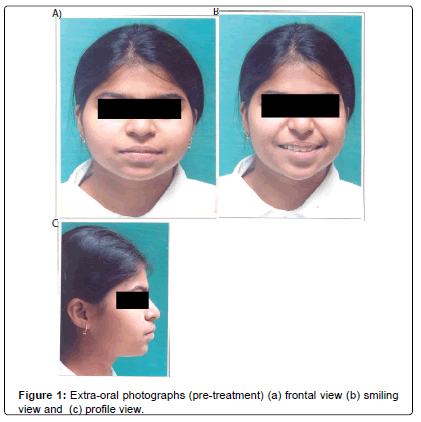
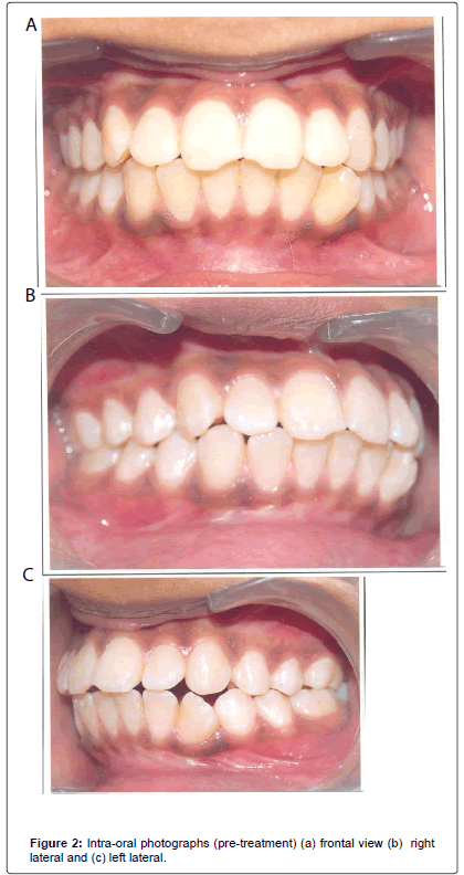
The patient, a 15-year-old female, had a convex profile and a Class I skeletal pattern with bialveolar protrusion (Figures 1 and 2). Cephalometric analysis showed an ANB angle of 4°, a mandibular plane angle (FMA) of 20° and facial angle of 88° (Table 1).
| Parameter | Pre-treatment | Post-treatment |
|---|---|---|
| SNA | 84° | 82° |
| SNB | 80° | 80° |
| ANB | 4° | 2° |
| FMA | 20° | 18° |
| PFH/AFH | 72.89% | 74.54% |
| U1-SN | 132° | 111° |
| IMPA | 118° | 100° |
| Facial angle | 88° | 85° |
| Upper lip to E | 1.5mm | -2mm |
| Lower lip to E | 4mm | 0mm |
Table 1: Cephalometric analysis.
The overjet and overbite were 5 mm and 2.5 mm respectively. There were arch-length discrepancies of 12 mm in upper arch and 10 mm in the lower arch.
The canine and molar relationships were Class I, but the maxillary incisors and mandibular incisors were proclined (U1-SN 132°, IMPA 118°).
The treatment plan called for extraction of both the maxillary and mandibular first premolars, followed by fixed appliance (Roth prescription, 0.022 slot) treatment using maxillary and mandibular micro-implants for anchorage control.
Treatment Progress
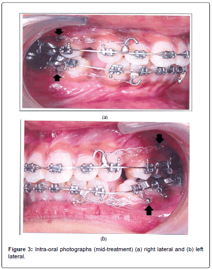
Brackets were bonded after extraction of upper and lower first premolar teeth and initial aligning and leveling of teeth was carried out in both the arches. The micro-implants (4 in number) were then inserted buccally in the interdental space between the second premolar and the first molar in both upper and lower arches. For the upper arch 8 mm long and for the lower arch 6 mm long microimplants [diameter of 1.2 mm] (Dentos Co., Taegu City, Korea) were used. Then en-masse retraction of six anterior teeth was carried out in both upper and lower arches on rectangular 19 × 25 stainless steel archwires with soldered hooks between lateral incisors and canines in each quadrant. Light forces (200 g) were used by applying power chains from the soldered hooks to the micro-implants in each quadrant for simultaneous upper and lower retraction (Figure 3).
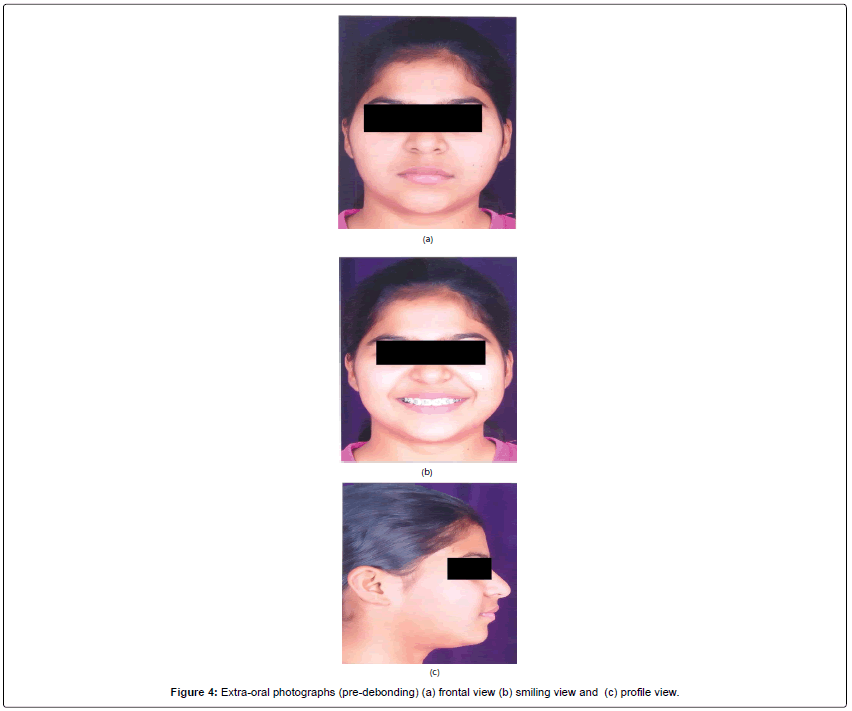
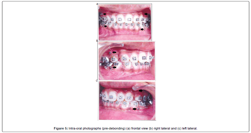
After about seven months after microscrew implantation, a Class I canine relationship had been achieved (Figures 4 and 5). Total active treatment time was 18 months.
Results
The patient showed good Class I skeletal and dental relationships after 18 months of total treatment time. The facial profile was improved with the retraction of the upper and lower lips. The ANB angle was reduced from 4° to 2°, the mandibular plane angle decreased from 20° to 18° in conjunction with the decrease in anterior facial height (Table 1). The proclined mandibular incisors were uprighted by 18° (from 118° to 100°).
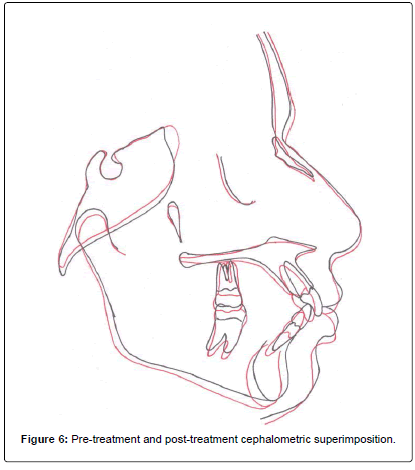
Cephalometric superimposition demonstrated a bodily retraction of the maxillary and mandibular anterior teeth. The maxillary posterior teeth moved slightly distally and showed a small amount of extrusion. The mandibular molars were uprighted and slightly intruded, causing the mandible to be rotated upward and forward (Figure 6).
Discussion
The microscrews used in this case were small enough to be implanted in the interseptal alveolar bone. To avoid any damage to the roots, however, the screws were implanted at a 30-40° angle between maxillary teeth and at a 20° angle for the mandibular teeth. Costa and colleagues confirmed that the buccal aspect of the alveolar process in the mandibular premolar and molar region is safe for miniscrew implantation [3]. There is no risk of root damage during the surgical procedure or from subsequent tooth movement.
Biomechanically, the maxillary force is applied near the center of resistance of the six anterior teeth, making it possible to achieve bodily intrusion and retraction.
Even though orthodontic force was applied just two weeks following implantation, none of the microscrews loosened during the treatment period. There is a possibility, however, of soft tissue impingement and inflammation around micro-implants. Such problems can be avoided by using a new micro-implant with a hook on its head for attaching elastics or a nickel titanium coil spring, and a smooth surface under the head where the screw contacts the soft tissue [5].
Conclusion
Micro-implant treatment has many advantages: As it does not depend on patient compliance with extraoral appliances, produces an early profile improvement giving the patient even more incentive to cooperate, shortens treatment time by retracting the six anterior teeth simultaneously and provides absolute anchorage for orthodontic tooth movement.
References
- Bae S,Park H, Kyung H, Kwon O (2002)Clinical Application of Micro-implant anchorage.J ClinOrthod36:298-302.
- Kanomi R (1997) Mini-implant for orthodontic anchorage. J ClinOrthod31:763-767.
- Costa A, Raffini M,Melsen B (1998)Microscrews as orthodontic anchorage. Int J Adult OrthodOrthogSurg 13:201-209.
- Lee JS, Park H, Kyung H. (2001)Micro-Implant Anchorage for Lingual Treatment of a Skeletal Class II Malocclusion. J ClinOrthod35:643-647.
- Sung J, Park H, Kyung H, Bae S (2001) Micro-Implant Anchorage for Treatment of Skeletal Class I Bialveolar Protrusion.J ClinOrthod 35:417-422.
Relevant Topics
- Advanced Bleeding Gums
- Advanced Receeding Gums
- Bleeding Gums
- Children’s Oral Health
- Coronal Fracture
- Dental Anestheia and Sedation
- Dental Plaque
- Dental Radiology
- Dentistry and Diabetes
- Fluoride Treatments
- Gum Cancer
- Gum Infection
- Occlusal Splint
- Oral and Maxillofacial Pathology
- Oral Hygiene
- Oral Hygiene Blogs
- Oral Hygiene Case Reports
- Oral Hygiene Practice
- Oral Leukoplakia
- Oral Microbiome
- Oral Rehydration
- Oral Surgery Special Issue
- Orthodontistry
- Periodontal Disease Management
- Periodontistry
- Root Canal Treatment
- Tele-Dentistry
Recommended Journals
Article Tools
Article Usage
- Total views: 12783
- [From(publication date):
July-2016 - Jul 02, 2025] - Breakdown by view type
- HTML page views : 11740
- PDF downloads : 1043

