Short Communication Open Access
Microglia: The Working Class Cell of the Brain
Livio Luongo* and Sabatino Maione
Department of Experimental Medicine, Division of Pharmacology, The Second University of Naples, Italy
- Corresponding Author:
- Livio Luongo
Department of Experimental Medicine
Division of Pharmacology
The Second University of Naples, Italy
Tel: +02 29520311
E-mail: livio.luongo@gmail.com
Received Date: May 27, 2016; Accepted Date: June 01, 2016; Published Date: June 08, 2016
Citation: Luongo L, Maione S (2016) Microglia: The “Working Class” Cell of the Brain. J Alzheimers Dis Parkinsonism 6:242. doi: 10.4172/2161-0460.1000242
Copyright: © 2016 Luongo L, et al. This is an open-access article distributed under the terms of the Creative Commons Attribution License, which permits unrestricted use, distribution, and reproduction in any medium, provided the original author and source are credited.
Visit for more related articles at Journal of Alzheimers Disease & Parkinsonism
The concept of microglia has been introduced by Pio del Rio- Hortega in the 1932.
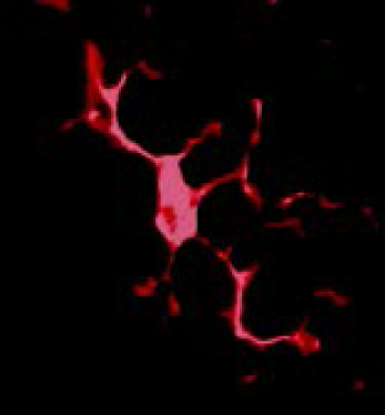
Microglias are the resident immune cells of the central nervous system (CNS). Their origin has been debated for a long time. At the beginning they were thought to become from neuroectoderm, whereas nowadays it is generally believed that they own peripheral origins and migrate into all regions of the CNS during the development and acquire a characteristic ramified shape called “resting microglia” (Figure 1) [1]. Unlike CNS macrophages found in the meninges, choroid plexus, and perivascular space, microglia derive from stem produced by primitive hematopoiesis in the yolk sac [2].
In the last decade several reports have shown the key role played by microglia cells in the brain diseases. Interestingly, these non-neuronal cells can acquire different phenotypes and perform various tasks in the brain. In particular, during the post-natal brain development microglia are in charge with the synaptic pruning necessary for the correct brain functions [3]. Thus, resting microglia is not “sleeping” cells. Indeed, it has recently become clear that microglias constantly patrol the brain environment and contact synapses. Activated microglia are able to remove damaged cells as well as dysfunctional synapses, a process defined “synaptic stripping” [4]. Therefore, it is now recognized that these cells are involved in the maintenance of CNS homeostasis.
The majority of the studies on microglia have been focused mainly on their functions in the injured brain or in neurological diseases associated with neuroinflammation including Alzheimer Disease, Parkinson Disease, neuropathic pain and others [5].
In particular, microglia can shift from a resting state to an activated pro-inflammatory (also called M1) phenotype in certain conditions. The activation process is triggered by several mediators released by other cells such as neurons, astrocytes and other in response to a peripheral or central insult. Indeed, it is possible to find the activation of microglia in several brain areas not only during CNS inflammatory conditions [5], but also in peripheral lesions or inflammatory/pathological conditions such as in the spinal cord of various form of neuropathic pain [6-9], in nucleus of solitary tract in the capsaicin-induced airway over-responsiveness [10] and in several brain areas of rodents with the myocardial infarction [11,12]. Beside this “dark side” of the microglia, that have been defined as “bad guys” in several pathological conditions [13], emerging evidence suggests also a second phenotype named alternatively activated (also called M2) microglia [14]. Interestingly, molecule that facilitates the phenotypic shift of microglia from M1 to M2 might represent potential pharmacological tools to treat chronic/ degenerative diseases. New theories highlight also the role of a third phenotype the so called “dystrophic/senescent” microglia. Intriguingly, the latest phenotype seems to be the major responsible for the neuronal damage in several pathological conditions such as Alzheimer Disease in human [15]. Recent reports also identified the presence of the dystrophic microglia in a transgenic mouse model of increased glutamatergic tone [16]. Therefore, microglia cells play several functions in the CNS in all the existing phenotypes. Indeed, several “nicknames” have been attributed to microglia such as scavengers, scapegoat, saboteur and gardeners. All these tasks justified the new appellation that we are going to attribute to microglia in this commentary: the working class cell of the brain.
References
- Kettenmann H, Hanisch UK, Noda M, Verkhratsky A (2011) Physiology of microglia. Physiol Rev 91:461-553.
- Alliot F, Godin I, Pessac B (1999)Brain Res Dev. Brain Res 117: 145-152.
- Paolicelli RC, Bolasco G, Pagani F, Maggi L, Scianni M, et al. (2011)Synaptic pruning by microglia is necessary for normal brain development. Science 333:1456-1458.
- Kettenmann H, Kirchhoff F, Verkhratsky A (2013) Microglia: New roles for the synaptic stripper. Neuron 77:10-18.
- Aguzzi A, Barres BA, Bennett ML (2013) Microglia: scapegoat, saboteur or something else? Science. 339:156-161.
- Luongo L, Sajic M, Grist J, Clark AK, Maione S, et al. (2008) Spinal changes associated with mechanical hypersensitivity in a model of Guillain-Barré syndrome. NeurosciLett 437:98-102.
- Luongo L, Palazzo E, Tambaro S, Giordano C, Gatta L, et al. (2010) 1-(2',4'-dichlorophenyl)-6-methyl-N-cyclohexylamine-1,4-dihydroindeno[1,2-c]pyrazole-3-carboxamide, a novel CB2 agonist, alleviates neuropathic pain through functional microglial changes in mice. Neurobiol Dis 37:177-185.
- Luongo L, Petrelli R, Gatta L, Giordano C, Guida F, et al. (2012) 5'-Chloro-5'-deoxy-(±)-ENBA, a potent and selective adenosine A(1) receptor agonist, alleviates neuropathic pain in mice through functional glial and microglial changes without affecting motor or cardiovascular functions. Molecules 17:13712-13726.
- Di Cesare Mannelli L, Pacini A, Corti F, Boccella S, Luongo L, et al. (2015) Antineuropathic profile of N-palmitoylethanolamine in a rat model of oxaliplatin-induced neurotoxicity. PLoS One 10: e0128080.
- Spaziano G, Luongo L, Guida F, Petrosino S, Matteis M, et al. (2015) Exposure to allergen causes changes in NTS neural activities after intratracheal capsaicin application, in endocannabinoid levels and in the glia morphology of NTS. Biomed Res Int2015:980983.
- Dworak M, Stebbing M, Kompa AR, Rana I, Krum H, et al.(2014) Attenuation of microglial and neuronal activation in the brain by ICV minocycline following myocardial infarction.AutonNeurosci185:43-50.
- Rinaldi B, Guida F, Furiano A, Donniacuo M, Luongo L, et al. (2015) Effect of prolonged moderate exercise on the changes of nonneuronal cells in early myocardial infarction. Neural Plast2015:265967.
- Watkins LR, Hutchinson MR, Ledeboer A, Wieseler-Frank J, Milligan ED, et al. (2007) Norman cousins lecture. Glia as the "bad guys": Implications for improving clinical pain control and the clinical utility of opioids. Brain BehavImmun 21:131-146.
- Stella N (2009)Endocannabinoid signaling in microglial cells. Neuropharmacology 56 Suppl 1:244-253.
- StreitWJ, Braak H, Xue QS, Bechmann I (2009) Dystrophic (senescent) rather than activated microglial cells are associated with tau pathology and likely precede neurodegeneration in Alzheimer's disease. Acta Neuropathol 118:475-485.
- Cristino L, Luongo L, Squillace M, Paolone G, Mango D, et al. (2015) d-Aspartate oxidase influences glutamatergic system homeostasis in mammalian brain. Neurobiol Aging 36:1890-1902.
Relevant Topics
- Advanced Parkinson Treatment
- Advances in Alzheimers Therapy
- Alzheimers Medicine
- Alzheimers Products & Market Analysis
- Alzheimers Symptoms
- Degenerative Disorders
- Diagnostic Alzheimer
- Parkinson
- Parkinsonism Diagnosis
- Parkinsonism Gene Therapy
- Parkinsonism Stages and Treatment
- Stem cell Treatment Parkinson
Recommended Journals
Article Tools
Article Usage
- Total views: 11195
- [From(publication date):
June-2016 - Apr 02, 2025] - Breakdown by view type
- HTML page views : 10310
- PDF downloads : 885

