Medial Patellar Plica Syndrome
Received: 09-Nov-2021 / Accepted Date: 23-Nov-2021 / Published Date: 30-Nov-2021 DOI: 10.4172/2167-7964.1000349
Abstract
Introduction: Synovial plicae are embryonic remnants of the knee that sometimes become symptomatic. Mediopatellar plica (MPP) is the most commonly symptomatic and may cause pain on the medial part of the knee. Magnetic resonance (MR) imaging and MR arthrography are useful methods in the evaluation of synovial plicae and helps in the pre-operative assessment.
Case description: We report a case of a 38-year-old man presenting a right knee pain caused by a Mediopatellar plica.
Conclusion: Synovial plicae of the knee are normal embryonic remnants which may occasionally be symptomatic. MRI is useful non-invasive tool for the diagnosis of synovial plicae, thanks to its high resolution in the knee assessment of knee pathologies. It also helps in the pre-operative assessment.
Introduction
Synovial plicae are embryonic remnants of the knee that sometimes become symptomatic, causing knee pain, which is called the plica syndrome. Common clinical symptoms such as knee pain or buckling are not specific and can lead to misdiagnosis [1].
Three types of synovial plicae are described according to the anatomical localization: Suprapatellar, infrapatellar and Mediopatellar. Medial or Mediopatellar plica (MPP) is the most commonly symptomatic, and its incidence varies between 18% and 92%. The earlier Japanese studies of Sakakibara reported rates of less than 55% [2]. MPP may cause medial pain on physical exertion in the medial part of the knee, or it may give mechanical symptoms.
The diagnosis of MPP is based on a detailed clinical examination and on specific tests. The diagnosis has previously been made only by invasive methods such as arthroscopy or arthrography. Recently MR images have been reported to yield useful information for knee assessment [3,4].
Magnetic resonance (MR) imaging and MR arthrography are useful methods in the evaluation of synovial plicae and allow differentiation of these entities from other etiologies of knee pain.
Case Description
We report case of a 38-year-old man, with no history of trauma, practicing an irregular physical activity, presented with pain and increased volume of the right knee, progressively evolving over the previous 6 months. He presented with pain on the medial surface of the patella above the joint space with a thickened and painful band on palpation. As there was no improvement under symptomatic treatment (anti-inflammatory agents), the orthopedist prescribed an MRI of the knee.
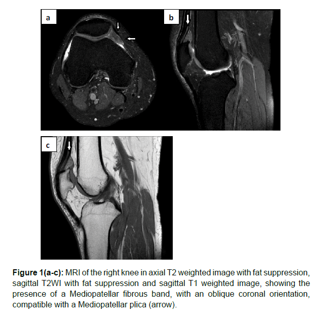
MRI of the right knee revealed a Mediopatellar fibrous band with an oblique coronal orientation, of low intensity in T1 WI and T2WI with fat suppression (Figure 1). Arthroscopic resection of the plica was indicated with a favorable outcome.
Discussion
Synovial plicae are an embryonic remnant that may be present in the joint capsule of the knee [4]. In the form of a synovial fold, thin, vascularized and has no known function [5].
The synovial plicae can be symptomatic as a result of chronic inflammation and fibrosis secondary to direct blow, repetitive sports activities, and a twisting force stretching the plica. This results in a secondary mechanical synovitis and erosion about the margins of the condyle and patellar cartilage [5].
The most commonly encountered plicae of the knee: suprapatellar, infrapatellar, and the Mediopatellar plica [6]. The Mediopatellar plica (MP) is considered the most likely to be symptomatic when it is thickened or fibrotic [5]. The Mediopatellar (MP) plica arises from the medial wall of the knee joint, has a downward orientation relative to the patella, and inserts into the synovium overlying the infrapatellar fat pad [4].
Sakakibara suggested a classification of Mediopatellar plicae based on arthroscopy [7].
• Type A: Elevation in the synovial wall
• Type B: Appear shelf-like, but not covering the anterior surface of the medial femoral condyle
• Type C: Large, shelf-like appearance and covering the anterior surface of the medial femoral condyle
• Type D: Fenestrated plica with a central defect
Clinically, Plica syndrome is defined as a painful impairment of knee function in which the only finding that helps explain the symptoms is the presence of a thickened and fibrotic plica. In Mediopatellar plica, the onset of symptoms after violent trauma to the knee is typical. The pain is located medial to the patella above the joint line. Nonspecific signs include crepitation, cracking, snapping, catching, pseudo blocking, and effusion. Clinical findings resemble those of a medial meniscus rupture or patella misalignment. A palpable, painful cord medial to the patella is almost pathognomonic for this pathologic condition [5].
Arthroscopy is the reference examination for both diagnostic and therapeutic purposes in the management of synovial plicae. MRI, being a non-invasive method of exploration, has a major interest in the detection of plicae because of high resolution and increased tissue characterization, and aid in the evaluation of pathological synovial plicae and the diagnosis of plica syndrome and the elimination of other differential diagnoses [2].
The most valuable sequences for the detection of plicae are fluid-sensitive ones including T2-weighted gradient-echo (T2*GRE) and intermediate-weighted proton density (PD) images with or without fat suppression (FS) [2]. The Mediopatellar plica can be seen as band-like structures of low signal intensity within the high-signal-intensity joint fluid and it is easily identified with some degree of joint distention [2, 5]. MR arthrography performed with fat suppression, T1 weighting, and intra-articular injection of gadolinium-based contrast material is a useful technique when there is insufficient joint fluid and a clinically significant plica is suspected. The contrast agent highlights joint surfaces and distends the capsule, allowing excellent visualization of the plicae [5].
Once the diagnosis has been made, nonsurgical treatment is always preferable initially combining rest, and non-steroidal anti-inflammatory, also, massage, cryotherapy, ultrasonography, and hamstring stretching. When symptomatic treatment fails, the indication for arthroscopic treatment arises, which consists of an excision of the plica [5].
References
- Paczesny Å, Kruczynski J (2009) Medial plica syndrome of the knee: diagnosis with dynamic sonography. Radiol 251: 439-446.
- Vassiou K, Vlychou M, Zibis A, Nikolopoulou A, Fezoulidis I, et.al  (2015) Synovial plicae of the knee joint: the role of advanced MRI. Postgrad Med J 91:35-40
- Stubbings, Nicholas, Toby Smith (2014) "Diagnostic test accuracy of clinical and radiological assessments for medial patella plica syndrome: a systematic review and meta-analysis." The Knee 21:486-490.
- Nakanishi, K, Inoue M, Ishida T, Murakami T, Tsuda K, et.al (1996) MR evaluation of Mediopatellar plica. Acta Radiologica 37:567-571.
- GarcÃa-Valtuille R, Abascal F, Cerezal L, GarcÃa-Valtuille A, Pereda T (2002) Anatomy and MR imaging appearances of synovial plicae of the knee. Radiographics 22 :775-784.
- Bellary S S, Lynch G, Housman B, Esmaeili E, Gielecki J (2012) Medial plica syndrome a review of the literature Clinical Anatomy 25:423-428.
Citation: Imrani K, Oubaddi T, Billah NM, Nassar I (2021) Medial Patellar Plica Syndrome. OMICS J Radiol 10: 349. DOI: 10.4172/2167-7964.1000349
Copyright: © 2021 Imrani K, et al. This is an open-access article distributed under the terms of the Creative Commons Attribution License, which permits unrestricted use, distribution, and reproduction in any medium, provided the original author and source are credited.
Share This Article
Open Access Journals
Article Tools
Article Usage
- Total views: 2300
- [From(publication date): 0-2021 - Apr 26, 2025]
- Breakdown by view type
- HTML page views: 1725
- PDF downloads: 575

