ISSN: 2168-9652
Biochemistry & Physiology: Open Access
Make the best use of Scientific Research and information from our 700+ peer reviewed, Open Access Journals that operates with the help of 50,000+ Editorial Board Members and esteemed reviewers and 1000+ Scientific associations in Medical, Clinical, Pharmaceutical, Engineering, Technology and Management Fields.
Meet Inspiring Speakers and Experts at our 3000+ Global Conferenceseries Events with over 600+ Conferences, 1200+ Symposiums and 1200+ Workshops on Medical, Pharma, Engineering, Science, Technology and Business

Measurement of Pancreatic Polypeptide and Its Peptide Variant in Human Serum and Plasma by Immunocapture-Liquid-Chromatography-Tandem Mass Spectrometry. Reference Intervals and Practical Assay Considerations.

Hernando Escobar1, Mark M Kushnir1,2, Julie A Ray1, Miles A Merrell1, Gwen Gomez1, Rebecca Fietkau1, Alan L Rockwood1,2* and Wayne Meikle A1,2,3
1ARUP Institute for Clinical and Experimental Pathology®, Salt Lake City, UT 84108, USA
2Department of Pathology, University of Utah School of Medicine, Salt Lake City, UT 84112, USA
3Department of Medicine, University of Utah School of Medicine, Salt Lake City, UT 84112, USA
*Corresponding Author : Alan L Rockwood, PhD
ARUP Laboratories, 500 Chipeta Way
Salt Lake City, Utah 84132, USA
Tel: 801-213-2934
Fax: 801.584.5109
E-mail: alan.rockwood@aruplab.com
Received July 25, 2014; Accepted August 14, 2014; Published August 21, 2014
Citation: Escobar H, Kushnir MM, Ray JA, Merrell MA, Gomez G, et al. (2014) Measurement of Pancreatic Polypeptide and its Peptide Variant in Human Serum and Plasma by Immunocapture-Liquid-Chromatography-Tandem Mass Spectrometry. Reference Intervals and Practical Assay Considerations. Biochem Physiol 3:140. doi:10.4172/2168-9652.1000140
Copyright: © 2014 Escobar H, et al. This is an open-access article distributed under the terms of the Creative Commons Attribution License, which permits unrestricted use, distribution, and reproduction in any medium, provided the original author and source are credited.

Visit for more related articles at Biochemistry & Physiology: Open Access

Abstract

Pancreatic polypeptide (PP) is secreted by pancreatic islets after ingestion of food. Meal composition is associated with concentration of PP released in circulation. PP can be elevated in blood of patients with endocrine pancreatic tumors, supporting its use as a biomarker. We developed an Immunocapture-Liquid Chromatography- Tandem Mass Spectrometry (IC-LC-MS/MS) assay to quantify Pancreatic Polypeptide (PP) and its metabolite, PP3-36, in human serum or plasma. PP and PP3-36 were enriched from serum or plasma using immunopurification and the samples were analyzed using LC-MS/MS (AB SCIEX 5500) Total imprecision of the method was less than 20%; LOD and LOQ for PP and PP3-36 were 5 pg/mL and 10 pg/mL, respectively. Reference intervals in healthy subjects were <265 pg/mL (fasting); and 20 to 639 pg/mL (postprandial). Regression analysis for the comparison with a commercial RIA method showed poor agreement. Different types of meal increase the peptides levels differently; enhanced secretion of PP and PP3-36 was observed after meals rich in either protein and fat, or carbohydrates and fat, as compared with meals containing only carbohydrates. No relationship was observed between PP and body mass index or body fat. In summary, a direct LC-MS/MS method for determination of PP and PP3-36 in serum and plasma was developed and validated. The only endogenous truncated form of PP detected along with the intact PP in patient samples was, PP3-36, Our data suggest association of the meal type with secretion of PP; associations between concentrations of PP and body mass index or body fat percentage were not observed.

Abstract

Pancreatic polypeptide (PP) is secreted by pancreatic islets after ingestion of food. Meal composition is associated with concentration of PP released in circulation. PP can be elevated in blood of patients with endocrine pancreatic tumors, supporting its use as a biomarker. We developed an Immunocapture-Liquid Chromatography-Tandem Mass Spectrometry (IC-LC-MS/MS) assay to quantify Pancreatic Polypeptide (PP) and its metabolite, PP3-36, in human serum or plasma. PP and PP3-36 were enriched from serum or plasma using immunopurification and the samples were analyzed using LC-MS/MS (AB SCIEX 5500) Total imprecision of the method was less than 20%; LOD and LOQ for PP and PP3-36 were 5 pg/mL and 10 pg/mL, respectively. Reference intervals in healthy subjects were <265 pg/mL (fasting); and 20 to 639 pg/mL (postprandial). Regression analysis for the comparison with a commercial RIA method showed poor agreement. Different types of meal increase the peptides levels differently; enhanced secretion of PP and PP3-36 was observed after meals rich in either protein and fat, or carbohydrates and fat, as compared with meals containing only carbohydrates. No relationship was observed between PP and body mass index or body fat. In summary, a direct LC-MS/MS method for determination of PP and PP3-36 in serum and plasma was developed and validated. The only endogenous truncated form of PP detected along with the intact PP in patient samples was, PP3-36, Our data suggest association of the meal type with secretion of PP; associations between concentrations of PP and body mass index or body fat percentage were not observed.

Keywords

Pancreatic polypeptide; Peptide hormones; Tandem Mass Spectrometry; Cancer biomarker; Food intake regulation; Reference intervals

Abbreviations

LC, Liquid Chromatography; MS/MS, Tandem Mass Spectrometry; MRM, Multiple Reaction Monitoring; LLOQ, Lower Limit of Quantification; ULOQ, Upper Limit of Quantification; PPIS, Internal Standard For PP; PP, Pancreatic Polypeptide; PP2-36, N-terminus Truncated Fragment of Pancreatic Polypeptide Hormone without the Alanine Residue; PP3-36, N-terminus Truncated Fragment of PP without Alanine and Proline; PP4-36, N-terminus Truncated Fragment of PP without Alanine, Proline and Leucine; NPY, Neuropeptide Y; PYY, Peptide YY; DPP-IV, Dipeptidyl Peptidase-IV Enzyme; IR, Immunoreactive; RIA, Radioimmunoassay; BMI, Body Mass Index.

Introduction

Human Pancreatic Polypeptide (PP) is a peptide expressed mainly in endocrine F-cells of the pancreatic islets [1], and to a lesser extent diffused throughout the exocrine parenchyma [2]. PP is a member of NPY family along with Neuropeptide Y (NPY) and Peptide YY (PYY), which act in the gut�brain axis. PP contains 36 amino acids and share with NPY and PYY theU-shape folding tertiary structure [3]. Enzymes as DPP-IV and neprilysin yield truncated fragments from NPY and PYY [4,5], however there were no reports of enzymatic action on PP. Five receptors for this peptide family include Y1, Y2, Y4, Y5 and Y6 located along the gut-brain axis; PP was reported to have high affinity to Y4 and Y5 receptors [3,4]. Studies with obese human subjects suggest that PP and PYY have a direct effect on weight control, satiety, reduction of food intake, delay of gastric emptying and regulation of energy expenditure by the body [1], while conflicting results have been reported on PP concentrations in obese and slender subjects [6].

In response to food intake PP is released from PP-cells (pancreatic polypeptide producing cells in the pancreas), increasing the PP concentration in plasma [7,8]. In the gastrointestinal tract, PP inhibits gastric emptying rate, exocrine pancreatic secretion and gallbladder contraction [9]. The composition of the meal was reported to be crucial for stimulating PP secretion, with some claiming that proteins and fat increase PP concentrations in plasma [8], while others indicated only proteins influence the release of PP [10].

With elevated levels reported in diverse malignant diseases predominantly in pancreatic endocrine neoplasms, PP has been proposed as a potential tumor marker [11-13], but PP can also be elevated in other clinical conditions [14]. Tumors with only PP-secretion have been reported [15]. In patients with endocrine pancreatic tumors PP concentrations in excess of 1,000 pg/mL were found in 15-74% of patients [13].

Using immunoprecipitation Gingerich et al. [16] identified in human plasma multiple immunoreactive truncated forms of PP of clinical significance, and demonstrates a strong affinity of the N-terminus truncated fragments, PP2-36, PP3-36, and PP4-36, towards receptors extracted from dog intestine. Fragment PP5-36 cross-reacted poorly, and PP 1-35 was incapable to bind. Presence of multiple immune-reactive fragments likely leads to overestimation of bioactive PP when biological samples analyzed by RIA methods [17].

The aim of this work was to develop a sensitive and robust method for identification and quantification of intact PP and its endogenous variants in serum and plasma, evaluate method performance, determine reference intervals in healthy individuals, evaluate relationship between PP with anthropomorphic measurements, and investigate association of the PP secretion with intake of meals of different composition.

Materials and Methods

Standards, reagents and apparatus

Human PP (APLEPVYPGDNATPEQMAQYAADLRRYINMLTRPRY-NH2, ≥95%) was acquired from ANASPEC (Fremont, CA). Anti-PP polyclonal antibodies were purchased from Bachem (Torrance, CA), Phoenix Pharmaceuticals, Inc. (Burlingame, CA) and Miravista/SSI products (Indianapolis, IN). Dynabeads® M-280 Tosyl activated magnetic beads were purchased from Life Technologies (Carlsbad, CA). Sitagliptin was acquired from Biovision (San Francisco, CA) and phosphoramidon disodium salt from Sigma-Aldrich (St. Louis, MO). Serum quality control samples were pooled human serum or plasma samples depleted from PP with Anti-PP antibody. Stable isotope labeled PP was used as internal standard (PPIS): APL*EPVYPGDNATPEQMAQYAADL*RRYINML*TRPRY-NH2 (L*=[13 C6, 15 N]) presenting a mass shift of 21 Da.

The N-terminus cleaved PP (PP3-36) LEPVYPGDNATPEQMAQYAADLRRYINMLTRPRY-NH2 was used as standard. PPIS and PP3-36 were synthesized at United Peptide (Herndon, VA) acquired with a certificate of amino acid analysis and net weight per vial. All other reagents were of highest purity commercially available. Solvents were of HPLC grade, purchased from JT Baker (Phillipsburg, NJ).

Sample preparation

Detailed description of the method is provided in the Supplementary Materials. All studies using human samples were approved by the Institutional Review Board (IRB) of the University of Utah.

Sample preparation was performed on a liquid handler Janus (Perkin Elmer, Waltham, MA). Quality control samples and calibrators for PP and PP3-36 were prepared in serum depleted from PP using anti-PP antibody capture. Coupling of the antibody to beads was performed according to the manufacturerâÂ?Â?s recommendations (see section 1.5 of the Supplementary Materials). 450 μL of serum or plasma was aliquoted and mixed with 100 pg of PPIS, and 250 μL of Trizma buffer 3X (150 mM Trizma, 300 mM NaCl, NaN3 0.09%, pH 7.6) into a lobind™ 96 well plate (Eppendorf, Hauppauge, NY). 1 μg antibody (Phoenix Pharmaceuticals) linked to beads was added to samples, quality controls and calibrators, and incubated with shaking for 1 h at 4°C. Beads were washed three times with Trizma buffer 1X (50 mM Trizma, 100 mM NaCl, NaN3 0.09%, pH 7.6). Peptides were eluted with 40 Ã?µL of buffer (5% acetic acid in 0.0025% BSA); the elution was transferred to a PCR 96-well plate, and analyzed by LC-MS/MS.

Liquid chromatography-Mass spectrometry

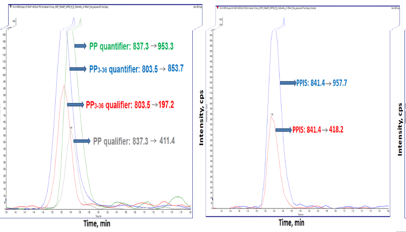
Detailed descrition of the method is provideded in section 1.1 of the Supplementary Materials. In brief, the HPLC system consisted of binary 1260 series HPLC pump (Agilent Technologies, Santa Clara, CA), and a HTC PAL auto sampler (LEAP Technologies, NC). Chromatographic separation was performed at 70°C on a Poroshell 300 SB-C18, 75 mmx2.1 mmx2.7 μm column with attached guard cartridge (Agilent, Santa Clara, CA). Mobile phases (A) 10 mM acetic acid in water and (B) 10 mM acetic acid in acetonitrile where delivered at 0.7 mL/min. Analysis was performed on an AB SCIEX TripleQuad™ 5500 Mass Spectrometer in positive ion mode using electrospray ionization (ESI) and MRM mode of acquisition (Supplementary Table 1). Data were processed using software Analyst® 1.6.1 (AB SCIEX). A parent ion was selected (Supplementary Figure 1) and two mass transitions were monitored for each compound as quantifier and qualifier respectively: m/z 837.3→953.0 and m/z 837.3→411.3 for PP; m/z 803.7→853.7 and m/z 803.7→197.2 for PP3-36 and m/z 841.3→953.0 and m/z 841.3→411.3 for the PPIS (Supplementary Table 2 and Figure 2).

Assay performance characteristics

1. Presence of the PP variants in serum samples was assessed using theoretical MRM transitions corresponding to immunoreactive PP fragments previously reported in literature: PP2-36, PP3-36 and PP4-36 [17]; in addition, mass transitions corresponding to the PP with two oxidized methionines were monitored [18].

Presence of the truncated PP was assessed using theoretical MRM transitions of the expected peptides (Supplementary Table 2) by analysis of 24 plasma samples of healthy donors (fasting and postprandial); theoretical MRM transitions were determined using Protein Prospector software v 5.12.0 (http://prospector.ucsf.edu/prospector/mshome.htm). Potential enzymatic action of DPP-IV or naprilysin during blood processing was evaluated using plasma samples of five donors by assessing degradation of PP and PP3-36 untreated and treated with sitagliptin and phosphoramidon (DPP-IV and naprilysin inhibitors, respectively), a detailed description of these enzymatic assays has been included in the Supplementary material, section 1.2.

2. Method validation experiments were performed according to The Clinical Laboratory Improvement Amendments (CLIA) guidelines. Performance of the assay was established based on evaluation of imprecision, Lower Limit Of Quantification (LLOQ), Limit Of Detection (LOD), Upper Limit Of Linearity (ULOL), recovery, carryover, ion suppression, interference, accuracy, evaluation of the anti-PP antibodies from commercial companies. Within-run, between run per day and total imprecision of the method was evaluated by analyzing triplicates of 5 samples containing 30, 50, 100, 500 and 1000 pg/mL of PP and PP3-36 repeated over a period of 5 days. Plasma pools containing 30 and 500 pg/mL of PP and PP3-36 were used as quality control samples. Pools of human plasma patient samples depleted from PP and PP3-36 using PP antibody were prepared spiking known standard concentration of PP and PP3-36 to contain 5, 10, 20, 30 and 40 pg/μL of each analyte to evaluate the LLOQ and LOD of the method analyzed over a 4 days period in three replicates each per day. The lowest concentration for which precision was within 15% and accuracies were within 20% of the expected value, was set as the lower limit of quantitation. Limit of detection was determined as the lowest concentration at which the peaks of the analytes were present in both mass transitions at the expected retention time and signal to noise ratio for the quantitative mass transition was ≥ 5. Samples for linearity test were pools of serum samples (remaining aliquots of patient samples submitted to ARUP laboratories for testing) depleted from PP using anti-peptide antibody and spiked with a defined concentration of PP and PP3-36 for seven levels total: 50, 250, 1000, 2500, 5000, 50000 pg/mL. The samples were aliquoted in tubes, stored at âÂ?Â?70°C and thawed prior to the analysis. They were analyzed in duplicates over a period of 5 days. The highest concentration at which precision was within 10% and accuracy within 20% of the expected values was considered to be the Upper Limit of Linearity (ULOL) of the method. Method recovery was determined with 11 serum samples analyzed as is and spiked with 650 pg/mL of PP and PP3-36. Samples were tested in duplicate. Difference between the observed and expected concentrations gave a measure of recovery of the sample preparation. Ion suppression was evaluated using post column infusion method [19]. A serum sample spiked with 50 pg of PP and other with PP3-36 were processed and injected into a flow of analyte with 1000 ng/mL at flow rate of 7 μL/min. Presence of potential interferences was evaluated by monitoring ratios of primary and secondary MRM transitions for PP and PP3-36; the acceptability range for the ratio was Ã?± 30% (0.7-1.3) based on comparison of the quantitative results using the two MS/MS transitions. Effects of icterus (bilirubin), hemolysis and lipemia were assessed in serum spiked with PP and PP3-36 mixed with the interfering substances: red blood cells (86 mg/L), lipids (320 mg/L) and bilirubin (173 mg/L). Method accuracy was evaluated by comparing the IC-LC-MS/MS method with a RIA [20]. Regression analysis was performed for 61 plasma samples using Deming regression. Commercial antibodies were purchased from three Vendors: Bachem (Torrance, CA); Phoenix Pharmaceuticals, Inc. (Burlingame, CA) and Miravista/SSI Products (Indianapolis, IN). Suitability of the antibodies for the method was assessed by analysis of serum samples containing 30 pg/mL of PP and PP3-36. The samples were analyzed in triplicate and concentrations were calculated based on calibration curve generated with calibration standards analyzed with the set of samples. The observed concentrations were evaluated using a one way ANOVA. The antibody incubation time was evaluated with one antibody selected by analysis of serum pooled from patients with a concentration of PP and PP3-36 at 100 pg/mL with incubation times of 1, 2, 3, 4 and 5 hours. Samples were analyzed in duplicate. The Incubation time corresponding to the maximum recovery was selected for the method. Carryover of the method was evaluated using experimental design outlined in EP Evaluator (Data Innovations, Burlington, VT). The method was evaluated by analyzing serum samples containing low (L) level (30 pg/mL) and high (H) level (100000 pg/mL) concentrations of PP and PP3-36 in the following sequence: L1, L2, L3, H1, H2, L4, H3, H4, L5, L6, L7, L8, H5, H6, L9, H7. Interferences were evaluated as described in the Supplementary material section 1.2.2

Sample stability and specimen suitability

Serum samples spiked with PP and PP3-36 were evaluated for storage stability at room temperature, refrigerator (4°C) and freezer (-20°C) after 1, 3, 7, 14, 21 and 28 days. Serum controls were stored in -70°C freezer and analyzed over 28 days. A within day evaluation was also performed for 2 h, 4 h, 8 h and 24 h at room temperature, refrigerator (4°C), and on ice (0°C). Samples were placed in -70°C freezer and analyzed in a single batch. Freeze-thaw cycles were evaluated. Blood collection tubes-type suitability evaluation was performed using potassium EDTA, lithium-heparin, serum, Serum Separator (SST) and PPACK (thrombin inhibitor) following manufacture guidelines.

Reference interval and body composition measurement

Reference intervals for PP total PPT (PP plus PP3-36) were established using serum or plasma from 118 self-reported healthy adults, ages 20-60, with no known current or past pancreatic medical conditions. Ratio of the concentrations of PP and PP3-36 in the circulation fasting and postprandial were calculated. A detailed description of the methodology is included in section 1.3.1 of the Supplementary Materials.

Association of PP concentrations with Body Fat Percentage (BF %) and Body Mass Index (BMI) was performed on 104 participants. A description of the BMI and BF% measurements methodology is described in Supplementary material, section 1.3.2

Comparison of meal composition on PP release

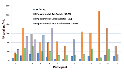
Blood samples were collected from 13 adult volunteers (ages 20-59, fasting and postprandial on three separate days). Participants did not have previous pancreatic medical condition and were advised avoid any strenuous physical activity 24 h before blood collection. Effect of the following meals was evaluated: first day rich in proteins and fat, second day rich in carbohydrates, and third day five subjects from the group consumed a meal rich in carbohydrates and fat. A detailed description of the methodology of analysis is described in the section 1.4 of the Supplementary Materials.

Results and Discussion

Results

Figure 1 shows a chromatogram with primary and secondary mass transitions of the analytes and the PPIS.

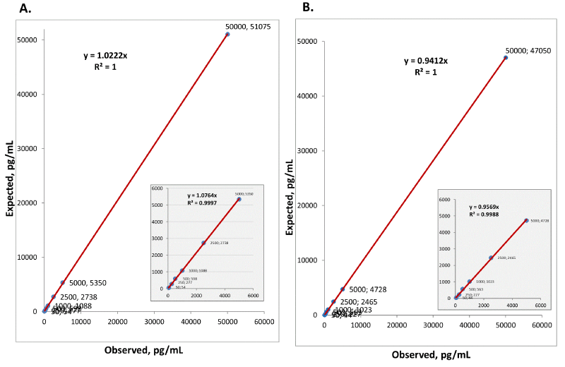
The search for the truncated fragments showed that PP3-36 was present in all analyzed samples in addition to the intact PP; while peaks of other variants were not detected (Supplementary Figure 3). The LLOQ and LOD for PP and PP3-36 was 10 and 5 pg/mL, respectively; the ULOQ was 1,000 pg/mL (Figure 2 and Supplementary Table 3). Data on the imprecision of the assay are listed in Tables 1 and 2.

We found PP and PP3-36 enrichment efficiency was comparable among three antibodies assessed for the method (Supplementary Table 5). One hour was selected as time of incubation (Supplementary Table 6). Recovery of PP was 83-98% and 85-119% for PP3-36 (Supplementary Table 7). No ion suppression was observed at retention time of both peptides.

Storage stability showed no significant degradation in samples stored frozen for a month at -20°C. Approximately 50% degradation of both peptides was observed after 24 h storage at 4°C. Complete degradation was observed after 24 h storage at room temperature. In brief, acceptable storage conditions for samples are: room temperature up to 2 h, refrigerated at 4°C for up to 8 h, on ice up to 24 h, and frozen (at -20°C or below) more than one month (Supplementary Figure 4). Controls kept at -70°C during 28 days and one freeze-thaw cycle showed no significant effect in peptide degradation.

PP and PP3-36 were stable in frozen serum and plasma samples collected in Na-Heparin, SST, PPACK or EDTA tubes (Supplementary Table 8). The results for PPACK also demonstrated no improvement in stability of PP and PP3-36.

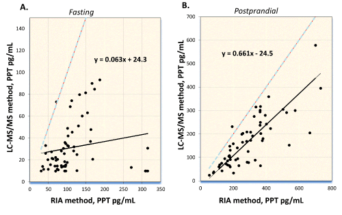
No interference was observed in samples icteric, hemolyzed and lipidemic (Supplementary Table 9). Method comparison between this IC-LC-MS/MS method and a commercial RIA method (n= 61) showed poor agreement between the methods using Deming regression (Figure 3): fasting, LC-MS/MS=0.063 RIA+24.3, R2=0.027; postprandial, LC-MS/MS=0.661 RIA � 24.5, R2=0.630.

Ratio of PPT concentrations for the same samples analyzed by RIA and LC-MS/MS ranged between 1.0 -79.0 and 0.8 - 6.0 in samples from individuals fasting and postprandial, respectively.

The nonparametric reference intervals established as central 95% are: fasting: <265 pg/mL of PPT and postprandial PPT (30 minutes after meal): 20-639 pg/mL. Supplementary Figure 5 shows chromatograms for PP and PP3-36 in a fasting healthy donor and postprandial donor, and in a negative control. The ratio PP/PP3-36 fasting was 0.08-6.45 and postprandial 0.19-2.01 (Supplementary Table 10). No differences in concentration of PP and PP 3-36 were found between collected blood samples treated and untreated with inhibitors for DPP-IV and neprilysin, enzymes involved in the fragmentation of other peptides of the NPY family (Supplementary Table 11). BMI and percent fat were calculated and compared with the PPT using a simple regression analysis (Supplementary Figure 6). Associations of PPT with BMI and body fat index in women and man were evaluated, no correlation between PPT and both indexes was observed.

Discussion

We describe a direct method for measuring intact PP in serum and plasma samples. Using theoretically predicted MRM transitions we identified in patient samples endogenous peptide PP3-36, a truncated form of PP which was present in all analyzed patient samples. The initial MS settings for PP3-36 were the same as those used for PP, once the variant was detected, a synthetic PP3-36 peptide was obtained and the conditions were reoptimized. Similar procedure was followed to detect the presence of other variants previously identified as having affinity to dog PP receptors, PP2-36 and PP4-36 [15], but the variants were not detected in plasma of healthy donors (n=48). MRM transitions used to explore those PP variants are summarized in Supplementary Table 2. Supplementary Figure 3 shows chromatograms of PP and PP3-36 in human plasma sample, while other potential variants were absent, including oxidized methionines. The PP carboxy-terminally fragment PP1-35, reported as incapable of binding to the PP receptors [14], was also not detected.

The presence of PP3-36 in blood together with the intact hormone PP1-36 follow the same expression of PYY and NPY, members of the peptide family circulating with two major forms: intact (PYY1�36 and NPY1-36) and truncated (PYY3�36 and NPY3-36). The truncated form is the product of cleavage of the amino terminus Tyr-Pro by the enzyme dipeptidyl peptidase-IV (DPP-IV) able to target proline-rich proteins and polypeptides [5,21] cleaving the peptide bond following a penultimate N-terminus proline, suggesting similar mechanism could generate the fragment PP3-36 from the intact PP hormone. We found enzyme inhibitors for DPP-IV, the most probable generator of PP3-36, and neprilysin, a metallopeptidase which cleave peptides at the amino side of hydrophilic residues did not exerts action on PP during sample processing demonstrating that the variant PP3-36 is of endogenous origin.

We used BSA solution as a blocking agent to passivate active sites in the flow path reducing peptide losses in the flow path of the HPLC; the HPLC column also required a preliminary treatment with BSA to reach an appropriate performance. Other approaches evaluated separately to reduce peptide losses in the HPLC system were addition of 5% of DMSO in mobile phases [22] and addition of 0.001% of Polyethylene glycol [23] or 0.03% CHAPS [24] in the acid elution buffer. The above approaches led to adverse results with undetectable peptides analytes. DMSO can enhance number of charges in the mass spectrometer electrospray ionization chamber [25] decreasing the selected charged precursor ion. Additional enhancement of the sensitivity was achieved through use of acetic acid (versus formic acid) as mobile phase additive modifiers (Supplementary Figure 7).

The method was linear (for both PP and PP3-36) in the range of 10 to 50000 pg/mL (Figure 2).

However the signal of PPIS at PP concentrations >1000 pg/mL was reduced (Supplementary Figure 8), suggesting a signal suppression by PP. The assay precision is acceptable for diagnostic purposes, and the LOQ afforded was six times lower than the commercial RIA method. No interference was observed in over 150 patient samples analyzed during the method evaluation.

We compared the new method with the commercial RIA (performed at ARUP laboratories [20]). The developed method quantitatively showed a poor agreement with the commercial RIA. The comparison showed extremely poor correlation for fasting donors with a bias 0.063 and R2=0.027. With the postprandial sample set agreement showed a better correlation with a proportional bias of 0.661 and R2=0.630, suggesting difference in the RIA method�s specificity in fasting and postprandial state.

A plausible explanation of this lack of correlation between methods resides in the unspecific nature of the RIA which can allow a PP overestimation caused by other analysts present in serum or plasma cross reacting with the antibody. The performance characteristics observed during the validation of the LC/MS/MS method suggest it has higher specificity compared to the RIA and would provide greater accuracy of measurements compared to immune-based methods.

As an alternative to the regression analysis we assessed agreement between the methods based on positivity and negativity rates using method-specific decision thresholds; in terms of classifying results as positive or negative based on the cutoff values of each method; the agreement between both methods was reasonably good (Supplementary Figure 9).

Meal composition results indicated that consumption of carbohydrates alone provides weak stimuli for secretion of PP, compared to the meals rich in protein and fat, or a combination of carbohydrate and fat (Figure 4).

We did not observe a relationship with release of PP with obesity [26], BMI, and body fat percentage; PPT concentrations were comparable in the fasting and postprandial samples of obese and non-obese subjects (Supplementary Figure 6).

Conclusions

In summary, we have developed a direct LC-MS/MS method for determination of PP and PP3-36 in serum and plasma using affinity immunoenrichment without enzyme digestion. The fragment PP3-36 is endogenously produced and present in circulating blood. Other earlier reported truncated forms of PP were not detected. The method has been fully validated according to CLSI guidelines and applied to the analysis of clinical samples. No relationship between PP levels with body size and adiposity was found. The results of method comparison with a RIA method show that differences exist between methodologies, emphasizing a need of standardization in PP testing. Association of PP determined by this method with pancreatic clinical conditions needs to be evaluated.

Acknowledgements

We thank ARUP Institute for Clinical and Experimental Pathology®. We also thank John Palmer (Agilent Technologies), Jesse Seegmiller and Russell Waugh (AB SCIEX). We are grateful of J. Warren Hunt and Heidi J. Carlisle for guidance in method implementation; Terry P. Vayo, Kimberly Kalp and Kisha C. Diamse for assistance with method development.


References

--

Tables and Figures at a glance

Table icon Table icon
Table 1 Table 2


Figures at a glance

Figure Figure Figure Figure
Figure 1 Figure 2 Figure 3 Figure 4
Post your comment

Share This Article

Article Usage

  • Total views: 14661
  • [From(publication date):
    December-2014 - Nov 21, 2024]
  • Breakdown by view type
  • HTML page views : 10165
  • PDF downloads : 4496
Top