Case Report Open Access
Maxillary First Molar with One C-Shaped Fused Root Diagnosed with Cone-Beam Computed Tomography: A Case Report
Xiujun Tan, Xueyang Liao, Ling Ye and Chenglin Wang*
West China Hospital of Stomatology, Sichuan University, China
- Corresponding Author:
- Chenglin Wang
State Key Laboratory of Oral Diseases
West China Hospital of Stomatology
Sichuan University, No. 14, Section 3
Renmin South Road, Chengdu, 610041, China
E-mail: wxonet@163.com
Received Date: January 28, 2016; Accepted Date: February 29, 2016; Published Date: March 08, 2016
Citation: Tan X, Liao X, Ye L, Wang C (2016) Maxillary First Molar with One C-Shaped Fused Root Diagnosed with Cone-Beam Computed Tomography: A Case Report. J Interdiscipl Med Dent Sci 3:190. doi: 10.4172/2376-032X.1000190
Copyright: © 2016 Tan X, et al. This is an open-access article distributed under the terms of the Creative Commons Attribution License, which permits unrestricted use, distribution, and reproduction in any medium, provided the original author and source are credited.
Visit for more related articles at JBR Journal of Interdisciplinary Medicine and Dental Science
Abstract
Introduction:The root canal system of maxillary first molar is complicated. It is hard to master its variations. This case report showed a rare root canal configuration diagnosed with cone-beam computed tomography (CBCT).
Methods :The dental operating microscope (DOM) and CBCT were used to search root canal orifices and confirm the rare root canal configuration.
Results: CBCT images showed the maxillary first molar having 5 root canals in one C-shaped root with fusion of the mesiobuccal, distobuccal and palatal roots.
Conclusions: The DOM and CBCT facilitated the diagnosis and treatment of a rare C-shaped fused root canal configuration in a maxillary first molar.
Keywords
Maxillary first molar; Cone-beam computed tomography; C-shape
Introduction
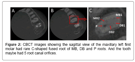
The maxillary first molar has complicated root canal system. The most common configuration has 3 roots and 3 or 4 root canals varying in the mesiobuccal root [1]. More and more variations were reported in the number of roots ranging from 1-5 and root canals ranging from 1-8 [2]. One rare anatomic configuration is C-shaped root canal. De Moor et al. [3] reported only 2 cases in 2175 root-filled maxillary first molars by evaluated radiographically, and the incidence of C-shapes in maxillary first molar was 0.091%. Cleghorn et al. [4] reviewed the literature including 2480 teeth, then found conical and C-shaped roots and canals in maxillary first molars were rarely 0.12%. Zheng et al. [5] studied the morphology of maxillary first molars in a Chinese subpopulation using cone-beam computed tomography (CBCT), and then found the incidence of fused roots was 2.71% and one fused root was 0.48%. From a few case reports [2,3,6-9] about C-shaped maxillary first molar, Martins et al. [2] found three different types of pulp chamber anatomies: Fusion between palatal (P) and distobuccal (DB) canals (type A), between mesiobuccal (MB) and DB canals (type B), and between 2 P canals (type C). Besides, Shin et al. [10] reported a maxillary first molar with an O-shaped root morphology. The purpose of this report is to present another unreported rare configuration in a maxillary first molar. CBCT scanning showed it was C-shaped fused root of the MB, DB and P roots with 5 root canals.
Case Report
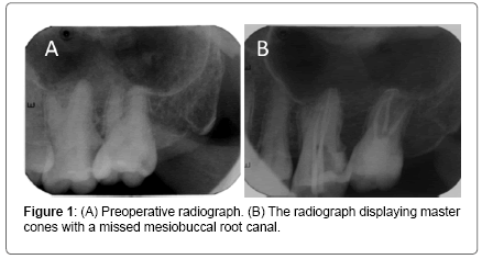
A 30-year-old male patient was ready to treat his maxillary left first and second molars. The two teeth were chronic pulpitis caused by carious lesion (Figure 1A). Root canal therapy (RCT) was proposed and accepted. The maxillary left second molar with 3 root canals was successfully processed in one-visit RCT.
One week later, the maxillary left second molar was asymptomatic. Then the maxillary left first molar was routinely instrumented 3 root canals with rubber dam and dental operating microscope (DOM) (Carl Zeiss, Jena, Germany). While the working length radiograph showed a missed mesiobuccal root canal (Figure 1B). After carefully probing under DOM, the orifice was still difficult to find, then a CBCT scan (Morita, Kyoto, Japan) of the left maxilla was performed. CBCT images showed the sagittal view of the maxillary left first molar had one rare C-shaped fused root (Figure 2A). And the tooth maybe had 5 root canal orifices with MB2 and DB2 close to P orifice (Figure 2B and C). The canals were dried and filled with calcium hydroxide (ApexCal, Ivoclar Vivadent, Schaan Liechtenstein) as an intracanal medicament for one week.
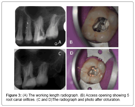
At the third appointment, with rubber dam and DOM, the pulp calcifications were thoroughly cleaned by ET ultrasonical tip (Satelec, Merignac, France), then the MB2 and DB2 root canal orifices were located following the CBCT images. Cleaning and shaping the MB2 and DB2 root canals were performed with hand and Niti rotary ProTaper files (Dentsply Maillefer, Ballaigues, Switzerland), meanwhile combined with 1.0% sodium hypochlorite and ethylenediaminetetraacetic acid (EDTA) (RC-Prep, Premier, PA, USA). The working length was determined using an apex locator (Dentaport ZX; Morita, Tokyo, Japan) and confirmed by diagnostic radiography (Figure 3A). 2% chlorhexidine gluconate was used as the final rinse solution coupled with ultrasonic agitation. The 5 canals were dried with absorbent paper points and then obturated with AH plus (Dentsply, Konstanz, Germany) and gutta-percha master cones added warm gutta-percha obturator (Suredent, Gyeonggi-do, Korea) by vertical condensation. Radiograph were taken to establish the quality of the obturation (Figure 3C), and the photo showed five orifices as same as shown in the CBCT image (Figure 3B and D).
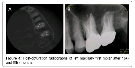
After 1 month, the teeth remained asymptomatic and CBCT showed root filling in 5 root canals (Figure 4A). At the 6-month recall examinations, the teeth had already restored with coronal restoration (Figure 4B). The prognosis was favourable.
Discussion
The C-shaped root canal system is usually referred as the root and root canal shaped like the letter C in any arbitrary cross section. Plenty of document information studied its incidence, morphology and clinical impact [11,12]. It is reported that the prevalence of C-shaped root canals in mandibular second molars was 39% in the Chinese population [13]. Although the occurrence of C-shaped root canals in the maxillary first molars is far less than it in the mandibular second molars, the basic principles are similar when clinicians encounter such cases.
The diagnosis consciousness of potential anatomic variations is essential for successful treatment. Combination of radiography and clinical examination under the microscope could encounter a higher incidence in the recognition of C-shaped canals [14]. Preoperative radiographs from different angle will contribute to identification of the root canal distribution [3]. The characteristic C-shaped roots are conical or square configuration by fused roots which may display an occlusoapical longitudinal groove on the buccal or lingual surface. Such narrow grooves may make the tooth prior to localize periodontal disease, which may be a diagnostic indication [15]. Access cavity preparation can obey the principle of orifice location. Adequate straight-line exposure of pulp-chamber floor is very important to find all of the root canal orifices, before you used ultrasonic tips to clean pulp chamber and small size files to explore root canal orifices. And some skills are advised such as looking for hemorrhagic spots, doing champagne or bubble test with sodium hypochlorite, modifying the conventional outline form to include the extra canals [16]. The connecting isthmus may be closure to the buccal or lingual side resulting in uncommon root canal orifices distribution [17]. In the present case, the DB and MB orifices are closer to the buccal, while DB2 and MB2 orifices close to P orifice. When the preoperative radiograph and above characteristic clinical examinations under the DOM still cannot make it clear, CBCT should be taken into consideration. It can provide 3-D image and help the clinicians to evaluate the root canal system at different levels [18]. The difficulty of cleaning, shaping and obturation should be the narrow structures such as isthmus, trough and fin. At first, clinicians should know root canal system in advance which would help to prevent excessive preparation the thin dentinal walls causing strip perforation [12]. Secondly, the common methods still include instruments, irrigation solutions and chemical agents. For C-shaped root canal system, the role of the latter two was emphasized. Thus in this case, hand and NiTi rotary instruments, EDTA, sodium hypochlorite and ultrasonic irrigation were combined to obtain efficacious debridement and shaping. Furthermore, calcium hydroxide paste was used as an intracanal medicament in order to further disinfection. Thirdly, obturation was done with AH plus and master gutta-percha cones added with warm gutta-percha vertical condensation.
Conclusion
The C-shaped root canal with fusion of the mesiobuccal, distobuccal and palatal roots in the maxillary first molar is a rare anatomic configuration. Knowing the diagnosis and treatment difficulty of these cases and using the DOM and CBCT can improve the treatment success.
References
- Walton R, Torabinejad M (2002) Principles and Practice of Endodontics, 3rd ed. Philadelphia: Saunders
- Martins JNR, Quaresma S, Quaresma MC, Frisbie-Teel J (2013) C-shaped maxillary permanent first molar: a case report and literature review. J Endod 39: 1649-1653.
- De Moor RJG (2002) C-shaped root canal configuration in maxillary first molars. International Endodontic Journal 35: 200-208.
- Cleghorn BM, Christie WH, Dong CCS (2006) Root and root canal morphology of the human permanent maxillary first molar: a literature review. J Endod 32: 813-821.
- Zheng QH, Wang Y, Zhou XD, Wang Q, Zheng GN et al. (2010) A cone-beam computed tomography study of maxillary first permanent molar root and canal morphology in a Chinese population. J Endod 36: 1480-1484.
- Newton CW, McDonald S (1984) A C-shaped canal configuration in a maxillary first molar. J Endod 10: 397–399.
- Dankner E, Friedman S, Stabholz A (1990) Shape configuration in maxillary first molars. J Endod 16: 601–603.
- Yilmaz Z, Tuncel B, Serper A, Calt S (2006) C-shaped root canal in a maxillary first molar:a case report. Int Endod J 39:162–166.
- Kottoor J, Velmurugan N, Ballal S, Roy A (2011) Four-rooted maxillary first molar having C-shaped palatal root canal morphology evaluated using cone-beam computerized tomography: a case report. Oral Surg Oral Med Oral Pathol Oral Radiol Endod 111: e41–45.
- Shin Y, Kim Y, Roh B D (2013) Maxillary first molar with an O-shaped root morphology: report of a case. Int J Oral Sci 5: 242-244.
- Kato A, Ziegler A, Higuchi N, Nakata K, Nakamura H, et al. (2014) Aetiology, incidence and morphology of the C-shaped root canal system and its impact on clinicalendodontics. Int Endod J 47: 1012-1033.
- Fernandes M, de Ataide I, Wagle R (2014) C-shaped root canal configuration: A review of literature. J Conserv Dent 17: 312-319.
- Zheng Q, Zhang L, Zhou X, Wang Q, Wang Y et al. (2011) C-shaped root canal system in mandibular second molars in a Chinese population evaluated by cone-beam computed tomography. Int Endod J 44: 857-862.
- Wang Y, Guo J, Yang HB, Han X, Yu Y (2012) Incidence of C-shaped root canal systems in mandibular second molars in the native Chinese population by analysis of clinical methods. Int J Oral Sci 4: 161-165.
- Melton DC, Krell KV, Fuller MW (1991) Anatomical and histological features of C-shaped canals in mandibular second molars. J Endod 17: 384-388.
- Karthikeyan K, Mahalaxmi S. New nomenclature for extra canals based on four reported cases of maxillary first molars with six canals. J Endod 36: 1073-1078
- Weine F S (1998) The C-shaped mandibular second molar: incidence and other considerations. J Endod 24: 372-375.
- Patel S (2009) New dimensions in endodontic imaging: Part 2. Cone beam computed tomography. Int Endod J 42: 463-475.
Relevant Topics
- Cementogenesis
- Coronal Fractures
- Dental Debonding
- Dental Fear
- Dental Implant
- Dental Malocclusion
- Dental Pulp Capping
- Dental Radiography
- Dental Science
- Dental Surgery
- Dental Trauma
- Dentistry
- Emergency Dental Care
- Forensic Dentistry
- Laser Dentistry
- Leukoplakia
- Occlusion
- Oral Cancer
- Oral Precancer
- Osseointegration
- Pulpotomy
- Tooth Replantation
Recommended Journals
Article Tools
Article Usage
- Total views: 12634
- [From(publication date):
April-2016 - Apr 07, 2025] - Breakdown by view type
- HTML page views : 11596
- PDF downloads : 1038


