Case Report Open Access
Management of a Large Periocular Infantile Hemangioma with Oral Propranolol and Surgical Excision: A Case Report and Review
| Michelle Y.Y. Wong1, Gordon S.K. Yau1*, Jacky W.Y. Lee1, Aaron T. K. Chu1,2, Victor T.Y. Tam1 and Can Y.F. Yuen1 | |
| 1Department of Ophthalmology, Caritas Medical Centre, Hong Kong Special Administrative Region, China | |
| 2Eye and Face Specialty Clinic, Rm. 905A, 26 Nathan Road, Tsimshatsui, Hong Kong Special Administrative Region, China | |
| Corresponding Author : | Gordon S. K. Yau Department of Ophthalmology Caritas Medical Centre 111 Wing Hong St. Kowloon, Hong Kong, China Tel: 852 34087911 Fax: 852 23070582 E-mail: skyau0303@gmail.com |
| Received June 19, 2014; Accepted July 26, 2014; Published August 02, 2014 | |
| Citation: Wong MYY, Yau GSK, Lee JWY, Chu ATK, Tam VTY, et al.(2014) Management of a Large Periocular Infantile Hemangioma with Oral Propranolol and Surgical Excision: A Case Report and Review. J Pregnancy and Child Health 1:103 doi: 10.4172/2376-127X.1000103 | |
| Copyright: © 2014 Wong MYY, et al. This is an open-access article distributed under the terms of the Creative Commons Attribution License, which permits unrestricted use, distribution, and reproduction in any medium, provided the original author and source are credited. | |
Visit for more related articles at Journal of Pregnancy and Child Health
Abstract
Hemangioma is a common vascular tumor of childhood. Management of large periocular infantile hemangiomas is challenging as it may result in deprivational amblyopia, squint and severe disfigurement. Timely and multidisciplinary management is required when complications arise. The purpose of the study was to report a case of a large periocular capillary hemangioma that was successfully treated with oral propranolol followed by surgical excision. A 2 month old girl presented with a large upper eyelid infantile capillary hemangioma complicated by dense amblyopia. The hemangioma failed to regress despite a course of oral steroid and 3 intra-lesional steroid injections. She was subsequently treated with oral propranolol followed by surgical excision of the hemangioma. At 1 year after treatment, her periocular hemangioma regressed with good cosmetic and visual outcome. Visually threatening periocular infantile hemangioma can be successfully treated with a combination of oral propranolol followed by surgical excision.
| Keywords |
| Infantile hemangioma; Intralesional steroid injection; Oral propranolol; Surgical excision |
| Abbreviations |
| PHACES syndrome: Posterior fossa malformations, Hemangiomas, Arterial anomalies, Cardiac defects, Eye abnormalities, Sternal cleft and supraumbilical raphe syndrome; PELVIS syndrome: Perineal hemangioma, External genitalia malformations, Lipomyelomeningocele, Vesicorenal abnormalities, Imperforate anus and Skin tag; MAHA: Micro-Angiopathic Hemolytic Anemia; RUL: Right Upper Lid; MRI: Magnetic Resonance Imaging; VEGF: Vascular Endothelial Growth Factor |
| Background |
| Infantile hemangioma is the most common benign vascular tumor of infancy characterized by early proliferation and subsequent spontaneous involution [1]. Infantile capillary hemangiomas affects up to 1-2% of neonates and it occurs more frequently in preterm infants [2]. More than 50% of all capillary hemangiomas occur in the head and face regions [3]. About one third of infantile hemangiomas are present at birth and the majority present by 6 months of age [4]. |
| Most capillary hemangiomas are clinically insignificant. Occasionally infantile hemangiomas may lead to significant structural abnormalities or disfigurement [5]. Large facial infantile hemangiomas can be associated with underlying congenital anomalies (PHACES syndrome) [6] while visceral hemangiomas may be associated with PELVIS syndrome [7] and systemic complications such as Kasabach- Merritt syndrome and microangiopathic hemolytic anemia (MAHA) [8,9]. |
| Periocular involvement is common and it may result in significant amblyopia in up to 60% of patients as a result of visual deprivation, anisometropic astigmatism, or strabismus [10]. The majority of capillary hemangiomas regress spontaneous and do not warrant any treatment. However, when it results in significant ocular complications, treatment should be initiated promptly. Conventional treatment modalities include intralesional steroid injections, oral steroid, pulsed-dye laser, and surgical excision. Other options include immunomodulators such as interferon alfa-2a, vincristine, and cyclophosphamide [11-13]. Christine et al. was the first to report in 2008, a successful treatment using oral propranolol. Since then, the use of oral propranolol has become the standard practice worldwide for the treatment of infantile hemangiomas [14]. Koay et al. recommended the use of a combination of oral propranolol and steroid as the first line treatment for periocular infantile hemangioma [15]. Recently, Guo et al. reported success in the use of topical non-selective beta-blocker (Timolol) in treatment of periocular hemangioma, with minimal systemic side effects [16]. |
| Case Report |
| A 2 month old baby girl was referred to our department in 2009, presenting with a progressively enlarging vascular tumour on the right upper eyelid resulting in mechanical ptosis. The lesion persisted despite a course of oral steroid for 4 weeks (prednisolone 2 mg/kg/day in 2 divided dose) as prescribed by the pediatrician. Her antenatal, birth, and family history were unremarkable. |
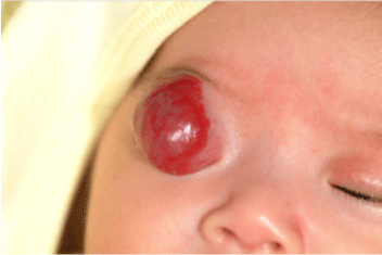
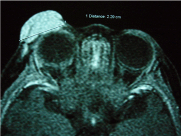
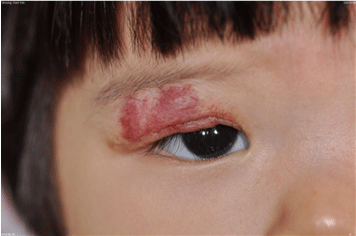
| On physical examination, there was a large capillary hemangioma over the right upper eyelid obscuring the visual axis (Figure 1). There was no obvious proptosis. The anterior and posterior segment examinations of her eyes were normal. MRI brain and orbit with contrast revealed a T2-enhanced right upper lid pre-septal mass compatible with an infantile capillary hemangioma. There was no intraorbital or intra-cranial extension (Figure 2). General examination was performed by a pediatrician. Her body weight on presentation was 6.2 kg. Her developmental milestones were up to date and there was no feature of systemic involvement including the absence of heart failure or organ dysfunctions. |
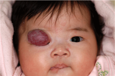
| She was initially treated with 3 intralesional steroid injections on a monthly interval (0.2 mL of 4mg/mL dexamethasone was injected superficially and 0.3 mL of 40 mg/mL triamcinolone was injected into the deeper part of the lesion). However, the hemangioma continued to grow in size (Figure 3). |
| She then received a combined course of oral propranolol (2 mg/kg/ day in 3 divided doses) and steroid (oral prednisolone 2 mg/kg/day) for 6 months. She was initially followed up daily for 3 days, weekly for 4 weeks, then bi-weekly for 5 months. |
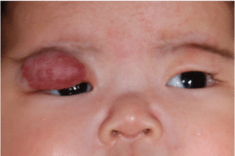
| She did not experience any side effect. At 4 weeks after the initiation of oral medications, the lesion showed significant reduction in size, it became lighter in color, and softer in consistency (Figure 4). Her ptosis improved and she was able to lift up her right upper lid spontaneously. |
| Three months later, there was still significant ptosis and deprivative amblyopia. Her visual acuity was 20/360 and 20/130 (Sheridan Garner test) over her right and left eye respectively. In view of her condition, surgical excision of the right upper eyelid hemangioma was performed. De-bulking of the vascular tumor was attempted via the transcutaneous approach under general anesthesia. Histology of the excised lesion was compatible with infantile capillary hemangioma. |
| At 3 months post-operatively, the right eyelid hemangioma was near complete resolution (Figure 5). There was no significant ptosis and the visual acuity of her right eye improved to 20/190. |
| Discussion |
| Large periocular hemangioma can result in significant mechanical ptosis of the involved eyelid, as well as inducing anisometropic astigmatism; both of which may lead to amblyopia [2,4,5]. Treatment is indicated when the lesion results in ocular or systemic complications. Several treatment modalities exist, including systemic and intralesional steroid injection, laser, or surgical excision. Other newer treatment options include oral non-selective beta-blocker or topical beta-blocker [11-14]. |
| Systemic steroid has been used since 1967 for large capillary hemangioma and may be the first-line treatment for managing large capillary hemangiomas [17]. The usual dose is oral prednisolone 1-2 mg/kg/day. Regression however is variable, and occasionally requires a long duration due to rebound growth of the tumor. The prolonged use of systemic steroid in children should be exercised with care due to its potential side effects including growth retardation, Cushing’s syndrome, adrenal insufficiency, poor wound healing, and infections. In view of the potential complications of systemic steroid, intra-lesional steroid injection is commonly used. Commonly, a few injections are needed to achieve resolution. Local complication of steroid injections including skin depigmentation, skin necrosis, fat atrophy may be encountered, and rarely, central retinal artery occlusion may occur [18,19]. |
| Systemic non-selective beta-blocker is a newer treatment modality and has revolutionized the management of capillary hemangiomas. In 2008, Christine et al. reported the rapid effects of oral propranolol in reducing the color and size of hemangiomas [14]. The underlying mechanisms are unclear, but possibly related to vasoconstriction, inhibition of stimulatory factors such as VEGF, or the induction of capillary endothelial cells apoptosis. The suggested dosage of oral propranolol is 2-3 mg/kg/day. This treatment modality has become an increasingly popular modality due to its efficacy and safety profile. Rarely, side effects of systemic beta-blockers can occur including hypotension, bradycardia, bronchospasm, and hypoglycaemia. Recently, the use of topical beta-blocker application for superficial hemangioma has been reported. Chambers et al. reported that the use of topical timolol gel 0.25% was effective in treating superficial capillary hemangiomas while reducing the systemic side effects [16,20]. |
| Laser photocoagulation has also found to be useful in superficial hemangiomas. Recent reports on pulsed dye laser has been found to be effective in improving colour, preventing further growth or inducing regression of the tumor, and achieving good cosmetic outcome [21]. It may also be effective as an adjunctive measurement to other treatment modalities. Surgical excision is often reserved for large hemangiomas that are not responsive to conservative treatments [22]. It is technically difficult to perform as the lesion is vascular and usually not encapsulated, thus bleeding may be difficult to control and intra-operative transfusion may be necessary. However, for large and persistent lesions that threaten vision despite conservative treatment, adjunctive surgical excision may be mandatory for complete regression to salvage the child’s vision. |
| Conclusion |
| We report a case of a 2 month old girl presenting with a large periocular infantile hemangioma refractory to intralesional and oral steroid. She was successfully treated by oral propranolol followed by surgical excision, resulting in good functional and cosmetic outcome. To the best of our knowledge, this was the first case report of combining oral propranolol and surgical excision for huge infantile upper lid capillary hemangiomas. |
References
- Bowers RE, Graham EA, Tomlinson KM (1960) The natural history of the strawberry nevus. Arch Dermatol 82: 667
- Amir J, Metzker A, Krikler R, Reisner SH (1986) Strawberry hemangioma in preterm infants. PediatrDermatol 3: 331-332.
- Warner M, Suen JY (1999) The natural history of hemangiomas. In: Hemangiomas and Vascular Malformations of the Head and Neck 13-45
- Drolet BA, Esterly NB, Frieden IJ (1999) Hemangiomas in children. N Engl J Med 341: 173-181.
- Callahan AB, Yoon MK (2012) Infantilehemangiomas: A review. Saudi J Ophthalmol 26: 283-291.
- Frieden IJ, Reese V, Cohen D. PHACE syndrome. The association of posterior fossa brain malformations, hemangiomas, arterial anomalies, coarctation of the aorta and cardiac defects, and eye abnormalities. Arch Dermatol. 1996;132(3):307-11.
- Girard C, Bigorre M, Guillot B, Bessis D (2006) PELVIS Syndrome. Arch Dermatol 142: 884-888.
- Hall GW (2001) Kasabach-Merritt syndrome: pathogenesis and management. Br J Haematol 112: 851-862.
- Haik BG, Karcioglu ZA, Gordon RA, Pechous BP (1994) Capillary hemangioma (infantile periocular hemangioma). SurvOphthalmol 38: 399-426.
- Stigmar G, Crawford JS, Ward CM, Thomson HG (1978) Ophthalmicsequelae of infantile hemangiomas of the eyelids and orbit. Am J Ophthalmol 85: 806-813.
- Bennett ML, Fleischer AB Jr, Chamlin SL, Frieden IJ (2001) Oral corticosteroid use is effective for cutaneous hemangiomas: an evidence-based evaluation. Arch Dermatol 137: 1208-1213.
- Ezekowitz RA, Mulliken JB, Folkman J (1992) Interferon alfa-2a therapy for life-threatening hemangiomas of infancy. N Engl J Med 326: 1456-1463.
- Frieden IJ, Haggstrom AN, Drolet BA, Mancini AJ, Friedlander SF, et al. (2005) Infantile hemangiomas: current knowledge, future directions. Proceedings of a research workshop on infantile hemangiomas, April 7-9, 2005, Bethesda, Maryland, USA. PediatrDermatol 22: 383-406.
- Léauté-Labrèze C, Dumas de la Roque E, Hubiche T, Boralevi F, Thambo JB, et al. (2008) Propranolol for severe hemangiomas of infancy. N Engl J Med 358: 2649-2651.
- Koay AC, Choo MM, Nathan AM, Omar A, Lim CT (2011) Combined low-dose oral propranolol and oral prednisolone as first-line treatment in periocular infantile hemangiomas. J OculPharmacolTher 27: 309-311.
- Guo S, Ni N (2010) Topical treatment for capillary hemangioma of the eyelid using beta-blocker solution. Arch Ophthalmol 128: 255-256.
- Zarem HA, Edgerton MT (1967) Induced resolution of cavernous hemangiomas following prednisolone therapy. PlastReconstrSurg 39: 76-83.
- Chowdri NA, Darzi MA, Fazili Z, Iqbal S (1994) Intralesional corticosteroid therapy for childhood cutaneous hemangiomas. Ann PlastSurg 33: 46-51.
- Shorr N, Seiff SR (1986) Central retinal artery occlusion associated with periocular corticosteroid injection for juvenile hemangioma. Ophthalmic Surg 17: 229-231.
- Chambers CB, Katowitz WR, Katowitz JA, Binenbaum G (2012) A controlled study of topical 0.25% timolol maleate gel for the treatment of cutaneous infantile capillary hemangiomas. OphthalPlastReconstrSurg 28: 103-106.
- Kwon SH, Choi JW, Byun SY, Kim BR, Park KC, et al. (2014) Effect of early long-pulse pulsed dye laser treatment in infantile hemangiomas. DermatolSurg 40: 405-411.
- Walker RS, Custer PL, Nerad JA (1994) Surgical excision of periorbital capillary hemangiomas. Ophthalmology 101: 1333-1340.
Figures at a glance
 |
 |
 |
 |
 |
| Figure 1 | Figure 2 | Figure 3 | Figure 4 | Figure 5 |
Relevant Topics
Recommended Journals
Article Tools
Article Usage
- Total views: 16250
- [From(publication date):
October-2014 - Dec 18, 2024] - Breakdown by view type
- HTML page views : 11827
- PDF downloads : 4423
