Make the best use of Scientific Research and information from our 700+ peer reviewed, Open Access Journals that operates with the help of 50,000+ Editorial Board Members and esteemed reviewers and 1000+ Scientific associations in Medical, Clinical, Pharmaceutical, Engineering, Technology and Management Fields.
Meet Inspiring Speakers and Experts at our 3000+ Global Conferenceseries Events with over 600+ Conferences, 1200+ Symposiums and 1200+ Workshops on Medical, Pharma, Engineering, Science, Technology and Business
Case Report Open Access
Lymphoepithelioma-like Carcinoma of the Renal Pelvis–A Case Report and Literature Review
| Liu A*, Cui W, Sun M, Wang H and Liu J | |
| Department of Radiology, First Affiliated Hospital of Dalian Medical University, China | |
| Corresponding Author : | Liu A Department of Radiology the First Affiliated Hospital of Dalian Medical University, China Tel: 818 964 6624 E-mail: cjr.liuailian@vip.163.com |
| Received July 21, 2014; Accepted August 25, 2014; Published September 02, 2014 | |
| Citation: Liu A, Cui W, Sun M, Wang H, Liu J (2014) Lymphoepithelioma-like Carcinoma of the Renal Pelvis–A Case Report and Literature Review. OMICS J Radiol 3:167. doi:10.4172/2167-7964.1000167 | |
| Copyright: © 2014 Liu A, et al. This is an open-access article distributed under the terms of the Creative Commons Attribution License, which permits unrestricted use, distribution, and reproduction in any medium, provided the original author and source are credited. | |
Visit for more related articles at Journal of Radiology
Abstract
Lymphoepithelial-like carcinoma of the renal pelvis is a rare entity. Only seven cases involving the renal pelvis have been previously reported in the world. We report the eighth case of lymphoepithelioma-like carcinoma of the renal pelvis.
| Keywords |
| Renal pelvis carcinoma; Renal pelvis; Lymphy |
| Introduction |
| The term ‘Lymphoepithelial-like carcinoma’ (LELC) is used for tumors that show morphologic similarity to lymphoepithelioma of the nasopharynx and relates to Epstein-Barr virus infection. This tumor also occurs in other parts of the body, such as parotid gland, lung, thymus, gastrointestinal tract [1-6]. However, it regards occurring at the upper urinary tract rarely, [7] only seven cases for renal pelvis have been reported. To our knowledge, this is the eighth case of lymphoepithelioma-like carcinoma of the renal pelvis. Zukerberg firstly reported 5 cases of bladder LELC in 1991 [8]. So far, 8 cases of kidney of LELC have been reported [7,9-15], except one in the kidney, another seven cases in the renal pelvis. Reports with complete CT images at present in abroad are only three [10,13,16]. |
| Case Report |
| An old man, 61-years. He was enrolled with left waist pain more than 10 years intermittently. He went to the hospital for gross hematuriaincreased 1 week. He had a history of left pyelolith. There was no tenderness in left kidney area. No recurrence has been seen for 3 months. |
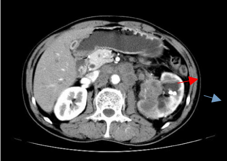
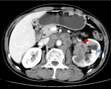
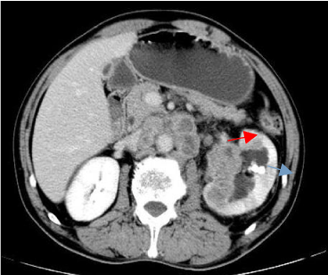
| It was demonstrated as a hypoechoic tumor with dotted blood flow signals in left ureteropelvic junction on color doppler ultrasound examination. Computerized Tomography (CT) Plain scanning demonstrated left kidney and left renal pelvis enlarged locally. There was a solid mass with clear boundary and irregular edge. Crosssectional maximum dimension is about 43.1 mm×37.2 mm. The CT value on plain scan was 23-49 HU (Figure 1). Enhanced CT revealed that mild-to-moderate enhancement in cortical phase and delayed enhancement in medulla and renal pelvis periods. Intensive degree of this lesion in three phases was lower than that of the renal parenchyma. And the CT values in three phases were 34-56 HU, 40-69 HU, 39-67 HU, respectively. There were left pyelectasis and multiple nodular high density shadow near the left pelvic. Part of abdominal aorta, whole left renal artery and vein were surrounded by the mass. Multiple nodules were found in the left renal hilum and retroperitoneal space. Part of nodules were fused, with the biggest size was 21.8 mm×18.2 mm. These nodules showed a mild peripheral enhancement in arterial phase (Figures 2-4). The right kidney is normal. Diagnosis of CT: Left renal pelvis carcinoma with left renal essence invasion and lymph nodes metastases in left renal hilum and retroperitoneal space. |
| Radical nephrectomy was performed. There was a gray solid, very brittle mass with clear boundary in the left renal pelvis involving the renal parenchyma and renal capsule. Its size was about 45 mm×30 mm×40 mm. |
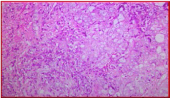
| Microscopic examination showed sheets of large polygonal tumor cells with large round or oval nuclei with large nucleoli and indistinct cytoplasmic membrane. There were large polygonal tumor cells with large undifferentiated round or spindle cell carcinoma with large nucleoli and indistinct cytoplasmic membrane. The stroma demonstrated prominent lymphoid infiltrates surrounding individual tumor cells consisting of small lymphocytes and plasma cells including T and B lymphocytes, plasma cells, histiocytes, occasionally neutrophils or acidophilus granulocyte, similar to that of nasopharyngeal lymphatic epithelial tumor carcinoma (Figure 5). Immunostaining revealed cell keratin protein 20 (CK20), Epstein-Barr virus (EBV) were negative. Cell Keratin 7 (CK7) and Carcino Embryonic Antigen (CEA) was focal positive. Pathological diagnosis: Lymphatic Epithelioma Carcinoma of the Left Renal Pelvis. |
| Discussion |
| Lymphoepithelioma usually presents as undifferentiated carcinoma of the nasopharynx. Tumors that show similar features but arise at different sites are called ‘lymphoepithelioma-like carcinoma’ (LELC). Yuta Yamada et al. [10] reported a case of kidney LELC, which was located in the middle of the left kidney and sized about 37 mm×30 mm. Epstein–Barr virus encoded RNA (EBER) in situ hybridization study was carried out using fluorescein labeled oligo probes, but failed to demonstrate the presence of Epstein-Barr virus (EBV) in the tumor or adjacent stromal lymphoid cells negative [10]. Cohen [13] reported Ultrasound demonstrated a right-sided hypoechoic mass and CT scanning confirmed a 4 cm tumor arising from the posterio-medial aspect of the right kidney. There was no hilar lymphadenopathy or renal vein involvement. The tumor extended through the pelvic muscle into the surrounding perinephric fat, but did not extend to surgical margins (Gerota’s Fascia). No evidence of metastatic disease was noted. In situ hybridization was performed for EBV (PNA, Dako) but failed to demonstrate the presence of EBV in the tumor cells or adjacent renal epithelial cells [13]. Elzevie et al. [16] reported Computerized tomography (CT) demonstrated a 3.5 cm×3 cm. tumor in the left kidney with positive lymph nodes. A tumor in the lower pole measuring 5.5 cm greatest diameter reached the renal pelvis and infiltrated the perirenal hilar fatty tissue. Immunostaining for Epstein-Barr virus was weakly positive [16]. |
| Differential Diagnosis: (1) Renal Pelvis Transitional Epithelial Carcinoma is the most frequent tumor in the pelvis. Its main symptom is painless hematuria. CT scanning demonstrates the multiple solid nodules with irregular boundary and mild enhancement in the renal pelvis. It can involves renal parenchyma or renal blood vessels in late stage and be associated with lymph node metastasis. CT signs are difficult to identify the disease. It is difficult to differentiated it from LELC rely on CT imaging only. (2) Renal Clear Cell Carcinoma in the Renal Pelvis: CT plain scan shows kidney contours locally or mass is eccentric growth. The neoplasm is more uneven obvious enhancement in cortical phase and tumor reinforcement degree decline in the periods of medulla and renal pelvis. Tumor reinforcement is significantly higher than that of normal renal parenchyma. There are liquefaction necrosis and hemorrhage, visible psuedocapsule. There are invasion in advanced renal vein and can be associated with lymph node metastasis. The characteristics of the rich of blood supply can help identify with lymphatic epithelial carcinoma cell tumor sample. Above all, the pelvic lymphatic epithelial tumor carcinoma is a rare disease, CT manifestations of renal pelvis within mass isodensity, infringement of renal parenchyma, mild-to-moderate CT tumor enhancement is associated with lymph node metastasis. But its confirmed diagnosis depends on the pathological morphology and immunohistochemical diagnosis. Our case and previous reports shown in Table 1 suggest that we must be aware of the presence of this entity even when patients are associated with tumors at the renal pelvis. |
| Conclusion |
| LELC of the renal pelvis is a very rare disease. Recognizing it is important for diagnostic implications since only a few cases have been reported. More reports of LELC of the renal pelvis are needed to evaluate the character of this entity. |
References |
|
Tables and Figures at a glance
| Table 1 |
Figures at a glance
 |
 |
 |
 |
 |
| Figure 1 | Figure 2 | Figure 3 | Figure 4 | Figure 5 |
Post your comment
Relevant Topics
- Abdominal Radiology
- AI in Radiology
- Breast Imaging
- Cardiovascular Radiology
- Chest Radiology
- Clinical Radiology
- CT Imaging
- Diagnostic Radiology
- Emergency Radiology
- Fluoroscopy Radiology
- General Radiology
- Genitourinary Radiology
- Interventional Radiology Techniques
- Mammography
- Minimal Invasive surgery
- Musculoskeletal Radiology
- Neuroradiology
- Neuroradiology Advances
- Oral and Maxillofacial Radiology
- Radiography
- Radiology Imaging
- Surgical Radiology
- Tele Radiology
- Therapeutic Radiology
Recommended Journals
Article Tools
Article Usage
- Total views: 13743
- [From(publication date):
September-2014 - Jul 19, 2025] - Breakdown by view type
- HTML page views : 9154
- PDF downloads : 4589
