Lymphangioma in Childhood Case Report and Review of Literature
Received: 04-Mar-2021 / Accepted Date: 18-Mar-2021 / Published Date: 25-Mar-2021 DOI: 10.4172/jmir.s1.1000003
Introduction
Lymphangioma colli is relatively uncommon malformation that can cause a variety of complications depending on its precise location, type, and size. The most commonly used classification divides these lesions into 3 main Lymphatic malformations which occur in 1:2'000 -4'000 live births [1]. Cervical macrocystic lymphatic malformations are also called cystic hygromas. Predominantly, they are located in the neck and axillary regions (95 %) and are found in the skin, mucosa, soft tissue and rarely in internal organs [2,3]. Lymphangiomas are mostly congenital malformations resulting from erroneous embryogenesis [4].They can also develop after lymphatic obstruction, inflammation or trauma. These acquired forms are much less common in children and are not discussed further in this article.
Lymphangiomas are malformations of the lymphatic system characterized by lesions that are cysts with thin walls [1,2]. These cysts can be macroscopic, as in cystic hygroma, or microscopic. Lymphangiomas are rare, hamartomatous, congenital malformations of the lymphatic system that involve the skin and subcutaneous tissues. The classification of lymphangiomas does not have a standard clear definition and universal application, in part because of the nature of the lymphangiomas that constitute the clinicopathological continuum.
Discussion
Groups based on the depth and size of these abnormal lymphatic vessels
Lymphangioma circumscriptum - the common form of cutaneous lymphangioma, is characterized by persistent, numerous clumps of translucent vesicles that usually contain clear lymphatic fluid. These vesicles represent superficial sac-like extensions of the underlying lymphatic vessels that occupy the papillae and exert pressure upward against the proper epidermis. These vesicles may be translucent or range from pink to dark red due to hemorrhage. These vesicles are often associated with vertical changes that give them a warty appearance.
Cavernous lymphangioma - usually present at birth, but can also occur later in the baby's life. These protuberant masses occur deep under the skin, usually on the neck, tongue and lips, and range in size, ranging from one to several centimeters centimeter in diameter. In some cases, they can affect the entire limb. These lesions are found deep in the dermis, resulting in painless swelling or thickening of the skin, mucous membranes and subcutaneous tissue.
Cystic hygroma shares many common features with cavernous lymphangiomas. Many authors agree that cystic hygroma is a form of cavernous lymphangioma, in which the degree of involvement and character is determined by its location [3-5]. These congenital lesions are located deep within the areola or loose connective tissue areas. They appear early in life as large masses of soft tissue, usually in the axilla, neck, or groin. These lesions are soft, varying in size and shape, and tend to grow extensively if not excised. Typical lesions are multilocular cysts filled with clear or yellow lymphatic fluid.
These malformations can occur at any age and can include any part of the body, but 90% occur in children under 2 years of age and include the head and neck. These malformations are either congenital or acquired. Congenital lymphangiomas are often associated with chromosomal abnormalities such as Turner's syndrome, although they may also exist in isolation. Lymphangiomas are usually diagnosed before birth with an ultrasound examination of the fetus. Acquired lymphangiomas can be the result of trauma, inflammation or lymphatic obstruction.
The direct cause of lymphangioma is a blockage of the lymphatic system during pre-natal development, although the symptoms can become visible only after the baby is born. The reason remains unknown. Why embryonic lymphatic sacs remain unconnected with the rest of the lymphatic system is also unknown.
A thick layer of muscle fibers that cause rhythmic contractions line the isolated primitive sacs. Rhythmic contractions increase internal pressure, leading to enlarged ducts. Surveys [6] support Wister’s observations. Said studies reveal that large cisterns extend deep into the skin and beyond clinical lesions. Lymphangiomas that are deep in the dermis show no communication with the lymphatic vessels.
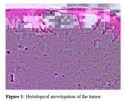
Microscopically, vesicles in lymphangioma circumscriptum are much-enlarged lymphatic channels that cause enlargement of the papillary dermis (Figure 1). They may be associated with acanthosis and hyperkeratosis. There are many channels at the top of the dermis that often extend to the subcutaneous area. The deeper vessels have large, thick-walled calibers that contain smooth muscle. The lumen is filled with lymph fluid, but often contains red blood cells, lymphocytes, macrophages and neutrophils. The channels are lined with flat endothelial cells. The interstitium has many lymphoid cells and shows evidence of fibroplasia (formation of fibrous tissue).
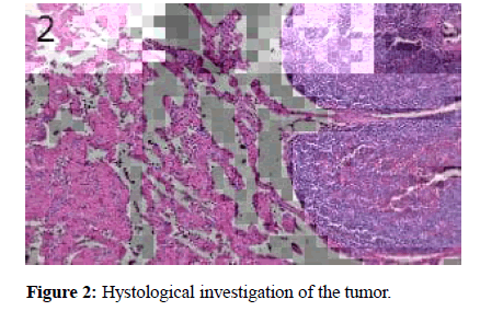
Nodules in cavernous lymphangiomas are characterized by large, irregular channels in the reticular dermis and subcutaneous tissue that are lined with a single layer of endothelial cells (Figure 2). The incomplete layer of smooth muscle often lines the walls of these irregularly shaped channels. The surrounding stroma consists of free or fibrous connective tissue with a number of inflammatory cells. These tumors often penetrate the muscles.
Cystic lymphangioma, which occurs in the first three months of pregnancy, is associated with genetic conditions such as Noonan syndrome and trisomy 13, 18 and 21. Chromosomal aneuploidy such as Turner syndrome or Down syndrome is found in 40% of cases of cystic hygroma.
In 1976, Wimster studied the pathogenesis of circumscriptum lymphangiomas, finding that lymphatic cisterns in the deep subcutaneous plane are separated from the normal network of lymphatic vessels. They communicate with the superficial lymphatic vesicles through vertical, enlarged lymphatic channels. Cystic hygroma is no different from cavernous lymphangiomas in histology.
Most lymphangiomas are benign lesions that lead only to a soft, slowly growing mass. Because they have no chance of becoming malignant, lymphangiomas are usually treated for cosmetic reasons only. Rarely, entry into critical organs can lead to complications, such as respiratory distress, when lymphangiomas compress the airways. Treatment includes aspiration, surgical excision, laser and radiofrequency ablation and sclerotherapy.
Case Study
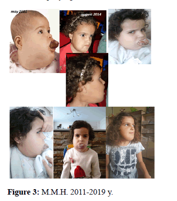
We present hereby case of congenital lymphangioma of the face, floor of the mouth, throat and tongue, hyperplasia of the gums and Frey's Syndrome. M.M.H is a girl, born in 2010. The diagnosis was made at birth and treatment began at the age of 7 months at the Pediatric surgery Clinic in Austria with immunosuppressant, and gradually the size of the lymphangioma began to decrease in size. At age 5, lymphangioma restarts with extreme growth and cannot be retracted into the child's mouth, which results in partial resection of the tongue - at the tip and base of the tongue. After 4 months, a new resection of the lymphangioma on the right is made - from the cheek to the neck. After another 4 months, a new operation was performed with resection of the lymphangioma under the chin and left with Bleomycin injection into the tongue. Following injection of Bleomycin into the residual lymphangioma and tongue, laser treatment of gum hypertrophy was performed in the right jaw area.
Beard plastic is performed at the age of 8 with bone and volume reduction of lymphangioma. The last procedure so far in September 2018 is sclerosis of the residual malformation of the cheek and tip of the tongue with Bleomycin. In April 2019 (Figure 3), a new sclerotherapy is coming after sclerotherapy of the right parotid area and the left corner of the jaw. In common viral and bacterial infections, the lymph nodes respond and the lymphangioma increases in size. The objective state beyond described indicates a normal size of the thyroid gland. The status of the chest and abdomen is normal, without accompanying abnormalities [6-8].
Conclusion
Diagnosis of lymphangioma is not difficult. The clinical picture is quite characteristic, nevertheless, lymphography is performed to clarify the diagnosis, and more often to clarify the anatomical location options. US and CT or MRI must be used for diagnosis.
Differential diagnosis of lymphangioma is performed with brachyogenous cysts of the neck, cysts from the remains of the thyroid-sublingual duct, dermoids, spinal hernias, lipomas, teratomas, and neck lymphadenitis. A close examination of the patient helps to distinguish between these diseases. The cyst puncture is followed by active aspiration and sclerotherapy based on two interconnected processes. Firstly, it allows achieving adhesion of the walls of the cyst followed by their scar lowering, and decreasing lymph production; secondly, the resulting lymph is immediately evacuated, without violence cyst.
Surgical treatment is carried out by the abatement of the inflammatory process. Surgical interventions for lymphangiomas can be very long and difficult. In cases of absence of urgent indications it is therefore advisable for the children to be operated after the first six months of their life.
References
- Defnet AM, Bagrodia N, Hernandez SL, Gwilliam N, Kandel JJ. (2016) Pediatric lymphatic malformations: evolving understanding and therapeutic options. Pediatr Surg Int 32: 425-433.
- Levy AD, Cantisani V, Miettinen M. (2004) Abdominal lymphangiomas: imaging features with pathologic correlation. Pictorial essay. AJR Am J Roentgenol 182:1485-1491.
- Lugo-Oliveieri CH, Taylor GA. (1993) CT differentiation of large abdominal lymphangioma from ascites. Pediatr Radiol 23:129-130.
- Rautio R, Keski-Kisula L, Laranne J, Laasonen E. (2003) Treatment of lymphangiomas with OK-432 (Picibanil). Cardiovasc Intervent Radiol 26:31-33.
- Harrison MR, Adzick NS, Flake AW, Vanderwall KJ, Bealer JF, Howell LJ, et al. (2015) Correction of congenital diaphragmatic hernia in utero VIII: Response of the hypoplastic lung to tracheal occlusion. J Pediatr Surg 31:1339-1348.
- Olutoye OO, Olutoye OA. (2012) EXIT procedure for fetal neck masses. Curr Opin Pediatr 24:386-393.
- Balakrishnan K, Menezes MD, Chen BS, Magit AE, Perkins JA. (2014) Primary surgery vs primary sclerotherapy for head and neck lymphatic malformations. JAMA Otolaryngol Head Neck Surg 140:41-45.
- Poddubnyi IV, Ryabov AB, Abramyan MA, Trunov VO, Kozlov MY, et al. (2016) Surgical Treatment of Lymphangiom in Children: Case Series. Oncopediatrics. 6:53-64.
Citation: Kalinova K, Georgiev KI (2021) Lymphangioma in Childhood Case Report and Review of Literature. J Mucosal Immunol Res S1: 003. DOI: 10.4172/jmir.s1.1000003
Copyright: © 2021 Kalinova K, et al. This is an open-access article distributed under the terms of the Creative Commons Attribution License, which permits unrestricted use, distribution, and reproduction in any medium, provided the original author and source are credited.
Select your language of interest to view the total content in your interested language
Share This Article
Recommended Journals
Open Access Journals
Article Tools
Article Usage
- Total views: 2792
- [From(publication date): 0-2021 - Dec 03, 2025]
- Breakdown by view type
- HTML page views: 1903
- PDF downloads: 889


