Lumbar Radiculopathy Caused by Degenerative Osteoarthritis and Epidural Lipoma
Received: 08-Dec-2018 / Accepted Date: 05-Jan-2019 / Published Date: 14-Jan-2019 DOI: 10.4172/2167-0846.1000337
Abstract
The patient we report is a 59-year-old male with lumbar radiculopathy due to epidural mass lipoma associated with unilateral posterior facet joint ostheoarthritis unresponsive to conservative treatment. Elective surgical intervention with right laminectomy, facetectomy, and foraminotomy for resection of the mass was performed. An encapsulated lipomatous lesion was excised. Pathohistological findings revealed true lipomatous tissue. Our case report increase suspicion for this rare spinal pathology on patients with lumbar radiculopathy and epidural mass diagnosed by MRI and histopathology. We strongly suggest operative treatment for this kind of symptomatic spinal epidural lesions.
Keywords: Degenerative osteoarthritis; Lumbar radiculopathy; Lipoma
Introduction
An extradural spinal lipoma is an extremely rare pathological entity. Usually it goes with myelospinal dysraphism or with history of endocrinopathy or corticosteroid use in obese patients [1,2]. Predilection site is thoracic spine and it can cause myelopathy. Facet osteoarthritis with fibrolipoma as epidural mass in lumbosacral region is extremely rare. According to published articles, there exists only one case report on solitary epidural lipoma with osteoarthritis in the literature [3]. The gold standard in diagnostic imaging for lumbar compression neuropathy is magnetic resonance imaging (MRI). MRI of a spinal epidural lesion will reveal hyperintensity on T1-weighted sequences and decreased to intermediate intensity on T2-weighted sequences.
Here we report a rare case of 59-year-old male with lumbar radiculopathy caused by epidural mass lipoma associated with unilateral ostheoarthritis of the posterior facet joint.
Case Report
History and examination
Our patient is a 59-year-old male, presented to our practice concerning refractory left L5 radiculopathy manifesting as persistent weakness, paresthesia, and severe radiating pain. The pain was refractory to 3 months of conservative medical. On examination, he was not obese. Severe limitation in straight leg raise test and motor weakness and paresthesia in L5 dermatome were observed on examination. Plain radiographs showed no evidence of instability and other pathologic findings.
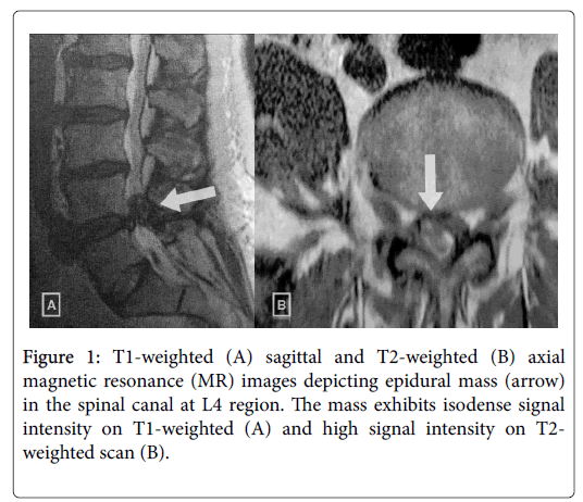
An epidural mass posterior to the L4 vertebral body that was isosignal to subcutaneous fat was revealed on magnetic resonance imaging (MRI) expressing compression asymmetrically on the right side of the cauda equine and the exiting left L5 nerve root on sagittal T1 and axial T2 weighted images (Figure 1). Degenerative arthritis of the right facet joint was also seen. Leg pain (measured by visual analogue scale [VAS] showed no response to physiotherapy and conservative therapy. Patient was highly motivated for surgical intervention to relieve pain.
Operation
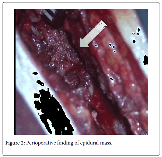
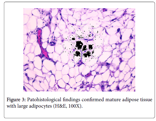
Elective right laminectomy, medial facetectomy, foraminotomy, and resection of the epidural mass were performed. The lesion was excised, and interval decompression of the spinal canal and nerve root was achieved. Gross examination of the lesion revealed a lobulated epidural mass encapsulated by a thin layer of brows tissue (Figure 2). Histopathology showed no evidence of vascular atypia and was consistent with a true adult extradural lipoma with degenerative osteoarthritis (Figure 3).
Postoperative course
There were no postoperative complications and the physical procedure was tolerated well by the patient. Two months after operation, there was a reduction in his low-back pain, right leg pain and paresthesia. Postoperatively, patient reported significant pain relief.
Discussion
Radicular symptoms of lumbar spine can be caused by pathologies such as disc herniation, spinal canal stenosis, or spinal tumors [4]. The possibility of lipomatous tissue in epidural space with mass effect should not be overlooked. They account for 0.4% of all spinal tumors when they are not associated with spinal dysraphism, obesity, chronic steroid treatment, or endocrinopathies [5]. Literature overview showed two case reports on solitary epidural lipoma. One of the cases describes a fall experienced by an obese (BMI 25 kg/m2), elderly female with osteoporosis that was considered to suffering of Paget's disease of the spine who experienced coincidental unilateral L5 lamina fracture. The second case described a patient with spontaneous onset of long lasting of symptoms. No one of these two case reports is associated with osteoarthritis. We found only one article of obese 55-years-old man suffering from radiculopathy and weakness of peroneal muscles caused by solitary epidural lipoma with facet arthritis.
Symptoms of epidural lipoma vary with his location. As they are slow growing lesions, clinical presentation usually includes gradually progressing symptoms, such as back pain or radiculopathy. Rarely this condition is associated with rapidly deteriorating neurologic deficits [5,6].
Our patient presented with refractory L5 radiculopathy manifesting as persistent peroneal weakness and paresthesia. Best to our knowledge it is first case with patient that was not obese and without history of endocrinopathy or corticosteroid use. We suggest that spinal epidural lipoma can be present also in people who are not obese. For better overview of bony architecture additional computed tomography (CT) scans can be used, but radiologic assessments mostly depends on magnetic resonance imaging (MRI). The gold standard in diagnostic imaging for lumbar compression neuropathy is MRI. MRI of a spinal epidural lipomatous lesion will reveal hyperintensity on T1-weighted sequences and decreased to intermediate intensity on T2-weighted sequences. MRI revealed an epidural mass posterior to the L4 vertebral body, compressing cauda equine and the exiting left L5 nerve root on sagittal T1 and axial T2 weighted images, isosignal with fat tissue.
We did not wanted to do CT of lumbar spine because it was already clear that with neurological and MRI finding there is an indication for operation, and we dint want to irradiate the patient because CT bring irreversible irradiation.
Depending on the exhibited symptoms and extent of neurologic deficit, treatment of spinal epidural lesions can be conservative or operative. Operative treatment is indicated in patients manifesting progressive neurological deficit or bladder dysfunction. Three previously reported cases of epidural lipoma were managed with surgical intervention, as all of them expressed progressive pain symptoms and neurologic deficit [1-3]. Our patient’s Initial symptoms were not relieved by physiotherapy and opioid analgesia. Severe radiculopathy followed by muscle weakness persisted for two months, before he underwent decompressive surgery. It is taught that facet joint degeneration causes abnormal movement between spinal segments and facet pain. In our opinion solitary epidural lipoma causing neurologic deficit should be excised at the time of diagnosis. The focus of our case report is solitary epidural lipoma associated with ipsilateral posterior facet joints degenerative arthritis.
Conclusions
Our case report increase suspicion for this rare spinal pathology on patients with lumbar radiculopathy and epidural mass diagnosed by MRI and histopathology. Histopathology is essential for exact diagnosis. Because of rarity of this entity, there is lack of definitive treatment guidelines. We strongly suggest operative treatment for this kind of symptomatic spinal epidural lesions.
References
- Schizas C, Ballesteros C, Roy P (2003) Cauda equina compression after trauma: An unusual presentation of spinal epidural lipoma. Spine 28: 148-151.
- Zevgaridis D, Nanassis K, Zaramboukas T (2008) Lumbar nerve root compression due to extradural, intraforaminal lipoma. J Neurosurg Spine 9: 408-10.
- Kim HK, Koh SH, Chung KJ (2012) Solitary epidural lipoma with ipsilateral facet arthritis causing lumbar radiculopathy. Asian Spine J 6: 203-206.
- Markovic M, Zivkovic N, Spaic M, Gavrilovic A, Stojanovic D, et al. (2016) Full-endoscopic interlaminar operations in lumbar compressive lesions surgery: Prospective study of 350 patients. J Neurosurg Sci 6: 1-15.
- Loriaux DB, Adogwa O, Gottfried ON (2015) Radiculopathy in the setting of lumbar nerve root compression due to an extradural intraforaminal lipoma: A report of 3 cases. J Neurosurg Spine 23: 55-58.
- López-González A, Giner RM (2008) Idiopathic spinal epidural lipomatosis: urgent decompression in an atypical case. Eur Spine J 2: 225-227.
Citation: Zivkovic N, Repac N, Janicijevic A, Nikolic I, Djurovic B (2019) Lumbar Radiculopathy Caused by Degenerative Osteoarthritis and Epidural Lipoma. J Pain Relief 8:337. DOI: 10.4172/2167-0846.1000337
Copyright: © 2019 Zivkovic N, et al. This is an open-access article distributed under the terms of the Creative Commons Attribution License, which permits unrestricted use, distribution, and reproduction in any medium, provided the original author and source are credited.
Share This Article
Recommended Conferences
42nd Global Conference on Nursing Care & Patient Safety
Toronto, CanadaRecommended Journals
Open Access Journals
Article Tools
Article Usage
- Total views: 5114
- [From(publication date): 0-2019 - Apr 25, 2025]
- Breakdown by view type
- HTML page views: 4274
- PDF downloads: 840


