Lown-Ganong-Levine (LGL) Syndrome in a 4 Years Old Girl with Abnormal Tricuspid Valve, Tricuspid Valve Regurgitation and Right Sided Heart Dilatation
Received: 17-Aug-2018 / Accepted Date: 29-Aug-2018 / Published Date: 06-Sep-2018 DOI: 10.4172/2572-4983.1000164
Keywords: Lown-Ganong-Levine (LGL) syndrome; Supraventricular tachycardia; Tricuspid valve
Introduction
LGL syndrome, includes a short PR interval, normal QRS complex, and paroxysmal tachycardia. The pathophysiology of this syndrome includes an accessory pathway connecting the atria and the atrioventricular (AV) node (James fiber), or between the atria and the His bundle (Brechenmacher fiber) [1]. A short A-V node refractory period and enhanced A-V conduction also has been suggested as a cause of this syndrome [2]. Other possible underlying mechanism is atrial myocarditis with possible causation of sudden death. [3] There are few case reports describing this syndrome in association with structural or functional heart disease [3-6]. Herein, we are reporting a case of LGL syndrome in a four years old girl with structurally and functionally abnormal tricuspid valve; to the best of our knowledge this is the first case in the literature describing such combination.
Case Report
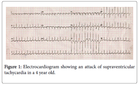
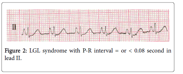
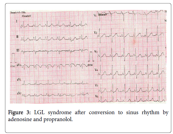
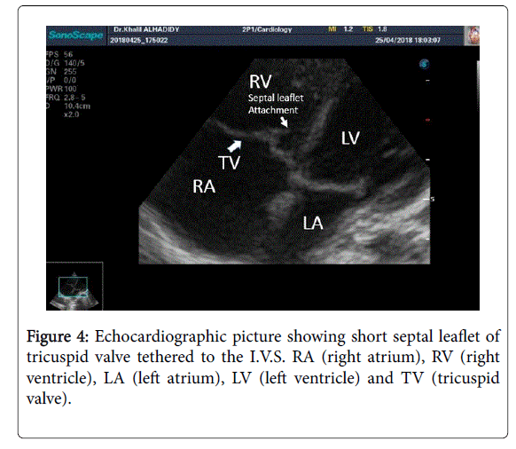
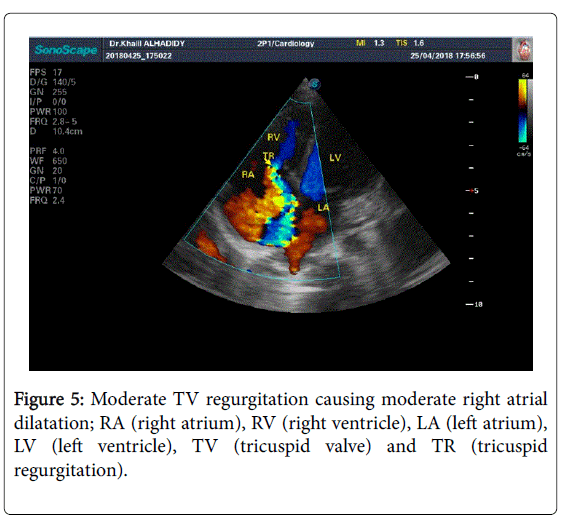
A 4 year old girl was presented to the author’s institute (Ibn ALAtheer pediatrics hospital in Mosul) with sudden onset of breathlessness, chest discomfort and palpitation for the first time. On physical examination she was frightened and irritable; her pulse rate was around 250 beats/minute; her blood pressure was 110/60 mmHg and there was grade 2-3/6 systolic murmur heard over the left lower sternal border on precordial auscultation. An electrocardiogram was obtained and revealed an attack of supraventricular tachycardia (SVT) (Figure 1). After a few trials of intravenous adenosine and beta adrenergic blocking drug, propranolol, the rhythm converted to sinus one but with PR interval shorter than the normal for the child age according to Davignon table (7] (Figures 2 and 3). An echocardiography examination was performed to evaluate the cardiac structure and functions and surprisingly we found abnormal structure of tricuspid valve (TV) with a septal leaflet that was tethered directly to the interventricular septum (I.V.S) without any visible septal papillary muscle (Figure 4). The tricuspid valve was not closing perfectly during systolic contraction causing tricuspid valve regurgitation of moderate severity with peak pressure gradient between right ventricle and right atrium of 35 mmHg which in turn caused mild to moderate right side dilatation (Figure 5).
Discussion
The tricuspid valve is the right-sided atrioventricular valve, and it is composed of three leaflets (i.e., anterior, posterior, and septal) attached to a fibrous annulus. In spite of the documented variations in the papillary muscles of this valve; the septal leaflet is the least likely one to be affected by the abnormalities of these papillary muscles and their related chordae tendineae. Echocardiography remains the best method of evaluation of TV structure and function [8,9].
Tricuspid regurgitation is functional and is a satellite of left-sided heart disease and/or elevated pulmonary artery pressure most of the time; and when progressive, it worsens the patient prognosis whatever the underlying etiology [10].
The gross cardiac structure is generally normal in patients with LGL syndrome and the problem is basically related to the structure and /or function of the cardiac conductive system [1,2]; however atrial myocarditis, isolated non compaction of the left ventricle, mitral valve prolapse and Fabry’s disease all have been reported in association with LGL syndrome without clear understanding of the role that these disorders may play in this syndrome [3-6].
The interesting point in our case is the finding of right side disease in association with LGL syndrome which has not been reported before; a point that should be considered in the future evaluation of LGL syndrome patients. Probably the main drawback in our case is the absence of electrophysiological study which, if were done, it could explain a new idea about the pathophysiology of this syndrome.
Conclusion
We conclude that the abnormalities of signal conduction described in association with LGL syndrome is probably liable to be affected by some cardiac structural disease and interestingly we are reporting right side heart disease in such problem.
References
- Hunter J, Tsounias E, Cogan J, Young M-L (2018) A Case of Lown-Ganong-Levine Syndrome: Due to an Accessory Pathway of James Fibers or Enhanced Atrioventricular Nodal Conduction (EAVNC). Am J Case Rep 19: 309-313.
- Benditt DG, Pritchett LC, Smith WM, Wallace AG, Gallagher JJ (1978) Characteristics of Atrioventricular Conduction and the Spectrum of Arrhythmias in Lown-Ganong-Levine Syndrome. Circulation 57: 454-465.
- Basso c, Corrado D, Rossi L and Thiene G (2001) Ventricular Preexcitation in Children and Young Adults: Atrial Myocarditis as a Possible Trigger of Sudden Death. Circulation 103: 269-275.
- Chen WW, Wong PH, Chow JS (1982) Mitral valve prolapse and pre-excitation. Pacing Clin Electrophysiol 5: 773-775.
- Shabanian R, Kiani A, Rad EM, Eslamiyeh H (2009) Lown-Ganong-Levine syndrome in a 3-month-old infant with isolated left ventricular noncompaction. Pediatr Cardiol 31: 274-276.
- Undas A , Rys D, Wegrzyn W, Musial J (2002) Atypical symptoms of Fabry's disease: sudden bilateral deafness, lymphedema and Lown-Ganong-Levine syndrome. Pol Arch Med Wewn 108: 1085-1090.
- Davignon A, Rautaharju P, Barselle E, Davignon A, Rautaharju P, et al. (1979) Normal EKG standards for infants and children. Pediatr Cardiol 1: 123-134.
- Demirbag R (2009) Management of the tricuspid valve regurgitation. Anadolu Kardiyol Derg 1: 43-49.
- Dolensky JR, Casa LD, Siefert AW, Yoganathan AP (2013) In vitro assessment of available coaptation area as a novel metric for the quantification of tricuspid valve coaptation. J Biomech 46: 832-836.
- Huttin O, Voilliot D, Mandry D, Venner C, Juillière Y, et al. (2016) All you need to know about the tricuspid valve: Tricuspid valve imaging and tricuspid regurgitation analysis. Arch Cardiovasc Dis 109: 67-80.
Citation: Alsuwayfee KI, Aldobooni R, Alfakee FM (2018) Lown-Ganong-Levine (LGL) Syndrome in a 4 Years Old Girl with Abnormal Tricuspid Valve, Tricuspid Valve Regurgitation and Right Sided Heart Dilatation. Neonat Pediatr Med 4: 164. DOI: 10.4172/2572-4983.1000164
Copyright: © 2018 Alsuwayfee KI, et al. This is an open-access article distributed under the terms of the Creative Commons Attribution License, which permits unrestricted use, distribution, and reproduction in any medium, provided the original author and source are credited.
Select your language of interest to view the total content in your interested language
Share This Article
Recommended Journals
Open Access Journals
Article Tools
Article Usage
- Total views: 11314
- [From(publication date): 0-2018 - Nov 05, 2025]
- Breakdown by view type
- HTML page views: 10388
- PDF downloads: 926

