Low Vision Rehabilitation Method in Children: An Update
Received: 17-May-2021 / Accepted Date: 31-May-2021 / Published Date: 07-Jun-2021
Abstract
The purpose of this article to evaluate the functional vision, early intervention, and types of low vision devices of the children who are visual impaired. And provide information related to the care, discuses advance technology including how these advance can help children to continue orientation and mobility with tunnel vision. Issue review Vision rehabilitation services can help children’s to the assessment of visual acuity, contrast sensitivity, visual field test who are visual impaired and also management the condition like USHER (USH) syndrome. The coordinated efforts of vision réhabilitations professional will make children with low vision maximize the residual vision, social, developmental and quality of life.
Keywords: Visual impairment; Usher syndrome; Low vision rehabilitation
Introduction
There are so many different definitions of visual impairment or even blindness in most countries that it is not possible to define a single one that can be universally adopted. According to World Health Organization (WHO), in 2002 there were more than 161 million visually impaired people. Thirty-seven million people were blind and one-hundred twenty four million people had low vision [1]. Childhood low vision refers to vision impairment in a person under 21 that cannot be corrected by medical or surgical treatments or conventional eyeglasses. A child can be born with low vision (congenital) or suffer vision loss during childhood. Vision loss can be caused by disease or damage to the eye or to visual areas of the brain [2-3].
Vision Rehabilitation Professionals
Your vision rehabilitation team may include:
• An ophthalmologist
• A optometrist (low-vision specialist)
• An occupational therapist
• A rehabilitation teacher
•An orientation and mobility specialist, who focuses on independent and safe travel
• A social worker
• A counselor
• An assistive technology professional
History Taking
Usher syndrome is a genetic condtions linking sensorineural hearing loss and the progressive eye disorder, retinitis pigmentosa. They are three most common cause of deaf-blindness wordwide and their combined prevalence is estimated to age from 3.5 to 6.2 in 1,00,000 persons. Clinical finding indicate the existence of at least three types. Patient with Usher type 1 are born deaf, do not develop intelligible speech, have vestibular problems and are thought to perceive night blindness in infancy or early childhood. Patients with Usher type 2 are born with hearing deficit but are able to develop intelligible speech and do not have balance problems. Night vision problems xand visual field changes are noted later in these patients. Patients with Usher type III are born with relatively good hearing that deteriorates over a decade or more. They can have progressive balance problems and they report night blindness in childhood or teens Six loci have been mapped so far for Usher type I [4-7], three genes for type II and one gene for type III [8,9]. To date, six of the above genes have been cloned [10].
Retinitis pigmentosa is a progressive visual loss caused by an impairment of the cells of the retina. Contraction of the visual fields (tunnel vision) and night blindness may precede objective evidence of retinitis pigmentosa as determined by ophthalmoscopic lesions characteristically peripheral in distribution. It is probable that more sensitive Electroretinogram (ERG) and the Eletrooculogram (EOG) would reveal abnormality at much earlier age, perhaps even at birth [11] (Figure 1).
The both of severe sight and hearing loss, such as in Usher syndrome, leads to a complex disability. Increased awareness of Usher syndrome amongst those who work with deaf children and young people is seen as essential since early identification of young people with Usher may enable them to adjust better to their ongoing visual loss (Vernon & Hicks) [12] (Figure 2).
There seems to be some overlap between types I and II of Usher syndrome in regard to the ophthalmologic findings. However, night blindness appears earlier in Usher type I [13,14].
Visual Acuity Testing
Visual acuity
Accurately measuring acuity is vital for determining best corrected acuity with refraction; monitoring the effect of treatment and progression of the disease, and estimate the dioptric power of optical devices necessary for reading regular size print (Figure 3) (Table 1).
| S.no | Visual skills | Functional assessment |
|---|---|---|
| 1 | Awareness and attention to object | It is the ability of the child to attend to an object |
| 2 | Visual tracking | Following moving objects |
| 3 | Visual scanning | The child is asked to look at one object and then to other object is turn |
| 4 | Visual discrimination | It is the ability to distinguish between the near and far objects |
| 5 | Visual figure | This refers to the ability of the child to isolate a particular picture/ object from the background |
| 6 | Visual memory | This refers to the child ability to store and recall past experiences |
| 7 | Visual closure | This activity should test the ability of the child to perceive a total picture or object when only a part is visible |
| 8 | For constancy | The ability to perceive the same object at different angles Picture such as tree, chair, spoon etc |
| 9 | Eye hand coordination | The child should be asked to tear the paper along the lines that you have marked |
| 10 | Eye foot coordination | It is the ability to perform a task Using eye and foot in accord [14] |
*Note: Functional vision is defined as vision that can be used to perform a task requiring vision, that is functional vision.
Table 1: Visual skills in functional assessment.
This can be done by organizing screening camps in rural areas targeting such individuals with low socioeconomic status and education. With recent advances in fundus imaging where good quality photographs can be taken in the undilated state, glaucoma screening can be much improved in camps. It has been shown that by using digital fundus photography in screening camps, there was a greater detection of posterior segment diseases [14]. Moreover, the IOP estimation can be performed quickly with newer devices like the rebound tonometry which gives objective data there by eliminating human errors in IOP estimation.
Detection acuity (Catford drum)
Estimates the minimum size visible. It’s tested in individuals who cannot perform two dimensional symbols or gratings. It helps to supply the knowledge about the dimensions and contrast of solid objects (Figure 4).
Resolution acuity
It tests with preferential looking cards, Cardiff acuity cards, Tellers acuity cards, LEA Gratings cards. It’s useful in individuals with cortical visual defect, delayed visual maturation, developmental disabilities and nerves opticus involvement (Figure 5).
Recognition acuity
It test with LEA symbol, HOTV, broken wheel acuity cards, letters or pictures It are often done by matching, naming, and pointing. It should be done binocularly first then uniocularly. Repeated testing is going to be required for more positive response [15,16].
Visual Field Testing
Visual Field Testing (confrontation test) field of vision assessment is administered binocularly to elicit homonymous defects. It refers to the sector which both the attentions easily see within the front the traditional field of vision is 180 degree ahead of the eye. The only method of testing is to bling snapping finger foam the side of the ear to the front move it up and down and marks the position where the person can see the finger (Figure 6).
Contrast Sensitivity
It is testing of youngsters with Mr Happy contrast sensitivity test, Hiding Heidi contrast test charts it helps to understand individual’s functional visual capabilities, choosing educational materials (Figure 7).
Refraction
Refraction is that the start line for all vision rehabilitation care. Always perform a cyclopegic refraction in individuals with unsteady fixation. Just in case where cycloplegic refraction isn’t possible then one can perform near retinoscopy or dynamic retinoscopy [17] (Table 2).
| S.no | Low vision devices | Examples |
|---|---|---|
| 1 | Optical devices | Telescopes, bi-optics, etc. |
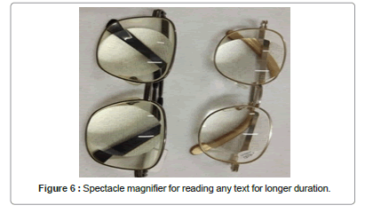
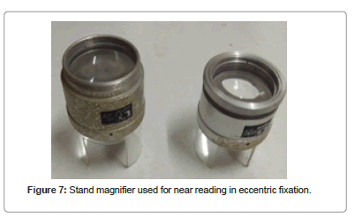
| 2 | Near optical devices | Spectacle magnifiers, high adds lenses, hand held magnifiers, stand magnifiers |
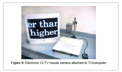
| 3 | Electronic magnification devices | Closed circuit television, hand held cctv magnifiers, adaptive |
| 4 | Peripheral vision enhancement | Scanning exercise, visual field expanders |
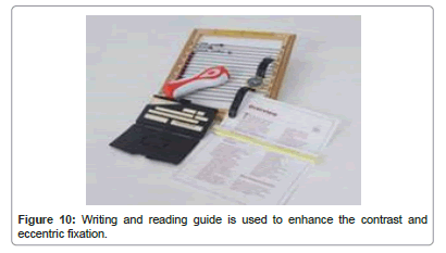
| 5 | Contrast enhancing device | Typo scopes, reading lamp, signature guide, bold line books, reading guide/writing guide, felt tipped pens, large print material |
| 6 | Glare control devices | Absorptive lenses for glare, tint, lens coatings, caps, visors |
| 7 | Postural correction devices | Writing aids/reading stand |
| 8 | Others | Vision stimulation charts and lights, vocal synthesisers etc. |
Table 2: Types of low vision devices.
Orientation and Mobility (O and M) with Tunnel Vision
Orientation means an awareness of position in space. It refers to the ability of a child to realize his surroundings, establishing body, space, and time relations with his environment through the senses of hearing, touch, smell, and residual vision (Figure 8).
Mobility means the capability of moving through the environment safely, efficiently, and independently. It refers to the ability of child to move and to react to various stimuli. This is often achieved through a teaching-learning process that involves the use of several features including a sighted guide, a long cane or a support cane, a protective arm, trailing, a guide dog, and an electronic aid (Figure 9).
O and M Training
Considering its complexity, O and M training must be stepwise and geared toward helping the low-vision child eventually move and navigate independently, O and M training is much more than simply teaching the technique on how to use the cane. Both the psychosocial and cognitive aspects relevant to teaching a person with disabilities must be considered, especially for them to get around with autonomy in locomotion and self-confidence, An O and M training program must be geared toward the difficulties of each low-vision child individually, A child who is born with low vision or who becomes visually impaired very early in life might have problems related to the development of concepts linked to position, location, direction, and distance [18].
Goals of Orientation and Mobility
The ultimate goal of orientation and mobility for a blind person is to move safely, independently efficiently and gracefully by utilizing a combination of orientation and mobility skills (Hill and Ponder).
Four of the most common ways for people with visual impairment to move around are by using
Humans guides
Here, a person with visual impairment travels with the help of human sighted guide. This becomes necessary when travelling in new places, busy streets and buildings not familiar to visually impaired person (Figure 10).
Humans guides
Certain species of dogs are trained to guide people with visual impairment. Once the dog is trained, the blind person and the guide dog need to be trained together. The training program includes walking down all the routes usually covered by the blind should know the route.
Walking canes
This is the most viable and affordable mode of travel for people with visual impairment in India. The blind person walks with the help of a long of folding cane.
Electric travel aids
With advancements in science and technology, many electronics gadgets have been developed to aid visually impaired people in travel, e.g. sonic guides, laser canes, etc. these help the user to detect obstacles before he/she comes into contact with them [19].
Types of Magnifications
Increase the size of the object by non-optical means.
Relative distance magnification
Moves the object closer to the eye. Children with visual impairment do this naturally.
Angular magnification
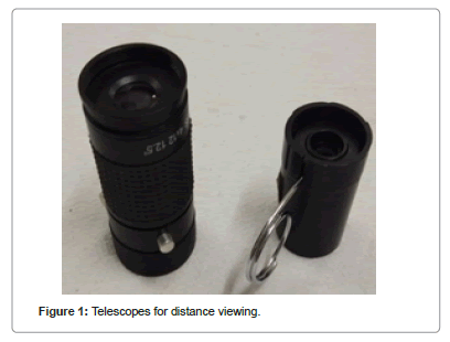
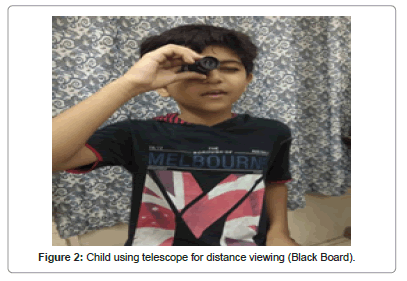
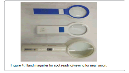
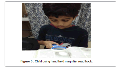
Increase the visual angle subtended by the object at the eye by optical means. Angular magnification Occurs when using a low vision device, such as a hand-held magnifier or telescope.
Electronic magnification
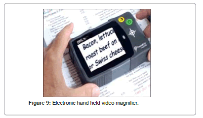
It is available in hand held; desk or arm mounted electronics magnification devices, computer software, as well as built in accessibility option on smart phone and tablets [20].
Technology
Braille system is that the child’s best medium of reading and therefore the role the teacher may be a challenging and changing one. The approaches utilized in teaching basic reading skills supported materials introduced alternative ways and therefore the child’s motivation in learning process. The varied sorts of instructional materials and equipment which are necessary as child progress school present unique opportunities for learning and reading experience. At an equivalent time, consideration of the physical aspects and formation of concepts is vital to success in reading. Problems affecting in child’s learning thanks to his lack of sight offer the teacher opportunities for experimental research and creativity. The ideas posses special nerve end which enable the touch reading. The world covered by light pressure of the finger recommendations on the paper gives the required information to the kid to discriminate between the various configurations of Braille letters which are written with Braille cell within the area of 6 mm × 3.6 mm approximately. For effective Braille reading a correct Braille mechanism must be developed By Braille mechanism, we mean the efficient movements of hands over the Braille line with proper hand position and finger position. Children who don’t develop better Braille mechanism just butterfly over the Braille sheet which contributes to the slow reading of them. For developing proper Braille mechanism certain tactual discrimination activities need to be provided.
Writing Braille
Students who read braille also usually write in braille, employing a sort of low or high tech devices. If your child writes in braille on a computer or Personal Digital Assistant (PDA), the teacher of scholars with visual impairments can use braille translation software, which converts the text and prints it out for you, the teacher, or anyone else who reads print.
Slate and Stylus
The slate and stylus are inexpensive portable tools wont to write braille-just the way paper and pencil are used for writing print. Slates are made from two flat pieces of metal or plastic held together by a hinge at one end. The slate exposes to carry paper. the highest part has rows of openings that are an equivalent shape and size as a braille cell. the rear part has rows of indentations within the size and shape of braille cells. The stylus may be a pointed piece of metal with a plastic or wooden handle. The stylus is employed to punch or emboss the braille dots onto the paper held within the slate. The indentations within the slate prevent the stylus from punching a hole within the paper when the dots are embossed. Slates and styluses are available many shapes and sizes. Tools for writing for youngsters who are blind or visually impaired.
Conclusion
Vision rehabilitation services allow children’s with visual impaired the power to realize greater control of their environment, which causes greater self confidence and an improved quality of life. Provision of low vision services can bring significant improvement in near and distance activities, social functioning and dependency. Therefore, it’s crucial that both pediatricians and ophthalmologists refer children with visual defect to vision rehabilitation to reinforce their learning abilities.
Conflict of Interest
The authors declared that there is no conflict of interest. Authors has given consent form.
References
- World Health Organization (WHO) (2009) Magnitude and causes of visual impairment.
- Turbert D, Gudgel D (2020) What is childhood low vision?
- Turbert D, Gudgel D (2020) Low vision rehabilitation teams and services.
- Kaplan J, Gerber S, Bonneau D, Rozet JM, Delrieu O, et al. (1992) A gene for Usher Syndrome type 1 (USH1A) maps to chromosome 14q. Genomics 14: 979-987.
- Kimberling WJ, Moller CG, Devenport S, Priluck IA, Beigtton PH, et al. (1992) Linkage of Usher Syndrome type I gene (USH1B) to the long arm of chromosome 11.Genomics 14: 988-994.
- Smith RJ, Lee EC, Kimberling WJ, Daiger SP, Pelias MZ, et al. (1992) Localization of two genes for usher syndrome type I to chromosome 11. Genomics 14: 995-1002.
- Wayne S, Lowry RB, McLeod DR, Knaus R, Farr C, et al. (1997) Localization of the Usher Syndrome type 1F (Ush1F) to chromosome 10. Am J Hum Genet Suppl 61: A300.
- Kimberling WJ, Weston MD, Moller C, Davenport SL, Shugart YY, et al. (1990) Localization of usher syndrome type II to chromosome 1q. Genomics 7: 245-249.
- Sankila EM, Pakarinen L, Kaariainen H, Aittomaki K, Karjalainen S, et al. (1995) Assignment of an Usher Syndrome type III (USH3) gene to chromosome 3q. Hum Mol Genet 4: 93-98.
- Tsilou ET, Rubin BI, Caruso RC, Reed GF, Pikus A, et al. (2002) Usher syndrome clinical types I and II: Could ocular symptoms and signs differentiate between the two types? Acta Ophthalmol Scand 80: 196-201.
- Reardon W (1994) Genetic factors and deafness. In: Pediatric otolaryngology. Adams DA, Cinnamond MJ, (ed). Butterworth Heinemann Ltd, Oxford, United Kingdom.
- Vemon M, Hicks W (1983) A group counselling and education programme for students with usher syndrome. J Vis Impair Blind 77: 62-66.
- Weil D, Blanchard S, Kaplan J, Guilford P, Walsh J, et al. (1995) Defective myosin VIIA gene responsible for usher syndrome type 1B. Nature 374: 60–61.
- Malini JS , Ragunathan R (2018) Training children with visual imapairment : Training programme for low vision and visual impairment. Chicago, Independtly Published, USA.
- Bailey IL, Lovie JE (1976) New design principles for visual acuity letter charts. Am J Optom Physiol Opt 53: 740-745.
- Sloan LL (1959) New test charts for measurement of visual acuity at far and near distances. Am J Ophthalmol 48: 807-813.
- Gur SL (2015) Pediatric low vision management. Delhi J Ophthalmol 26: 81-87.
- Vasconcelos G, Fernandes LC (2016) Low vision: Orientation and mobility. AAO
- Wiener WR, Welsh RL, Blasch BB (1980) Foundation of orientation and mobility.(3rd edn). New York, American Foundation for the Blind, USA.
- Wilkinson ME (2005) Low vision rehabilitation–who, when and how. Saudi J Ophthalmol 199: 117-124.
Citation: Kumar R, Parmar N, Saini R (2021) Low Vision Rehabilitation Method in Children: An Update. Optom Open Access 6: 147.
Copyright: © 2021 Kumar R, et al. This is an open-access article distributed under the terms of the Creative Commons Attribution License, which permits unrestricted use, distribution, and reproduction in any medium, provided the original author and source are credited.
Share This Article
Recommended Journals
Open Access Journals
Article Usage
- Total views: 1605
- [From(publication date): 0-2021 - Feb 22, 2025]
- Breakdown by view type
- HTML page views: 1125
- PDF downloads: 480

