Review Article Open Access
Long Lasting Disease: Leprosy
| Pramoda Earla* | ||
| Department of Microbiology, Aditya Degree College [PG Courses], Andhra University, India | ||
| Corresponding Author : | Pramoda Earla Department of Microbiology Aditya Degree College [PG Courses] Affiliated to Andhra University, Kakinada East Godavari District, Andhra Pradesh, India Tel: +91-7416948660 E-mail: pramodaearla@gmail.com |
|
| Received March 20, 2015, Accepted March 25, 2015, Published March 27, 2015 | ||
| Citation: Earla P (2015) Long Lasting Disease: Leprosy. J Infect Dis Ther 3:R1-001. doi:10.4172/2332-0877-R1-001 | ||
| Copyright: © 2015 Earla P. This is an open-access article distributed under the terms of the Creative Commons Attribution License, which permits unrestricted use, distribution, and reproduction in any medium, provided the original author and source are credited. | ||
Related article at Pubmed Pubmed  Scholar Google Scholar Google |
||
Visit for more related articles at Journal of Infectious Diseases & Therapy
Abstract
There are several infectious diseases from ancient times to which people got affected, suffered and died too. Leprosy can be regarded as the most infectious, transmittable and long lasting disease among all infectious diseases. The other name of leprosy is Hansen’s disease which was named after the physician Gerhard Armauer Hansen. The first causative agent of leprosy disease in humans is Mycobacterium leprae (M. Leprae) which has identified by microscopy technique. It is a rod-shaped gram-positive acid fast bacterium. The phenolic glycolipid 1 is the glycolipid material present in cell wall of this bacterium which generally shows immunological specificity in M. leprae. Survival of this acid fast bacterium in the host cell depends on the cell wall structure. Mycobacterium lepromatosis is a newly emerged leprosy-causing organism. It is emerging day by day as one of the major infectious diseases all over the world including developing countries. It is estimated that approximately 90% of the population develop protective immunity towards this disease, and, therefore, do not get sick after getting effected with this leprosy. Genetic and environmental factors are playing vital role in leprosy infection. The main symptoms are skin sores, bumps or lumps that will never go away after several weeks or months and which will become permanent if untreated foe a long time. It will mainly affect skin region. We cannot treat leprosy as a highly infectious disease. It is probably transmitting through droplets from the mouth and nose during close and frequent contacts with untreated patients. These bacteria mainly infect skin macrophages and Schwann cells in peripheral nerves. The involvement of autonomic fibers causes alteration in glandular functions. It will lead to dry mucous membrane and dry skin and which is responsible for the loss of tactile, thermal and pain sensibility. Incubation period of leprosy is usually two to four years with major manifestations. The Semmes-Weinstein technique is a widely used technique to evaluate plantar sensibility. Multi drug therapy (MDT) and early diagnosis are the key elements in eliminating the leprosy disease as a concern of public health. The ultimate aim is the development of a prophylactic vaccine, to protect against both drug-resistant and drug-susceptible strains. However, immunoprophylaxis for the leprosy disease continues to be largely speculative. The research in the area of leprosy remains an active area of scientific research.
| Abstract |
| There are several infectious diseases from ancient times to which people got affected, suffered and died too. Leprosy can be regarded as the most infectious, transmittable and long lasting disease among all infectious diseases. The other name of leprosy is Hansen’s disease which was named after the physician Gerhard Armauer Hansen. The first causative agent of leprosy disease in humans is Mycobacterium leprae (M. Leprae) which has identified by microscopy technique. It is a rod-shaped gram-positive acid fast bacterium. The phenolic glycolipid 1 is the glycolipid material present in cell wall of this bacterium which generally shows immunological specificity in M. leprae. Survival of this acid fast bacterium in the host cell depends on the cell wall structure. Mycobacterium lepromatosis is a newly emerged leprosy-causing organism. It is emerging day by day as one of the major infectious diseases all over the world including developing countries. It is estimated that approximately 90% of the population develop protective immunity towards this disease, and, therefore, do not get sick after getting effected with this leprosy. Genetic and environmental factors are playing vital role in leprosy infection. The main symptoms are skin sores, bumps or lumps that will never go away after several weeks or months and which will become permanent if untreated foe a long time. It will mainly affect skin region. We cannot treat leprosy as a highly infectious disease. It is probably transmitting through droplets from the mouth and nose during close and frequent contacts with untreated patients. These bacteria mainly infect skin macrophages and Schwann cells in peripheral nerves. The involvement of autonomic fibers causes alteration in glandular functions. It will lead to dry mucous membrane and dry skin and which is responsible for the loss of tactile, thermal and pain sensibility. Incubation period of leprosy is usually two to four years with major manifestations. The Semmes-Weinstein technique is a widely used technique to evaluate plantar sensibility. Multi drug therapy (MDT) and early diagnosis are the key elements in eliminating the leprosy disease as a concern of public health. The ultimate aim is the development of a prophylactic vaccine, to protect against both drug-resistant and drug-susceptible strains. However, immunoprophylaxis for the leprosy disease continues to be largely speculative. The research in the area of leprosy remains an active area of scientific research. |
| Keywords |
| Leprosy; Mycobacterium leprae; Mycobacterium lepromatosis; Acid fast bacterium; Microscopy; Clinical susceptibility; Pedigree analysis; Genetic marker analysis; Twin studies; Linkage studies; Association studies; Skin macrophages; Schwann cells; Peripheral nerves; Muscle atrophy; Anti-leprosy agents; Early diagnosis; Azathioprine; Cyclosporine A; Corticosteroids |
| Abbreviations |
| DLL: Diffuse Lepromatous Leprosy; MDT: Multi Drug Therapy; SNP: Single Nucleotide Polymorphism; T-lep: Tuberculoid leprosy; L-Lep: Lepromatous Leprosy; CMI: Cell Mediated Immunity; SW Test: Semmes-Weinstein Test |
| Introduction |
| There are several infectious diseases from ancient times to which people got affected, suffered and died too [1]. Leprosy can be regarded as the most infectious, transmittable and long lasting disease among all infectious diseases. It is a chronic and granulomatous disease, mainly caused by two bacteria named Mycobacterium leprae and Mycobacterium lepromatosis. The other name of leprosy is Hansen’s disease [2-11] which was named after the physician Gerhard Armauer Hansen. |
| Mycobacterium leprae |
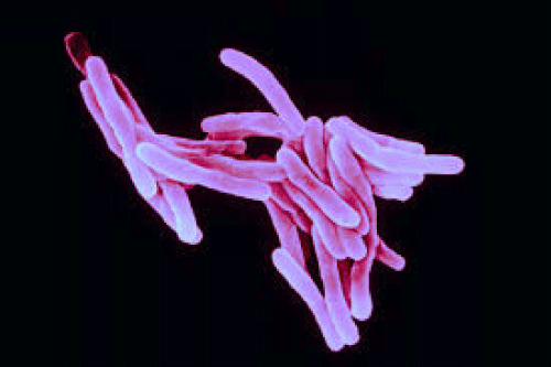
| The first causative agent of leprosy disease in humans is M. leprae which has identified by microscopy technique [12] (Figure 1). It is a rod-shaped gram-positive acid fast bacterium which lasts up to thirty six hours at room temperature and 11-16 days in the host system for multiplication which can be considered very slow. |
| M. leprae is having a cell envelope made up of plasma membrane and cell wall made up of lipid-rich outer layer when examined under electron microscope. The phenolic glycolipid 1 is the glycolipid material present in cell wall of this bacterium which generally shows immunological specificity in M. leprae. Survival of this acid fast bacterium in the host cell depends on the cell wall structure [3]. |
| Mycobacterium lepromatosis |
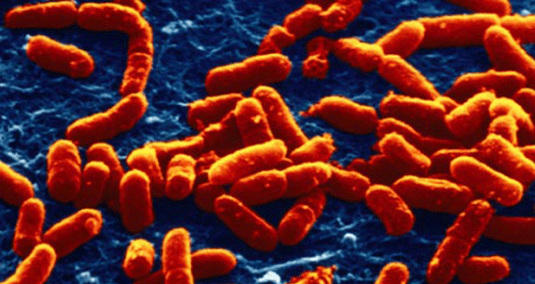
| Mycobacterium lepromatosis is a recently emerged leprosy-causing acid fast bacterium (Figure 2). Preliminary phylogenetic analysis of 16S rRNA and few other gene segments revealed significant divergence from Mycobacterium leprae is a well-known causative agent of leprosy that justifies the status of M. lepromatosis as a new emerged species [13]. M. lepromatosis caused not only all diffuse lepromatous leprosy (DLL) cases specifically but also more cases of lepromatous leprosy and other clinical forms of leprosy [14]. |
| It is emerging day by day as one of the major infectious diseases all over the world including developing countries. |
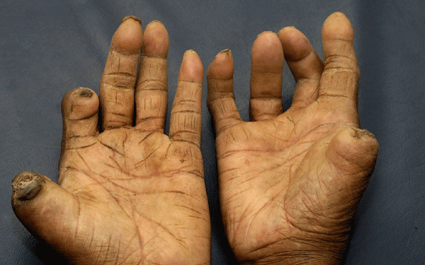
| M. lepromatosis is having the ability to infect Schwann cells of peripheral nervous system and it can be identified as neuropathogenic microbe [15]. The World Health Organization estimates that there are one million leprosy carriers and currently about two to three million people present with different types of physical disabilities due to leprosy. The ideal prevalence for leprosy disease should be of less than one case per ten thousand inhabitants [16]. It is estimated that approximately ninety percent of the population develop protective immunity towards leprosy disease and therefore do not get sick after getting effected with this leprosy (Figure 3). However, other individuals show clinical susceptibility to these pathogens which leads to the changes in immune response [3]. |
| Genetic and Environmental Factors |
| Genetic and environmental factors are playing vital role in leprosy infection. There are different approaches to investigate genetic mechanisms of resistance which includes pedigree analysis, genetic marker analysis, twin studies, investigation of particular genes, etc. Twin studies are playing vital role and they are showing higher incidence of leprosy in monozygotic when compared to dizygotic twins. This study is indicating the important role of genetics in causing different types of infectious diseases [3]. |
| Linkage studies and association studies are very useful in the investigation of different types of genetic diseases. Linkage studies are mainly dealing with gene mapping experiments, whereas association studies are dealing with allele frequencies comparison of a particular gene. This analysis generally occurs between patients and healthy individuals. Most of these studies are using single-base polymorphisms as markers for investing genetic disorders. SNP is a single nucleotide polymorphism which can lead to alterations in the functions and structures of a protein [3]. |
| Immunology of Leprosy Disease |
| Leprosy is a chronic disease caused by an acid fast bacterium named Mycobacterium leprae (M. leprae) which shows a broad spectrum of clinical features. Tuberculoid leprosy is at one end and lepromatous leprosy is at the other end of the spectrum. T-lep patients will generally show high levels of cell-mediated immune responses against M. leprae, which will results in resistance to leprosy infection with very less clinical manifestations. On the other hand, L-lep patients will show very low cell-mediated immune responses against the leprosy bacteria as well as the developed form of the disease [17]. |
| Symptoms |
| The main symptoms of leprosy disease include disfiguring of skin sores, bumps or lumps that do not go away after several months. These symptoms will become permanent if they left untreated. It will mainly affect skin region. There are three main types of the disease: Tuberculoid (a mild form of leprosy); Lepromatous (a more severe form of the disease, where kidneys and male reproductive organs may also affect); and Borderline (patients with symptoms of both tuberculoid and lepromatous forms) [18]. |
| Transmission |
| We cannot treat leprosy as a highly infectious disease. It is probably transmitting through droplets from the mouth and nose during close and frequent contacts with untreated patients. Nasal secretions from the lepromatous patients can yield ten million viable organisms per day. All the people who become infected will not develop leprosy disease (genetic factors are influential). People who are living in endemic areas are at highest risk with poor conditions such as insufficient diet, contaminated water and inadequate bedding. These people will also subject to other diseases that decrease or compromise immune function [19]. |
| Loss of Sensibility |
| These bacteria mainly infect skin macrophages and Schwann cells in peripheral nerves [16,20]. The involvement of autonomic fibers causes alteration in glandular functions. It will lead to dry mucous membrane and dry skin and which is responsible for the loss of tactile, thermal and pain sensibility. The damaged motor fibers are responsible for the abolition of muscular response or muscle atrophy. The detection of protective sensory losses becomes very important to identify peripheral neuropathy and also to avoid plantar ulcer development and eventual amputation of lower limbs. The injury of sensory fibers is not a unique feature of leprosy. Other diseases such as vascular disorders, diabetes and different neuropathies of trauma can also generate plantar sensory loss [19]. |
| Symptoms during Incubation period |
| Incubation period of leprosy is usually two to four years with major manifestations of skin lesions, muscle atrophy, numbness, photophobia, nasal stuffiness and blurred vision [21]. |
| Diagnosis |
| Semmes-Weinstein test |
| The Semmes-Weinstein technique is a widely used technique to evaluate plantar sensibility. This test consists of nylon wires of same size and of different diameters with a variation of strength 0.05 g to 300 g and the order of associated colors are: green, blue, violet, dark red, orange, magenta and red. The protective sensation loss in the feet and hands is primarily indicated by the lack of response to the stimulus of the violet, blue and green filaments [19]. |
| M. leprae which is infectious to human can be considered very critical microorganism as it cannot be cultured in artificial media. Resistance to anti-leprosy drugs such as rifampicin, dapsone and ofloxacin evolves by an amino acid substitution at the binding sites of these drugs [19]. There is an urgent requirement to discover new anti-leprosy agents. Although the available treatment for leprosy is effective, it is also quite expensive. Recent advances in biological sciences and computational approach are allowing our researchers to understand and also to design drug targets which are safe and inexpensive. Pharmacophore development, structure based optimization and virtual screening techniques are playing vital role in designing drugs [2]. |
| Treatment |
| One of the major problems encountered for reducing the incidence of leprosy disease is late diagnosis which will lead to active infectious diseases [3]. MDT and early diagnosis are the key elements in eliminating the leprosy disease as a concern of public health. The approach of MDT consists of three drugs: rifampicin, dapsone and clofazimine. Dapsone (diaminodiphenylsulfone) was the first developed leprosy drug and it remained effective for a long time until the bacteria develops resistance. MDT required to be taken over a twelve month period for the active treatment. In addition, to suppress the cellular immunological response, Immunosuppressants such as cyclosporine A and azathioprine can be used in association with corticosteroids [19]. MDT protocol depends on combinatorial antibacterial effect of three chemotherapeutic agents, clofazimine, dapsone and rifampicin. This combination treatment is administered for six to twenty four months under partial medical supervision (Table 1) [21]. |
| Conclusion |
| One strand of leprosy research is focused on detection of disease to enable earlier treatment and the other strand of research is focused towards the assessment of patients for post-treatment. The ultimate aim for the leprosy research is the development of a prophylactic vaccine which will protect against both drug-resistant and drug-susceptible strains. However, immunoprophylaxis for the leprosy disease continues to be largely speculative. This is due to problems with culturing the infectious agent. The research in the area of leprosy remains an active area of scientific research [18]. |
References
- Earla P (2014) Ancient Diseases-Microbial Impact. J Anc Dis Prev Rem 2: R1-001.
- Ganatra SH, Bodhe MN, Tatode PN (2013) Inhibition Studies of Pyrimidine Class of Compounds on Enoyl-AcpReductase Enzyme. J ComputSciSystBiol 6: 025-034.
- Tonelli-Nardi SM, Graça CR, Paschoal VD (2014) Update on Genetics of Leprosy. J Anc Dis Prev Rem 2: 109.
- Lyrio EC, Campos-Souza IC, Corrêa C, Lechuga G, Verícimo M, et al. (2015) Interaction of Mycobacterium leprae with the HaCaT human keratinocyte cell line: New frontiers in the cellular immunology of leprosy. ExpDermatol.
- Pinheiro RO, Salles JS, Sarno EN, Sampaio EP (2011) Mycobacterium leprae-host-cell interactions and genetic determinants in leprosy: an overview. Future Microbiol 6: 217-230.
- Lasry-Levy E, Hietaharju A, Pai V, Ganapati R, Rice ASC, et al. (2011) Neuropathic Pain and Psychological Morbidity in Patients with Treated Leprosy: A Cross-Sectional Prevalence Study in Mumbai. PLoSNegl Trop Dis 5: e981.
- Idema WJ, Majer IM, Pahan D, Oskam L, Polinder S, et al. (2010) Cost-Effectiveness of a Chemoprophylactic Intervention with Single Dose Rifampicin in Contacts of New Leprosy Patients. PLoSNegl Trop Dis 4: e874.
- Walsh GP, Dela Cruz EC, Abalos RM, Tan EV, Fajardo TT, et al. (2012) Limited Susceptibility of Cynomolgus Monkeys (Macacafascicularis) to Leprosy after Experimental Administration of Mycobacterium leprae. Am J Trop Med Hyg 87: 327-336.
- Jarduli LR, Sell AM, Reis PG, Sippert EA, Ayo CM, et al. (2013) Role of HLA, KIR, MICA, and Cytokines Genes in Leprosy. Biomed Res Int 2013: 989837.
- Duthie MS, Gillis TP, Reed SG (2011) Advances and hurdles on the way toward a leprosy vaccine. Hum Vaccin 7: 1172-1183.
- Lastória JS, MilanezMorgado de Abreu MA (2014) Leprosy: review of the epidemiological, clinical, and etiopathogenic aspects - Part 1. An Bras Dermatol 89: 205-218.
- Mantellini GG, Goncalves GG, Padovani CR (2012) Physical Disabilities in Leprosy: Some Contemporary Basic Aspects. J Mycobac Dis 2: 121.
- Han XY, Sizer KC, Thompson EJ, Kabanja J, Li J, et al. (2009) Comparative Sequence Analysis of Mycobacterium leprae and the New Leprosy-Causing Mycobacterium lepromatosis. J Bacteriol 191: 6067-6074.
- Han XY, Jessurun J (2013) Severe Leprosy Reactions Due to Mycobacterium lepromatosis. Am J Med Sci 345: 65-69.
- Singh P, Benjak A, Schuenemann VJ, Herbig A, Avanzi C, et al. (2015) Insight into the evolution and origin of leprosy bacilli from the genome sequence of Mycobacterium lepromatosis. ProcNatlAcadSci U S A pii: 201421504.
- Cordeiro TL, CiprianiFrade MA, Ana Regina SB Barros and Toraboschi Foss N (2014) Postural Balance Control of the Leprosy Patient with Plantar Sensibility Impairment. Occup Med Health Aff 2: 158.
- Ohyama H, Kato-Kogoe N, Takeuchi-Hatanaka K, Yamanegi K, Yamada N, et al. (2012) T-cell Responses Involved in the Predisposition to Periodontal Disease: Lessons from Immunogenetic Studies of Leprosy. J Clin Cell Immunol S1: 005.
- Sandle T (2013) Global Strategies for Elimination of Leprosy: A Review of Current Progress. J Anc Dis Prev Rem 1: e112.
- Kumar A, Iyer K, Shanthi V, Ramanathan K (2014) Extraction of BioactiveCompounds from Millingtoniahortensis for the Treatment of Dapsone Resistance in Leprosy. J MicrobBiochemTechnol R1: 006.
- Lockwood DNJ, Suneetha L, Sagili KD, Chaduvula MV, Mohammed I, et al. (2011) Cytokine and Protein Markers of Leprosy Reactions in Skin and Nerves: Baseline Results for the North Indian INFIR Cohort. PLoSNegl Trop Dis 5: e1327.
- Sieni AIA, Layati WZA, Abbasi FAA (2013) Temporal Adverse Effects in Leprosy Saudi Patients Receiving Multi Drug Therapy. ClinExpPharmacol 3: 141.
Tables and Figures at a glance
| Table 1 |
Figures at a glance
 |
 |
 |
||
| Figure 1 | Figure 2 | Figure 3 |
Relevant Topics
- Advanced Therapies
- Chicken Pox
- Ciprofloxacin
- Colon Infection
- Conjunctivitis
- Herpes Virus
- HIV and AIDS Research
- Human Papilloma Virus
- Infection
- Infection in Blood
- Infections Prevention
- Infectious Diseases in Children
- Influenza
- Liver Diseases
- Respiratory Tract Infections
- T Cell Lymphomatic Virus
- Treatment for Infectious Diseases
- Viral Encephalitis
- Yeast Infection
Recommended Journals
Article Tools
Article Usage
- Total views: 14427
- [From(publication date):
April-2015 - Nov 23, 2024] - Breakdown by view type
- HTML page views : 10062
- PDF downloads : 4365
