Locked Posterior Shoulder Dislocation with Impression Fracture Treated all Arthroscopically with the Use of an Allograft Bone Block: A Case Report
Received: 20-Oct-2022 / Manuscript No. JCEP-22-77847 / Editor assigned: 24-Oct-2022 / PreQC No. JCEP-22-77847 / Reviewed: 11-Nov-2022 / QC No. JCEP-22-77847 / Revised: 18-Nov-2022 / Manuscript No. JCEP-22-77847 / Published Date: 24-Nov-2022 DOI: 10.4172/2161-0681.22.13.430
Abstract
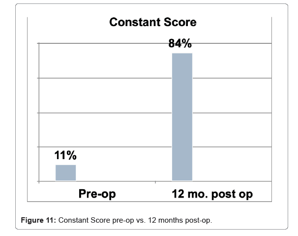
Locked posterior shoulder dislocations are uncommon and pose many difficulties in diagnosis. They are often overlooked during initial examination and delayed diagnosis adversely affects healing process. Apart from many open treatment options there are reports of single attempts to treat such cases arthroscopically. We present an original case of a posterior locked dislocation of the shoulder joint with a fracture of the lesser tuberosity followed by reverse Hill-Sachs fracture, treated in a novel fashion all-arthroscopically with the use of allogeneic bone graft. According to Constant Shoulder Score that tries to asses’ functional and subjective performance of the shoulder joint before the operation and after 12 months, we achieved a leap from 11% to 84%. The patient restored almost full range of motion and painless movement in activities of daily life as well as during sports. The use of an arthroscope reduces the invasiveness of the procedure, improves visualization of the joint and allows augmentation of the bone loss without performing an open approach. We believe that this is a promising method of treatment for selected cases of locked posterior shoulder dislocation.
Keywords: Locked posterior shoulder dislocation; Arthroscopy; Bone graft; Case report
Introduction
Locked posterior shoulder dislocations are uncommon and poses many Shoulder dislocations are most common among all joints of the human body. In the United States it is reported to occur 11.2 per 100 000 population per year. Posterior shoulder dislocations are rare and by various authors usually refer to 2 to 5% of all dislocations [1,2]. McLaughlin [3] in a group of 581 patients after shoulder dislocation described 22 cases of posterior dislocations (3.8%). Although the treatment of this disease is no more demanding than the treatment of anterior dislocation, late diagnosis, and prolonged period between injury and treatment adversely affects the final result. Major causes of posterior dislocations can be described os 3 ”E”, ie. Electric shock, Epilepsy or Extreme trauma [4]. Chronic posterior shoulder dislocations are those that were not recognized within three weeks after the injury [5]. According to literature time duration from injury to proper diagnosis and appropriate treatment varies from 3 weeks to 4.5 years [5]. Furthermore, in the series reported by McLaughlin [3] mean time between injury and diagnosis was 8 months. The standard procedure after diagnosing posterior shoulder dislocation accompanied by fracture of the lesser tuberosity is open reduction with internal fixation, often supplemented with McLaughlin procedure (subscapularis tendon tenodesis in the bone defect of the humeral head). Recently however, there have been reports of arthroscopic assisted treatment of dislocations and fractures, which seems to be applicable in some cases. We present a case of a patient with an overlooked shoulder dislocation followed by fracture of the lesser tuberosity treated with arthroscopic assisted closed reduction with reconstruction of the articular surface impression fracture and stabilization of the lesser tuberosity fracture.
Case Presentation
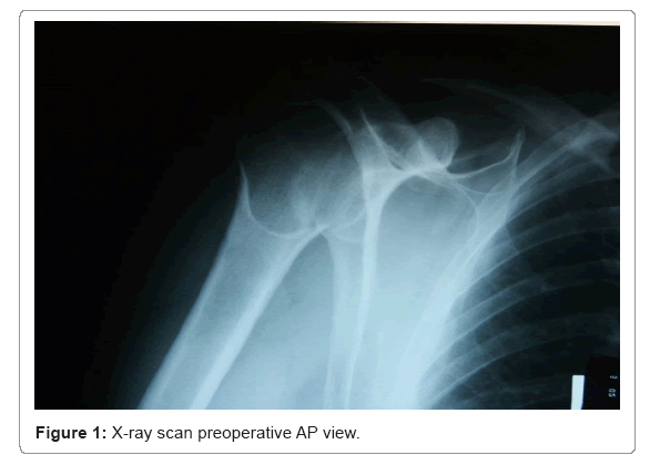
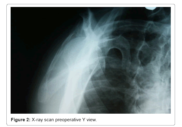
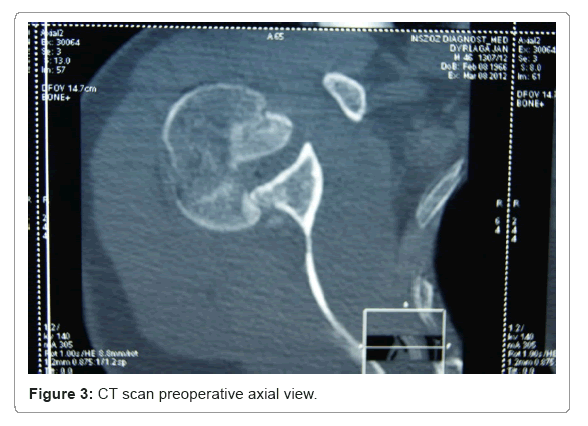
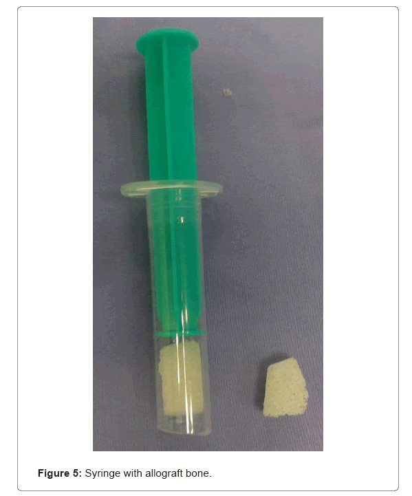
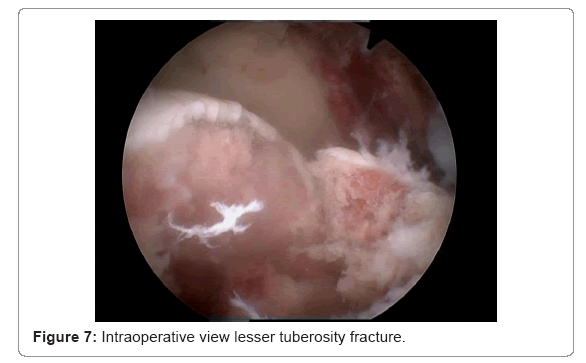
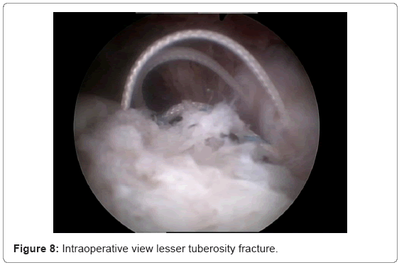
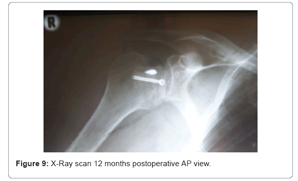
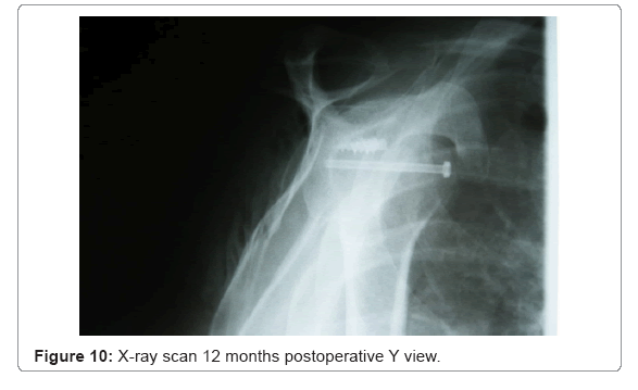
46 year old right-handed male patient (dominant hand) injured his right shoulder during a fall directly on the shoulder from a ladder- height of about 1 meter. He presented on Figures 1 and 2 the day of the injury to the emergency department complaining from shoulder pain and restricted mobility. The doctor performed classic AP and transthoracal X-ray, and found no abnormalities thus diagnosing shoulder contusion. The patient was referred for further evaluation and treatment to orthopedic clinic within two weeks. The patient presented to outpatients clinic 2 weeks after with significant range of motion restriction and moderate pain. The admitting physician (orthopedic surgeon) ordered an X-ray in 2 projections (AP and Y) and did not recognize the posterior dislocation, but consecutively ordered shoulder CT. 22 days after the initial trauma the patient with CT Figures 3 and 4 scans and a set of X-rays presented to our hospital again complaining from pain and severe restriction of Range Of Motion (ROM) with shoulder set in internal rotation. ROM was limited to 20 degrees of forward flexion, 20 degrees of internal rotation, and 20 degrees of abduction and 10 degrees of external rotation. The patient reported no neurovascular disorders peripherally. Based on imaging results the posterior dislocation of the scapulo-humeral joint with avulsion fracture of the lesser tuberosity and visible reverse Hill-Sachs lesion was diagnosed. In addition, taking into account a careful CT scans analysis, the extent of impression fracture of the articular surface of the humeral head was determined and assessed at about 25% after physical examination, considering patient’s age, the nature of his work, activity, expectations and imaging studies we decided to perform a reconstruction of the articular surface. Due to the extent of the injury and its chronic nature in preoperative planning we considered the following options: closed reduction of dislocation with arthroscopic stabilization of fractures, open reduction and stabilization of fractures. The patient was operated on in general anesthesia following an interscalenic block. The patient was then placed in the beach chair position. Axial traction was applied to upper extremity for several minutes and then with the arm in abduction and delicate traction gentle movements combined with internal and external rotation were done in order to relax the joint capsule. Then the standard arthroscopic posterior portal was made. The switching stick was introduced through this posterior portal into the shoulder joint, placed on the posterior-medial edge of the humeral head fracture in front of the posterior glenoid rim. Reposition was made with constant axial traction and external rotation and with the use of switching stick as a lever. Intraoperative radiological control was performed confirming reposition. Clinical control also confirmed reposition, but the joint was still unstable. The dislocation occurred each time the shoulder was internally rotated. While placing the patient in the beach chair position with atraction of 1.5 kg and maintaining the arm in neutral rotation there was no spontaneous dislocation, so the surgical field was set in this position. An arthroscope was then inserted through the standard posterior portal. After the initial inspection of the joint, the anterolateral portal was made (through the rotator interval above the biceps tendon). Blood clots and small pieces of cartilage and bone were rinsed out with plenty of irrigation fluid and then a thorough evaluation of intra-articular structures was made. In the next step deposits of fibrin were removed with a shaver. Extensive reverse Hill-Sachs fracture was found and so was a fracture of approximately 25% of the articular surface of the anterior part of humeral head, in the direct vicinity of the lesser tuberosity. It was an impression fracture involving articular surface with cancellous bone of the humeral head. The lesser tuberosity was not only fractured but also displaced medially, retracted by the subscapularis muscle. The tendon of the long head of the biceps was dislocated and jammed between the fracture surfaces of the humeral head and lesser tuberosity thus preventing reduction. Another arthroscopic access was made the anterior portal (through the rotator interval over the subscapularis tendon) and an attempt to reduce the biceps tendon was made using tissue grasper. Biceps tendon appeared to be in place, but because of its instability a consecutive dislocation was observed after releasing the grasper. Biceps tendon tenotomy was performed as further procedures within the shoulder joint would be otherwise impossible. The tenodesis of the LHB was not taken into consideration because the fracture pattern passed through the bicipital groove. Then with a delicate switching stick manipulation, the osteochondral fragment of the humeral head was elevated and reduced so as not to disrupt the continuity of cartilage surface of a fragment still being connected to the rest of the humeral head. A significant loss of cancellous bone of the humeral head was found under the surface of undamaged cartilage. We decided to augment the bone loss with cancellous bone allograft. First the intra and extra articular subscapularis tendon tenolysis was done. After the release of all adhesions full reposition of the lesser tuberosity was obtained. Using two K wires the lesser tuberosity was fixed temporarily to the humeral head while holding it in a place with tissue grasper. After temporary stabilization of the lesser tuberosity the anterolateral portal was expanded and 2 cancellous bone allografts measuring 10 × 10 × 10 mm and 10 ×10 ×15 mm were introduced into the fracture gap using a 5 ml end-cut syringe. This step of the procedure was intended to fill the loss of cancellous bones at the site of the impression fracture (Figures 5 and 6). Then, using K wires as guide the cannulated drill was used. With X-ray monitor control, a single cannulated screw was introduced compressing the fracture gap. The next stage of the surgery comprised double-loaded Figures 7 and 8 anchor introduction into the humeral head through lesser tuberosity fixing the upper part of the subscapularis tendon above the fracture gap. After removing K wires, reduction stability control was checked by means of delicate rotational movements of the shoulder joint. After surgery, the patient arm was unloaded with an orthosis with limited abduction and neutral rotation of the arm. Starting from the second post-operative day the patient was allowed to perform gentle exercises involving passive external rotation, elbow joint flexion and pendulum exercises. All exercises were carried out with assistance and to an extent limited by pain. After 6 weeks the orthosis was removed and active exercises begun to increase range of motion and muscle strength. The patient was allowed 6 months after surgery to return Figures 9,10 and 11 to manual labor. The results of the surgery were assessed 12 months after revealed a significantly increased range of motion and strength tests improvement assessed by means of Constant Score-a rise from 11% to 84%. The patient returned to painless manual labor and sports.
Discussion
Posterior shoulder dislocation is relatively rare, about 1.1 cases per 100 000 inhabitants per year [6], but very often initially misdiagnosed. According to some authors this percentage may reach 41%, and as Gerber stated such patients accounted for 59%. Physical examination is dominated by a significant reduction in joint mobility, the arm is fixed in internal rotation and has no active and passive external rotation, and the contour of the shoulder is altered, with a noticeable displacement of the humeral head backwards. The patient complains of pain and impairment of daily living tasks like combing hair or washing face [2]. We performed x-ray in two projections (true AP and Y), however, an alternative projection Y is quite sufficient. We perform a CT scan in doubtful cases which additionally allows us to specify the nature of McLaughlin fractures and humeral bone loss in percentage. One may often find accompanying injuries such as intra-articular damage to the posterior joint capsule and posterior labrum, rotator cuff tear or a surgical neck fracture or of the greater tuberosity [7]. Treatment depends on the extent and type of damage, associated dislocation and the time elapsed from trauma until treatment. In the presence of a reversed Hill-Sachs fracture performing reposition as the only treatment is not sufficient, because a concomitant fracture is a major risk factor of re-dislocation. A key element of treatment is to augment the bone loss and to restore proper biomechanics of the joint. McLaughlin as the first stated that the size of the loss determines the type of the procedure [3]. He is the author of the oldest and most well-known method of operating reverse Hill-Sachs’ fracture, comprising suturing the subscapularis tendon in the bone loss of humeral head [3]. Neer has modified this method of lesser tuberosity transferring with the attachment of the subscapularis muscle into the bone loss of humeral head, claiming that at the same time we can obtain good joint stabilization and fill the Hill-Sachs bone loss [8]. Some authors have criticized this method because of the limitation of internal rotation of the arm during postoperative rehabilitation, leading to development of osteoarthritis and non-anatomical relations in the shoulder joint. This makes it difficult to implant shoulder prosthesis in the future [7]. Hawkins proposed [9] treatment recommendation depending on the extent of the bone defect. He suggests a closed reposition in analgesia for defects to 20%, and after the reposition advises to immobilize the shoulder in external rotation for 4-6 weeks. In cases of defects from 20 to 45%, due to the expected instability he advocated for reconstructive operations (isolated transfer of subscapularis or lesser tuberosity fragment) and for defects over 45% with associated degenerative changes arthroplasty. However, the average age of patients with extensive defects of humeral head is around 45 years, [10] therefore, it would be better to perform joint preserving procedures rather than alloplasty [1,3,4]. In those cases Gerber suggests a segmental humeral head defect reconstruction using allograft.
Conclusion
This method has proven to be very convenient and effective in long term follow-up. This kind of surgery may be carried out with the use of allografts or auto graft. Most locked posterior shoulder dislocations are nowadays treated with open techniques, but few reports about arthroscopic treatment present encouraging results. We propose fracture reposition and reconstructive surgery without any open approach and encourage using our novel method to place the bone allografts into the joint in all-arthroscopic minimally invasive manner.
Funding
No external funding was received.
Competing Interests
There are no conflicts of interest.
Ethics Approval
Not applicable
Consent to participate
The patient has given his informed consent to participate.
“I hereby give my written and informed consent to participate in the study and follow-up examinations”
Consent for publication
The patient has given his informed consent for this case report to be published
“I hereby give my written and informed consent for my medical data to be published provided my personal and confidential data will be secured”
Availability of data and material
The data and material is fully available
Code Availability
Not applicable.
Author’s contributions
AB operated on the patient and made a case draft, MP wrote the final manuscript version and critically analyzed provided data, prepared the table and images. MS´ help to collect data. RB as the department chairman provided not only general support but also helped to concept and designs the case. He is the co-author of the manuscript. All authors read and approved the final manuscript.
Acknowledgments
No acknowledgments to be mentioned.
References
- Kayali C, Agus H, Kalenderer O, Turgut A, Imamoglu T (2009) Overlooked posterior shoulder dislocation: preoperative and postoperative CT studies (a case report). Ortop Traumatol Rehabil 11: 177-182.
[Google Scholar] [PubMed]
- Cicak N (2004) Posterior dislocation of the shoulder. J Bone Joint Surg Br 86: 324-332.
[Crossref] [Google Scholar] [PubMed]
- McLaughlin HL (1952) Posterior dislocation of the shoulder. J Bone Joint Surg Am 24: 584-590.
[PubMed]
- Gadek A, Slusarski J, Kasprzyk M, Ciszek E (2008) Locked posterior shoulder dislocation. Przeglad Lekarski 65: 299-303.
[PubMed]
- Rowe CR, Zarins B (1982) Chronic unreduced dislocations of the shoulder. J Bone Joint Surg Am 64: 494-505.
[PubMed]
- Robinson CM, Aderinto J (2005) Posterior shoulder dislocations and fracture-dislocations. The Journal of bone and joint surgery. J Bone Joint Surg Am 87: 639-650.
[Crossref] [Google Scholar] [PubMed]
- Assom M, Castoldi F, Rossi R, Blonna D, Rossi P (2006) Humeral head impression fracture in acute posterior shoulder dislocation: New surgical technique. Knee Surg Sports Traumatol Arthrosc 14: 668-672.
[Crossref] [Google Scholar] [PubMed]
- Diklic ID, Ganic ZD, Blagojevic ZD, Nho SJ, Romeo AA (2010) Treatment of locked chronic posterior dislocation of the shoulder by reconstruction of the defect in the humeral head with an allograft. The Journal of bone and joint surgery. British volume 92: 71-76.
[Crossref] [Google Scholar] [PubMed]
- Hawkins RJ, Neer CS, Pianta RM, Mendoza FX (1987) Locked posterior dislocation of the shoulder. J Bone Joint Surg Am 69: 9-18.
[Google Scholar] [PubMed]
- Dervin GF, Brunet JA, Healey DC (2002) A Modification of the McLaughlin procedure as salvage for missed locked posterior fracture-dislocation of the humeral head: A Case report. J Bone Joint Surg Am 84: 804-807.
[Crossref] [Google Scholar] [PubMed]
Citation: Błasiak A, Zhang J, Podsiadło M, Śliwa M, Brzóska R (2022) Locked Posterior Shoulder Dislocation with Impression Fracture Treated all Arthroscopically with the Use of an Allograft Bone Block: A Case Report. J Clin Exp Pathol 13: 430. DOI: 10.4172/2161-0681.22.13.430
Copyright: © 2022 Błasiak A, et al. This is an open-access article distributed under the terms of the Creative Commons Attribution License, which permits restricted use, distribution, and reproduction in any medium, provided the original author and source are credited.
Share This Article
Recommended Journals
Open Access Journals
Article Tools
Article Usage
- Total views: 1102
- [From(publication date): 0-2023 - Mar 12, 2025]
- Breakdown by view type
- HTML page views: 940
- PDF downloads: 162

