Lipid and Organelle Membrane Lipid Composition Mitochondria
Received: 02-Aug-2022 / Manuscript No. bcp-22-71257 / Editor assigned: 04-Aug-2022 / PreQC No. bcp-22-71257 / Reviewed: 10-Aug-2022 / QC No. bcp-22-71257 / Revised: 17-Aug-2022 / Manuscript No. bcp-22-71257 / Published Date: 24-Aug-2022 DOI: 10.4172/2168-9652.1000391 QI No. / bcp-22-71257
Introduction
Lipid in any of a diverse group of organic compounds including fats, oils, hormones, and certain components of membranes that are grouped together because they do not interact appreciably with water. One type of lipid, the triglycerides, is sequestered as fat in adipose cells, which serve as the energy-storage depot for organisms and also provide thermal insulation. Some lipids such as steroid hormones serve as chemical messengers between cells, tissues, and organs, and others communicate signals between biochemical systems within a single cell. The membranes of cells and organelles (structures within cells) are microscopically thin structures formed from two layers of phospholipid molecules. Membranes function to separate individual cells from their environments and to compartmentalize the cell interior into structures that carry out special functions. So important is this compartmentalizing function that membranes, and the lipids that form them, must have been essential to the origin of life itself [1].
Water is the biological milieu—the substance that makes life possible—and almost all the molecular components of living cells, whether they be found in animals, plants, or microorganisms, are soluble in water. Molecules such as proteins, nucleic acids, and carbohydrates have an affinity for water and are called hydrophilic (“water-loving”). Lipids, however, are hydrophobic (“water-fearing”) [2]. Some lipids are amphipathic—part of their structure is hydrophilic and another part, usually a larger section, is hydrophobic. Amphipathic lipids exhibit a unique behaviour in water: they spontaneously form ordered molecular aggregates, with their hydrophilic ends on the outside, in contact with the water, and their hydrophobic parts on the inside, shielded from the water. This property is key to their role as the fundamental components of cellular and organelle membranes [3].
Lipoproteins are lipid-protein complexes that allow all lipids derived from food or synthesized in specific organs to be transported throughout the body by the circulatory system. The basic structure of these aggregates is that of an oil droplet made up of triglycerides and cholesteryl esters surrounded by a layer of proteins and amphipathic lipids—very similar to that of a micelle, a spherical structure described in the section Fatty acids. If the concentration of one or another lipoprotein becomes too high, then a fraction of the complex becomes insoluble and is deposited on the walls of arteries and capillaries [4]. This buildup of deposits is called atherosclerosis and ultimately results in blockage of critical arteries to cause a heart attack or stroke. Because of the gravity of this condition, much research is focused on lipoproteins and their functions. The emphasis in the following discussion is therefore placed on human lipoproteins.
Classification and formation
There are four major classes of circulating lipoproteins, each with its own characteristic protein and lipid composition. They are chylomicrons, very low-density lipoproteins (VLDL), low-density lipoproteins (LDL), and high-density lipoproteins (HDL). Within all these classes of complexes, the various molecular components are not chemically linked to each other but are simply associated in such a way as to minimize hydrophobic contacts with water [5]. The most distinguishing feature of each class is the relative amounts of lipid and protein. Because the lipid and protein composition is reflected in the density of each lipoprotein (lipid molecules being less dense than proteins), density, an easily measured attribute, forms the operational basis of defining the lipoprotein classes. Measuring density also provides the basis of separating and purifying lipoproteins from plasma for study and diagnosis. The table gives a summary of the characteristics of the lipoprotein classes and shows the correlation between composition and density (Table 1).
| Human plasma lipoproteins | |||||
| chylomicron | VLDL | IDL | LDL | HDL | |
| Density (g/ml) | <0.95 | 0.950–1.006 | 1.006–1.019 | 1.019–1.063 | 1.063–1.210 |
| Components (% dry weight) | |||||
| protein | 2 | 7 | 15 | 20 | 40–55 |
| triglycerides | 83 | 50 | 31 | 10 | 8 |
| free cholesterol | 2 | 7 | 7 | 8 | 4 |
| cholesteryl esters | 3 | 12 | 23 | 42 | 12–20 |
| phospholipids | 7 | 20 | 22 | 22 | 22 |
| Apoprotein composition | A-I, A-II, | B-100, C-I, | B-100, C-I, | B-100 | A-I, A-II, |
| B-48, C-I, | C-II, C-III, | C-II, C-III, | C-I, C-II, | ||
| C-II, C-III | E | E | C-III, D, E | ||
Table 1: Human plasma lipoproteins.
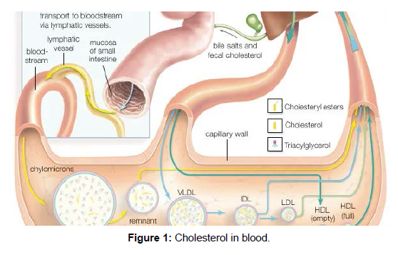
The principal lipid components are triglycerides, cholesterol, cholesteryl esters, and phospholipids. The hydrophobic core of the particle is formed by the triglycerides and cholesteryl esters [6]. The fatty acyl chains of these components are unsaturated, and so the core structure is liquid at body temperature. The table gives more details about the nine different protein components, called apoproteins, of the lipoprotein classes. With the exception of LDL, which contains only one type of apoprotein, all classes have multiple apoprotein components (Figure 1).
Synthesis of lipoprotein complexes in the small intestine, liver, and blood plasma and their delivery to peripheral tissues of the body [7].
Very low-density lipoproteins (VLDL)
VLDL is a lipoprotein class synthesized by the liver that is analogous to the chylomicrons secreted by the intestine. Its purpose is also to deliver triglycerides, cholesteryl esters, and cholesterol to peripheral tissues. VLDL is largely depleted of its triglyceride content in these tissues and gives rise to an intermediate-density lipoprotein (IDL) remnant, which is returned to the liver in the bloodstream. As might be expected (see table), the same proteins are present in both VLDL and IDL.
Low-density lipoproteins (LDL)
Low-density lipoproteins are derived from VLDL and IDL in the plasma and contain a large amount of cholesterol and cholesteryl esters. Their principal role is to deliver these two forms of cholesterol to peripheral tissues. Almost two-thirds of the cholesterol and its esters found in plasma (blood free of red and white cells) is associated with LDL [8].
High-density lipoproteins (HDL)
Lipoproteins of this class are the smallest, with a diameter of 10.8 nm and the highest protein-to-lipid ratio. The resulting high density gives this class its name. HDL plays a primary role in the removal of excess cholesterol from cells and returning it to the liver, where it is metabolized to bile acids and salts that are eventually eliminated through the intestine. LDL and HDL together are the major factors in maintaining the cholesterol balance of the body. Because of the high correlation between blood cholesterol levels and atherosclerosis, high ratios of HDL to cholesterol (principally as found in LDL) correlate well with a lower incidence of this disease in humans.
Functions, origins, and recycling of apolipoproteins
The nine classes of apoproteins listed in the table are synthesized in the mucosal cells of the intestine and in the liver, with the liver accounting for about 80 percent of production [9].
Chylomicrons are synthesized in the intestinal mucosa. The cells of this tissue, although able to make most apoproteins, are the principal source of apoB (the B-48 form) and apoA-I. The apoC-II component of chylomicrons is an activator for a plasma enzyme that hydrolyzes the triglyceride of these complexes. This enzyme, called lipoprotein lipase, resides on the cell surface and makes the fatty acids of triglycerides available to the cell for energy metabolism. To some degree, the enzyme is also activated by apoC-II, present in minor amounts in chylomicrons.
Lipids in biological membranes
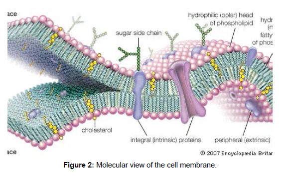
Biological membranes separate the cell from its environment and compartmentalize the cell interior. The various membranes playing these vital roles are composed of roughly equal weight percent protein and lipid, with carbohydrates constituting less than 10 percent in a few membranes. Although many hundreds of molecular species are present in any one membrane, the general organization of the generic components is known. All the lipids are amphipathic, with their hydrophilic (polar) and hydrophobic (nonpolar) portions located at separate parts of each molecule. As a result, the lipid components of membranes are arranged in what may be called a continuous bimolecular leaflet, or bilayer. The polar portions of the constituent molecules lie in the two bilayer faces, while the nonpolar portions constitute the interior of the bilayer. The lipid bilayer structure forms an impermeable barrier for essential water-soluble substances in the cell and provides the basis for the compartmentalizing function of biological membranes [10].(Figure 2)
Composition of the lipid bilayer
Most biological membranes contain a variety of lipids, including the various glycerophospholipids such as phosphatidyl-choline, -ethanolamine, -serine, -inositol, and -glycerol as well as sphingomyelin and, in some membranes, glycosphingolipids. (These compounds are described in the section Fatty acid derivatives. Cholesterol, ergosterol, and sitosterol (described in the section Cholesterol and its derivatives) are sterols found in many membranes. The relative amounts of these lipids differ even in the same type of cell in different organisms, as shown in the table on the lipid composition of red blood cell membranes from different mammalian species. Even in a single cell, the lipid compositions of the membrane surrounding the cell (the plasma membrane) and the membranes of the various organelles within the cell (such as the microsomes, mitochondria, and nucleus) are different, as shown in the table on various membranes in a rat liver cell (Table 2).
Organelle membrane lipid composition by weight percent of rat liver cells |
|||||
| Source: From Thomas E. Andreoli et al., Membrane Physiology, 2nd ed. (1987), Table II, chapter 27. | |||||
| membrane | |||||
| lipid | plasma membrane | microsome | inner mitochondria | outer | nuclear |
| mitochondria | |||||
| cholesterol | 28 | 6 | <1.0 | 6 | 5.1 |
| phosphatidylcholine | 31 | 55.2 | 37.9 | 42.7 | 58.3 |
| sphingomyelin | 16.6 | 3.7 | 0.8 | 4.1 | 3 |
| phosphatidylethanolamine | 14.3 | 24 | 38.3 | 28.6 | 21.5 |
| phosphatidylserine | 2.7 | — | <1.0 | <1.00 | 3.4 |
| phosphatidylinositol | 4.7 | 7.7 | 2 | 7.9 | 8.2 |
| phosphatidic acid and cardiolipin | 1.4 | 1.5 | 20.4 | 8.9 | <1.00 |
| lysophosphatidylcholine | 1.3 | 1.9 | 0.6 | 1.7 | 1.4 |
Table 2: Organelle membrane lipid composition by weight percent of rat liver cells.
Acknowledgement
We acknowledge all those who in one way or the other, contributed towards the success of this work. We greatly appreciate all the referenced authors for their intellectual contributions.
Conflict of interest
The authors declare that there is no conflict of interests regarding the publication of this paper.
References
- Wesp LM, Deutsch MB (2017)Hormonal and Surgical Treatment Options for Transgender Women and Transfeminine Spectrum Persons.Psychiatr Clin North Am40: 99-111.
- Dahl M,Feldman JL, Goldberg J, Jaberi A (2015) Endocrine Therapy for Transgender Adults in British Columbia: Suggested GuidelinesVancouver Coastal Health. Endocrine therapy 120-153.
- Bourns, Amy (2015) Guidelines and Protocols for Comprehensive Primary Care for Trans Clients.Sherbourne Health Centre 425-521.
- Murad Mohammad Hassan, Elamin, Mohamed B Garcia, Magaly Zumaeta, Mullan Rebecca J, et al. (2010)Hormonal therapy and sex reassignment: A systematic review and meta-analysis of quality of life and psychosocial outcomes.Clinical Endocrinology 72(2): 214-231.
- White Hughto, Jaclyn M, Reisner Sari L (2016) A Systematic Review of the Effects of Hormone Therapy on Psychological Functioning and Quality of Life in Transgender Individuals.Transgender Health 21-31.
- Amsler, Mark (1986) The Languages of Creativity: Models, Problem-solving, Discourse. University of Delaware Press.
- Astbury WT (1961) Molecular Biology or Ultrastructural Biology?.Nature 190(4781): 11-24.
- Ben-Menahem Ari (2009)Historical Encyclopedia of Natural and Mathematical Sciences.Historical Encyclopedia of Natural and Mathematical Sciences by Ari Ben-Menahem Berlin Springer Springer 29-82.
- Burton, Feldman (2001) The Nobel Prize: A History of Genius, Controversy, and Prestige.Arcade Publishing 12-30.
- Butler John M (2009) Fundamentals of Forensic DNA Typing. Academic Press 14-19.
Indexed at, Google Scholar, Crossref
Indexed at,Google Scholar,Crossref
Indexed at,Google Scholar,Crossref
Indexed at,Google Scholar,Crossref
Indexed at,Google Scholar,Crossref
Indexed at,Google Scholar,Crossref
Indexed at,Google Scholar,Crossref
Indexed at,Google Scholar,Crossref
Indexed at,Google Scholar,Crossref
Citation: Pujnk G (2022) Lipid and Organelle Membrane Lipid Composition Mitochondria. Biochem Physiol 11: 391. DOI: 10.4172/2168-9652.1000391
Copyright: © 2022 Pujnk G. This is an open-access article distributed under the terms of the Creative Commons Attribution License, which permits unrestricted use, distribution, and reproduction in any medium, provided the original author and source are credited.
Share This Article
Recommended Journals
Open Access Journals
Article Tools
Article Usage
- Total views: 1242
- [From(publication date): 0-2022 - Apr 26, 2025]
- Breakdown by view type
- HTML page views: 859
- PDF downloads: 383


