Case Report Open Access
Lessons Learnt in a 17 Year Old with Fever of Unknown Origin: Haemophagocytic Lymphohistiocytosis-The Most Fatal Outcome of EBV Infection
| Ibraheim H*, Yip K and Arami S | |
| Department of Medicine, Princess Royal University Hospital, London, UK | |
| Corresponding Author : | Ibraheim H Department of Medicine, Princess Royal University Hospital, London, UK Tel: +44 1689863000 E-mail: hajir.ibraheim@gmail.com |
| Received: September 8, 2015 Accepted: November 6, 2015 Published: November 12, 2015 | |
| Citation: Ibraheim H, Yip K, Arami S (2015) Lessons Learnt in a 17 Year Old with Fever of Unknown Origin: Haemophagocytic Lymphohistiocytosis-The Most Fatal Outcome of EBV Infection. J Infect Dis Ther 3:250. doi:10.4172/2332-0877.1000250 | |
| Copyright: © 2015 Ibraheim, et al. This is an open-access article distributed under the terms of the Creative Commons Attribution License, which permits unrestricted use, distribution, and reproduction in any medium, provided the original author and source are credited. | |
| Related article at Pubmed, Scholar Google | |
Visit for more related articles at Journal of Infectious Diseases & Therapy
Abstract
We report a young patient who had persistent fever as a result of the most fatal outcome of an Epstein-Barr Virus infection-haemophagocytic lymphohistiocytosis (EBV-HLH). A 17 year old girl with a background of IgG subclass deficiency presented with a history of fever, sore throat and vomiting. The impression was of EBV related infectious mononucleosis which was supported by a positive monospottest and high EBV DNA levels. Despite a decreasing level of EBV DNA levels, she remained febrile and tachycardic with worsening liver function, cytopenia and no improvement on broad spectrum antimicrobials. Repeated septic screens were negative.
By day 16 HLH was considered and a bone marrow aspirate showed haemphagocytosis. She was started on steroids with a rapid clinical and biochemical response. This case highlights the importance of having a low threshold of suspicion for EBV-HLH in patients with EBV that continue to deteriorate, particularly with a history of immunodeficiency.
| Background |
| Haemophagocytic Lymphohistiocytosis is a rare and aggressive syndrome characterized by hypercytokinemia resulting from excessive activation of antigen presenting cells (macrophages, histiocytes) and CD8+ T cells [1]. |
| Clinical features include fever, splenomegaly, cytopenia and hepatitis [1-5]. As a result of elevated cytokine activity, biochemical markers include high levels of serum ferritin, lactate dehydrogenase, triglycerides and soluble CD25 [2-5]. Haemophagocytosis is a characteristic finding although histological evidence of this may be present later [1-4]. |
| There are a number of slightly different diagnostic criteria used. The table below outlines the diagnostic criteria used by Henter et al. in the 2004 HLH-trial [4]. We refer to these criteria in our report (Table 1). |
| HLH can be primary (familial) or secondary, triggered by malignancy or infection-most commonly Epstein-Barr virus, as was the case in our patient [3]. The treatment in both cases involves early diagnosis and identification of potential triggers, initiation of steroids, chemotherapy (etoposide, ciclosporin) and in some cases only haematopoeitic cell transplantation is curative [6-8]. |
| EBV driven HLH has a high mortality rate secondary to multiorgan failure [1-6]. This is compounded by the challenge in its diagnosis as it is rare, the clinical and biochemical manifestations are not always apparent on presentation and there is a wide differential diagnosis. The prognosis is very poor if undiagnosed with survival up to several months. For this reason we were motivated to write up this case so that we could raise awareness of the signs and symptoms associated with this life threatening disorder that often affects children and young adults. |
| Case Presentation |
| A 17 year old Caucasian student presented to the emergency department with a 4 day history of fever, vomiting and a sore throat. |
| Her past medical history included IgG subclass deficiency (IgG2 and IgG4), previous pneumonia, viral meningitis and bronchiectasis. She was on regular three times weekly azithromycin prophylaxis and had occasional immunoglobulin replacement. There was no significant family history. |
| On examination she was tachycardic, had a temperature of 38.5°C and had a petechial rash on her soft palate. There were no findings on examination of the chest, abdominal, or neurological systems. |
| Her initial bloods showed neutropenia and thrombocytopenia, mildly raised CRP and elevated transaminases. The monospot test was positive with subsequent testing showing high EBV DNA levels (53,000 copies/ml). A viral throat swab was also done and was positive for EBV. |
| An abdominal ultrasound scan on day 2 (to investigate deranged livwww.omicsonline.org/open-access/relationship-between-the-status-of-blood-supply-in-the-nonhypervascularhepatocellular-nodules-among-chronic-liver-diseases-and-the-2167-0889-1000198.phper function) showed no abnormalities. |
| She was started on a broad spectrum penicillin to cover a possible underlying bacterial infection but she remained febrile. Blood cultures were negative. |
| By day 3 antibiotics were escalated and a single dose of immunoglobulins administered on the basis that this may enhance her immune response to the underlying infection given her background of IgG deficiency. She was started on valganciclovir to cover CMV but this was stopped when the CMV results were negative two days later. Other serology tests for hepatitis, Herpes simplex virus, (HSV) Anti streptolysin O titres and Parvovirus B19 were also negative. |
| Her liver function progressively worsened with a predominant transaminitis picture, and she became pancytopenic by day 5. The gastroenterology team impression was of EBV related hepatitis. |
| By day 8, despite the fall in the EBV DNA levels (5000 copies/ml), she continued to deteriorate clinically and had a persistent fever. She had low grade temperature most of the day (ranging between 37.8°C-38.5°C) but often had at least one high grade temperature towards the evening (ranging between 38.5°C-39.3°C). On day 9 she developed a non-allergic erythematous maculopapular rash on her cheeks, and limbs. The dermatologists suggested a possible auto immune aetiology. |
| By day 16 the haematologists raised the suspicion for EBV-HLH in the context of persistent fever, rash, EBV infection, cytopenia and abnormal liver function tests all of which were grossly out of proportion with a self-limiting infectious mononucleosis. Blood tests showed high serum ferritin, LDH and triglycerides and low fibrinogen levels, which were consistent with the laboratory findings of HLH. |
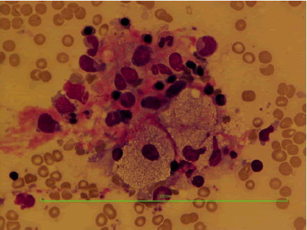
| A bone marrow aspirate confirmed florid haemophagocytosis (Figure 1). In order to exclude primary HLH, genetic studies were carried out (perforin mutation-later negative). An abdominal MRI was done to exclude lymphoma as a potential trigger. The imaging showed no enlarged lymph nodes but interestingly there was marked new hepatosplenomegaly (19 cm liver and 17 cm spleen) when there was no organomegaly on the ultrasound done earlier in her admission. |
| A 5 day course of IV immunoglobulins and high dose prednisolone dose was commenced on day 22 (as per 2004 HLH treatment protocol induction) [5] with a rapid improvement in fevers and wellbeing within 24 hours. She continued on high dose steroids for 10 days before tapering down, with complete resolution of clinical and laboratory features. Twelve months later the patient remains well with no evidence of relapse and not requiring any maintenance immunosuppression. |
| Our patient did not have all of the signs on presentation- the rash and splenomegaly evolved throughout her admission which highlights the need to have an open mind with regards to diagnosis especially in the immunocompromised. |
| Discussion |
| Epstein-Barr virus (EBV) is a B lymphotrophic human γ-herpes virus that can cause a range of clinical manifestations including selflimiting infectious mononucleosis, chronic EBV, lymphoproliferative disorders and HLH at its most extreme [9]. EBV-HLH can culminate in multi organ failure and death thus timely diagnosis is critical. |
| HLH-EBV is encountered mainly in children and adolescents, in adults HLH is more commonly secondary to an underlying malignancy [1,2,5]. Immunocompromised individuals, such as our patient, are at higher risk of acquired EBV-HLH [10]. |
| Clinical signs can include fevers, rash, splenomegaly, hepatitis. Laboratory features include cytopenia and elevated ferritin, triglycerides, LDH, soluble CD25 (Scd25), haemophagocytosis on biopsy. |
| Our patient was found to eventually have all the classical findings of HLH although the rash and splenomegaly developed later during her admission. |
| Our patient was suspected to have HLH by the haematologists on day 17 of admission, and this was confidently supported by the bone marrow biopsy and laboratory features of cytopenia, high LDH, ferritin, triglycerides and splenomegaly. This fulfilled 6 out of 8 of Henter et al HLH 2004 criteria. The remaining two criteria may have well been present but were not tested for ( sCD25>24,000 and absent or low Natural killer cell activity). |
| Whilst the EBV viremia was reducing, her symptoms persisted and liver function was worsening- this should act as a prompt that this was not a case of simple EBV infection. In an immunocompromised patient such as ours, EBV-HLH is more likely to occur and therefore important to identify early. |
| The delay in diagnosis was partly due to the similarity between of the initial presentation to a wide range of infectious and inflammatory conditions. |
| Another challenge to a timely diagnosis is related to the rare occurrence of EBV driven HLH syndrome [9]. The majority of clinicians that came into contact with our patient were not aware of HLH. |
| In terms of treatment the HLH-94 and HLH-2004 trial protocols of the Histiocyte society are both based on use of intravenous immunoglobulins, corticosteroids, etoposide and cyclosporine to induce remission [5,7]. HSCL may be needed to maintain remission although increasingly less patients are needing to resort to this In our patient, remission was induced with corticosteroids alone which is rare [7,8]. |
| Learning Points |
| • EBV-HLH can mimic other infections or inflammatory conditions and should be prompted by signs of unexplained fever, cytopenia, splenomegaly and deranged liver function especially on a background of pre-existing immune deficiency. |
| • HLH is the most fatal complication of EBV. |
| • High mortality rate in EBV-HLH is partly due to delayed diagnosis. |
| • Treatment includes immunoglobulins, steroids, chemotherapy and haematopoietic stem cell transplant. |
| • Early involvement of haematologists is paramount. |
References
- Fisman DN (2000) Hemophagocytic syndromes and infection. Emerg Infect Dis 6: 601-608.
- Freeman HR, Ramanan AV (2011) Review of haemophagocyticlymphohistiocytosis. Arch Dis Child 96: 688-693.
- Imashuku S (2002) Clinical features and treatment strategies of Epstein-Barr virus-associated hemophagocyticlymphohistiocytosis. Crit Rev OncolHematol 44: 259-272.
- Henter JI, Horne A, Aricó M, Egeler RM, Filipovich AH, et al. (2007) HLH-2004: Diagnostic and therapeutic guidelines for hemophagocyticlymphohistiocytosis. Pediatr Blood Cancer 48: 124-131.
- Janka GE, Schneider EM (2004) Modern management of children with haemophagocyticlymphohistiocytosis. Br J Haematol 124: 4-14.
- Imashuku S, Hibi S, Ohara T, Iwai A, Sako M, et al. (1999) Effective control of Epstein-Barr virus-related hemophagocyticlymphohistiocytosis with immunochemotherapy. Histiocyte Society. Blood 93: 1869-1874.
- Imashuku S, Naya M, Yamori M, Nakabayashi Y, Hojo M, et al. (1997) Bone marrow transplantation for Epstein-Barr virus-related clonal T cell proliferation associated with hemophagocytosis. Bone Marrow Transplant 19: 1059-1060.
- Lee JS, Kang JH, Lee GK, Park HJ (2005) Successful treatment of Epstein-Barr virus-associated hemophagocyticlymphohistiocytosis with HLH-94 protocol. J Korean Med Sci 20: 209-214.
- Cohen JI (2000) Epstein-Barr virus infection. N Engl J Med 343: 481-492.
- Fardet L, Lambotte O, Meynard JL, Kamouh W, Galicier L, et al. (2010) Reactive haemophagocytic syndrome in 58 HIV-1-infected patients: clinical features, underlying diseases and prognosis. AIDS 24: 1299-1306.
Tables and Figures at a glance
| Table 1 |
Figures at a glance
 |
| Figure 1 |
Relevant Topics
- Advanced Therapies
- Chicken Pox
- Ciprofloxacin
- Colon Infection
- Conjunctivitis
- Herpes Virus
- HIV and AIDS Research
- Human Papilloma Virus
- Infection
- Infection in Blood
- Infections Prevention
- Infectious Diseases in Children
- Influenza
- Liver Diseases
- Respiratory Tract Infections
- T Cell Lymphomatic Virus
- Treatment for Infectious Diseases
- Viral Encephalitis
- Yeast Infection
Recommended Journals
Article Tools
Article Usage
- Total views: 10858
- [From(publication date):
December-2015 - Nov 21, 2024] - Breakdown by view type
- HTML page views : 10157
- PDF downloads : 701
