Research Article Open Access
Langerhans Cells in Oral Mucosa from Patients with Acquired Immunodeficiency Syndrome
| Élia Cláudia de Souza Almeida1*, Renata Margarida Etchebehere2, Benito André da Silveira Miranzi3, Jannaína Grazielle Pacheco Olegário4, Vitorino Modesto Santos5, Mario Leon Silva-vergara6, Gabriela Jerônimo Dal Moro7 and Maria das Graças Reis8 |
||
| 1Doctor of Health Science, Institute of Biological and Natural Sciences, Brazil | ||
| 2Doctor of Surgical and Forensic Pathology, UFTM, Brazil | ||
| 3Doctor of Dentistry, UNIUBE, Brazil | ||
| 4Doctor in Health Science, Department of General Pathology, UFTM, Brazil | ||
| 5Doctor of Internal Medicine, Department of Armed Forces Hospital and Catholic University, Brasília-DF, Brazil | ||
| 6Doctor of Clinical Medicine, UFTM, Brazil | ||
| 7Student of Nutrition Graduation, UFTM, Brazil | ||
| 8Doctor of Science, Professor of Histology, UFTM, Brazil | ||
| Corresponding Author : | Élia Cláudia de Souza Almeida Institute of Biological and Natural Sciences Triângulo Mineiro Federal University, Rua Cazuza 160 apto 102 Uberaba-MG, Brazil Tel: 34-3318 5441 Fax: 34-3318 5462 E-mail: eliacsa@dcb.uftm.edu.br |
|
| Received July 03, 2014; Accepted August 26, 2014; Published August 30, 2014 | ||
| Citation: Almeida EC, Etchebehere RM, Miranzi BA, Pacheco Olegário JG, Santos VM, et al. (2014) Langerhans Cells in Oral Mucosa from Patients with Acquired Immunodeficiency Syndrome. J Infect Dis Ther 2:160. doi:10.4172/2332-0877.1000160 | ||
| Copyright: © 2014 de Souza Almeida EC, et al. This is an open-access article distributed under the terms of the Creative Commons Attribution License, which permits unrestricted use, distribution, and reproduction in any medium, provided the original author and source are credited. | ||
Related article at Pubmed Pubmed  Scholar Google Scholar Google |
||
Visit for more related articles at Journal of Infectious Diseases & Therapy
Abstract
Background: Oral manifestations are common in patients with acquired immunodeficiency syndrome (AIDS).
Objectives: Compare the number of Langerhans cells and intensity of anti-CD1a expression in the mucous membranes of the oral cavities of the same patients with AIDS and HIV-negative individuals.
Materials and methods: Sixteen autopsied adults were investigated, including 11 with AIDS, and 5 HIV negative. We identified Langerhans cells in three oral regions of the same subject using an anti-CD1a antibody and quantified them in cells/mm2. Were applied normality tests and Mann-Whitney.
Results: The numbers of Langerhans cells in the AIDS patient group were less than in the control group, but didn't differ significantly between the two groups. The intensity of anti-CD1a expression was lower in patients with AIDS. Of the three areas, the greater intensity of CD1a cells were found in the masticatory mucosa.
Discussion: We observed a reduction of Langerhans cells in the oral mucosa of patients with AIDS and this is the first report, to our knowledge, that evaluate three different oral mucosal membranes in the same subject.
Conclusion: Our study suggests that AIDS influences on the depletion of Langerhans’ cells, particularly in the specialized mucosa, and the intensity of expression of anti- CD1a regardless of the type of oral mucosa.
| Keywords |
| Acquired Immunodeficiency Syndrome; Langerhans cells; Mononuclear phagocyte system; HIV; CD1a antigen |
| Introduction |
| In individuals affected by acquired immune deficiency syndrome (AIDS), the digestive system is a frequent target of changes as a result of infection with human immunodeficiency virus (HIV) and several other pathogens. Although the mucosal surfaces are natural sites of penetration and probable reserves of HIV, the oral cavity is not primarily considered as a route of transmission of the virus, with exceptions of transmission through breastfeeding or oral sex. However, due to the diversity of infectious processes and perioral and oral manifestations related to this particular chronic infection, this site has been studied by some authors [1-6]. |
| The oral mucosa is made up of stratified squamous epithelial cells that vary morphologically by region, being called specialized mucosa in the tongue, masticatory mucosa in gingival tissues and hard palate, and lining mucosa in the cheeks, floor of the mouth, soft palate, the deep portion of the vestibule, the ventral portion of the lingual and internal lips [7]. Among the cell types found in the oral mucosa are Langerhans cells, which originate from bone marrow and reside in the stratum spinosum, where their cell bodies exhibit extensions that permeate surrounding epithelial cells. They belong to the mononuclear phagocyte system (MPS), which are processors and presenters of antigens, and they act as peripheral components of the immune system, expressing CD1a antigen molecules on their cell surfaces [8-13]. |
| The epithelium interfaces between the superficial oral tissues and deeper tissues and Langerhans cells are key components of the immune response in the oral mucosa, and people with AIDS show an impaired immune response. Therefore, this study investigated Langerhans cells in the different mucous membranes of the oral cavity of autopsied individuals with AIDS [6,14]. The aim of the present study was to compare the numbers Langerhans cells and the intensity of their CD1a expression in the masticatory, lining, and specialized of oral mucosal of individuals with AIDS versus those in HIV-negative individuals (controls). |
| Material and Methods |
| This study was approved by the Research Ethics Committee under protocol number 879. Specimens were obtained from autopsies performed at the Department of Surgical Pathology and Department of General Pathology at the Clinical Hospital of the Federal University of Triângulo Mineiro, Uberaba, MG or the Clinical Hospital at the School of Medicine of Ribeirão Preto, SP between April 2007 and July 2010. |
| The ages ranged from 25 to 54 years, with a mean age of 40.5 ± 7.9 years in the AIDS group and a mean age of 35.8 ± 6.9 years in the control group. The mean body mass index (BMI) for these groups was 22.3 and 27.6 kg/m2, respectively. The majority (54.5%) of patients in the AIDS group had malnutrition (BMI<18 Kg/m2). All patients were immunocompetent in control group, not undergoing chemotherapy or radiotherapy and were dentate at least in the region where the masticatory mucosa fragment was removed. All patients in the AIDS group had defining criteria of this syndrome [15]. Considering the inclusion criteria, was chosen by a non-probabilistic sample of convenience, according to the accessibility used the Mann-Whitney test [16]. Samples from three regions of the buccal mucosa (lining, masticatory, and specialized) were examined for each of the 16 subjects. The AIDS group included 11 individuals and the control group 5. Only 2/11 of the AIDS group subjects were receiving antiretroviral therapy and none were receiving chemotherapy or radiotherapy. Small fragments (~1 cm2) were harvested from each of the three regions of interest of the mucosa and were fixed in Carnoy's solution for 30 minutes, processed, and embedded in paraffin. Thereafter, the specimens were subjected to immunohistochemistry simultaneously. They were incubated with an anti-CD1a. Primary antibody (Cell Marque ®) at a dilution of 1:20 for 1 hour and 30 minutes Immunolabeling was enhanced with the streptavidin-biotin complex technique (LSAB+ System-HRP, DAKO ®) according to the manufacturer’s intructions. Images of Langerhans cells were captured at 400× magnification with a video camera attached to a brightfield microscope and a computer system with Q Win Capture software (Leica). Cells were identified and quantified throughout length of the fragments and expressed in cell numbers per mm2, considering only those with defined cell bodies and at least one identifiable dendritic extension. |
| The intensity of anti-CD1a expression was classified subjectively as absent, weak, moderate or intense. Statistical analysis was performed using Biostat ® version 5.0. A normality test and Mann-Whitney test were applied. Values were considered significant at p ≤ 0.05. |
| Results |
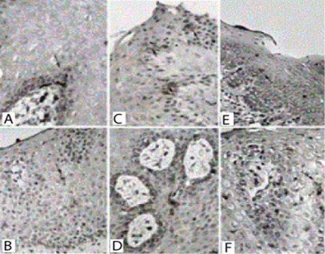
| Langerhans cells labeled with anti-CD1a were observed in the fragments of all three types of mucosa in individuals from both groups, and were always in the suprabasal location (Figure 1). The lining mucosa was the region with the highest number of Langerhans cells per mm2, while the masticatory mucosa was lower. Comparing groups AIDS and control, the distribution wasn’t normal and the statistical test used was the Mann-Whitney test. Groups lining mucosa and masticatory aren’t statistically significant different, since the specialized mucosa showed a statistically significant difference between groups (p=0.01) (Table 1). There was a wide variation in the number of Langerhans cells between individuals and the results were illustrated in Table 1. The intensity of anti-CD1a expression was greatest in the masticatory mucosa, although the number of cells was low in this region. Expression of anti-CD1a was lower in AIDS patients (weaker positivity) in all regions of the oral mucosa (Table 2). Anti-CD1a expression was illustrated in Figure 1. |
| A-B. Mucosal lining of HIV-negative control (A) and AIDS patients (B). C-D Masticatory mucosa from HIV-negative control (C) and an AIDS patient (D). E-F Specialized mucosa from HIV-negative control (E) and an an AIDS patient (F) (400×). |
| Of the 11 AIDS patients, 9 had viral load available and 4 of these over 50 000 viral copies/ml. Among the 9 patients with available viral load, only one had CD4 counts greater than 200 cells/mm3 and this was not on ART. |
| Discussion and Conclusion |
| Our observation of fewer Langerhans cells in AIDS patients, particularly in the specializated mucosa, fits with observations by other authors in the oral mucosa and gastrointestinal tract [2,17-22]. However, we found no prior reports in the literature evaluating three different mucous membranes of the oral cavity of the same individuals, as in the present study. |
| HIV have tropism of Langerhans cells, in addition to its effects on T lymphocytes, macrophages, dendritic cells, and endothelial cells. Chronic HIV infection is characterized by a progressive depletion of Langerhans cells, as evidenced in this study. HIV infection likely has direct effects on oral mucosal immunity as well as indirect effects related to its effects on the immune response [13]. |
| Regional variations in the distribution and density of Langerhans cells in the mucosa in different locations of the oral cavity with similar functions have been described previously, but from different individuals [23-25]. In the present study, we observed similar regional variations in the same individuals, with a greater number of Langerhans cells in the lining mucosa, followed by the specialized and the masticatory, in both groups. |
| It was described recently that more Langerhans cells are present in HIV-positive individuals with periodontitis, using anti-S100 immunohistochemistry, compared to HIV-negative individuals. The lower specificity of the anti-S100 antibody relative to the anti-CD1a antibody used in this study may have contributed to their observation of a larger effect [25,26]. Moreover, we evaluate patients with AIDS and, in the previous study, patients had an HIV infection, without immunosuppression declared. |
| Periodontitis is an inflammatory infectious disease, common in the general population, including among HIV-positive and SIDA individuals. Kato Segundo et al (2011) conducted a study in patients with AIDS, antiretroviral therapy (ART) and periodontal disease. These authors categorized the oral disease in severity. The authors observed that the use of ART decreases the viral load and consequently the destruction of Langerhans cells in gingiva of patients with periodontitis [27]. In the present study, only 2 of AIDS group subjects were receiving ART and most had high viral load and low CD4 count. |
| Another factor that could contribute to there being fewer Langerhans cells in patients with AIDS is malnutrition, which can impair the immune response to various inflammatory and infectious processes [28,29]. |
| In conclusion, by comparing different regions within the same individuals, our study provides evidence that AIDS probably influences the number of Langerhans cells in the oral mucosa, particularly in the specialized mucosa, and the intensity of expression of anti-CD1a regardless of the type of oral mucosa. However, further studies with a larger number of subjects are needed to confirm our findings. |
| Financial disclosure |
| Funding support was provided by the National Council for Scientific and Technological Development (CNPq), Coordination for the Improvement of Higher Education Personnel (CAPES), Minas Gerais State Research Foundation (FAPEMIG), and the Teaching and Research Foundation of Uberaba (FUNEPU); Hospital de Clinicas at the Faculdade de Medicina de Ribeirão Preto-USP, SP. |
| Disclosure of potential conflicts of interest: The authors had full freedom of manuscript preparation and there were no potential conflicts of interest. |
| Acknowledgment |
| Department of Pathology, Faculty of Medicine, University of São Paulo, Ribeirão Preto, São Paulo for allowing the collection of part of the research material. |
References
- Dias EP, Feijó EC, Polignano GAC (1998) Diagnósticoclínico e cito – histopatológico das manifestaçõesbucais da AIDS. J Bras Doenças Sex Transm 10: 10-16.
- Guimarães LC, Silva ACAL, Micheletti AMR, Moura ENM, Silva-Vergara ML, et al. (2012) Morphological changes in the digestive system of 93 human immunodeficiency virus positive patients: an autopsy study. Rev Inst Med Trop Sao Paulo 54: 89-93.
- Hocini H, Bomsel M (1999) Infectious human immunodeficiency vírus can rapidly penetrate a tight human epithelial barrier by transcytosis in a process impaired by mucosal immunoglobulins. J infect dis 179: 448-453.
- Liu X, Zha J, Chen H, Nishitani J, Camargo P, et al. (2003) Human immunodeficiency virus type 1 infection and replication in normal human oral keratinocytes. J Virol 77: 3470-3476.
- Souza LB, Pinto LP, Medeiros AMC, AraújoJr RF, Mesquita OJX (2000) Oral manifestations in patients with AIDS in a Brazilian population. PesqOdont Bras 14: 79-85.
- Boy SC, van Heerden MB, Wolfaardt M, Cockeran R, Gema E, et al. (2009) An investigation of the role of oral epithelial cells and Langerhans cells as possible HIV viral reservoirs. J Oral Pathol Med 38: 114-119.
- Gartner LP (1994) Oral anatomy and tissue types. SeminDermatol 13: 68-73.
- Banchereau J, Briere F, Caux C, Davoust J, Lebecque S, et al. (2000) Immunobiology of dendritic cells. Annu Rev Immunol 18: 767-811.
- Cutler CW, Jotwani R (2006) Dendritic cells at the oral mucosal interface. J Dent Res 85: 678-689.
- Lipscomb MF, Masten BJ (2002) Dendritic cells: immune regulators in health and disease. Physiol Rev 82: 97-130.
- Piguet V, Blauvelt A (2002) Essential roles for dendritic cells in the pathogenesis and potential treatment of HIV disease. J Invest Dermatol 119: 365-369.
- Jaitley S, Saraswathi T (2012) Pathophysiology of Langerhans cells. J Oral MaxillofacPathol 16: 239-244.
- Horewicz VV, Ramalho L, dos Santos JN, Ferrucio E, Cury PR (2013) Comparison of the distribution of dendritic cells in peri-implant mucosa and healthy gingiva. Int J Oral Maxillofac Implants 28: 97-102.
- Challacombe SJ, Naglik JR (2006) The effects of HIV infection on oral mucosal immunity. Adv Dent Res 19: 29-35.
- Brasil. Ministério da Saúde. Secretaria de VigilânciaemSaúde. ProgramaNacional de DST e AIDS (2003) Critérios de de?nição de casos de aids emadultos e crianças./ Ministério da Saúde, Secretaria de VigilânciaemSaúde, Programa Nacional de DST e Aids. Ministério da Saúde: Brasília.
- Oliveira, TMV (2001) Amostragemnãoprobabilistica: adequação de situaçõesparauso e limitações de amostrasporconveniência, julgamento e quotas. Administração Online [online] FECAP. 2.
- Brown KN, Trichel A, Barratt-Boyes SM (2007) Parallel loss of myeloid and plasmacytoid dendritic cells from blood and lymphoid tissue in simian AIDS. J Immunol 178: 6958-6967.
- Charton-Bain MC, Terris B, Dauge MC, Marche C, Walker F, et al. (1999) Reduced number of Langerhans cells in oesophageal mucosa from AIDS patients. Histopathology 34: 399-404.
- Myint M, Yuan ZN, Schenck K (2000) Reduced numbers of Langerhans cells and increased HLA-DR expression in keratinocytes in the oral gingival epithelium of HIV-infected patients with periodontitis. J ClinPeriodontol 27: 513-519.
- Rocha L, Silva R, Olegário J, Corrêa R, Teixeira V, et al. (2010) Esophageal epithelium of women with AIDS: thickness and local immunity. Pathol Res Pract 206: 248-252.
- Rocha LP, de Melo E Silva AT, Gomes NC, Faria HA, Silva RB, et al. (2011) The influence of gender and of AIDS on the immunity of autopsied patients' esophagus. AIDS Res Hum Retroviruses 27: 511-518.
- Tschachler E, Groh V, Popovic M, Mann DL, Konrad K, et al. (1987) Epidermal Langerhans cells--a target for HTLV-III/LAV infection. J Invest Dermatol 88: 233-237.
- Upadhyay J, Upadhyay RB, Agrawal P, Jaitley S, Shekhar R (2013) Langerhans cells and their role in oral mucosal diseases. N Am J Med Sci 5: 505-514.
- Chou LL, Epstein J, Cassol SA, West DM, He W, et al. (2000) Oral mucosal Langerhans' cells as target, effector and vector in HIV infection. J Oral Pathol Med 29: 394-402.
- Hussain LA, Lehner T (1995) Comparative investigation of Langerhans' cells and potential receptors for HIV in oral, genitourinary and rectal epithelia. Immunology 85: 475-484.
- Petersen PE, Ogawa H (2012) The global burden of periodontal disease: towards integration with chronic disease prevention and control. Periodontol 2000 60: 15-39.
- Kato Segundo T, Souto GR, Mesquita RA, Costa FO (2011) Langerhans cells in periodontal disease of HIV negative and HIV positive patients undergoing highly active antiretroviral therapy. Braz Oral Res 25: 255-260.
- Ambrus JL Sr, Ambrus JL Jr (2004) Nutrition and infectious diseases in developing countries and problems of acquired immunodeficiency syndrome. ExpBiol Med (Maywood) 229: 464-472.
- Gasparis AP, Tassiopoulos AK (2001) Nutritional support in the patien t with HIV infection. Nutrition 17: 981-982.
Tables and Figures at a glance
| Table 1 | Table 2 |
Figures at a glance
 |
| Figure 1 |
Relevant Topics
- Advanced Therapies
- Chicken Pox
- Ciprofloxacin
- Colon Infection
- Conjunctivitis
- Herpes Virus
- HIV and AIDS Research
- Human Papilloma Virus
- Infection
- Infection in Blood
- Infections Prevention
- Infectious Diseases in Children
- Influenza
- Liver Diseases
- Respiratory Tract Infections
- T Cell Lymphomatic Virus
- Treatment for Infectious Diseases
- Viral Encephalitis
- Yeast Infection
Recommended Journals
Article Tools
Article Usage
- Total views: 14055
- [From(publication date):
October-2014 - Apr 03, 2025] - Breakdown by view type
- HTML page views : 9412
- PDF downloads : 4643
