Kinematic Causes and Exercise Rehabilitations of Patients with Round Shoulder, Thoracic Kyphosis and Forward Head Posture (FHP)
Received: 25-Jul-2016 / Accepted Date: 12-Sep-2016 / Published Date: 19-Sep-2016 DOI: 10.4172/2161-1165.1000263
Abstract
Purpose: To analyze the cause in kinematics and corresponding exercise rehabilitation of round shoulder, thoracic kyphosis and forward head posture. Methods: We recruited 26 patients with obvious symptoms of round shoulder, thoracickyphosis and forward head posture, with a difference of 4 degree or more in positive rake angle. Patients include 16 males with an average age of 24 ± 3.4 (Mean ± SD) and 10 females with an average age of 25 ± 3.7, with an overall duration of pathological symptoms of 3.4 ± 1.3 years. The sports prescription effectiveness was evaluated base on positive rake angle, neck vertebrae mobility and glenohumeral mobility. Results: The positive rake angles were decreased from 5.4 ± 2.6 degrees (pretreatment) to 2.2 ± 1.2 degrees (post-treatment). Cervical joint mobility range bending from front to back was improved from 56.8 ± 2.6 degrees (pre-treatment) to 76.6 ± 2.2 degrees (post-treatment), the range bending from left to right improved from 46.1 ± 3.7 degrees (pre-treatment) to 80.1 ± 3.4 degrees (post-treatment), and the range rotating from left to right improved from 80.5 ± 9.2 degrees (pre-treatment) to 122.8 ± 9.6 degrees(post-treatment). The glenohumeral mobility was improved from 144.7 ± 7.5 degrees (internal bending), 43.4 ± 4.7 degrees (external stretching), 77.3 ± 5.7 degrees (internal rotation), 69.8 ± 5.3 degrees (external rotation) to 172.3 ± 6.8 degrees (internal bending), 54.5 ± 4.1 degrees (external stretching), 83.2 ± 6.6 degrees (internal rotation), 80.4 ± 7.3 degrees (external rotation). Significant difference between pre-treatment and post-treatment were observed based on evaluation of all three factors (p<0.05). Conclusion: Abnormal postures, such as round shoulder, thoracic kyphosis and forward head posture, could be resulted from long-time operation of computers, which induced unbalanced kinematics of muscles around the neck, chest and back. Underload resistive exercises and stretching of chest and arms have significant improving effect for correcting round shoulder, thoracic kyphosis and forward head posture.
Keywords: Round shoulder; Thoracic kyphosis; Forward head posture (FHP); Unbalanced kinematics; Sports prescription
164521Introduction
Normal human body displays a relaxing posture of head and shoulders, with chin tucked in, head sticked up and shoulders opened naturally. Yet in the Internet era, frequent usage of mobile device and electronic equipment resulted in abnormal postures such as round shoulder, thoracic kyphosis and forward head posture (FHP), which become more and more common for large populations.
Round shoulder, thoracic kyphosis and FHP are abnormal postures of human body, including excessive anterior positioning of cervical spine, over-bending of thoracic vertebra, increased positive rake of scapula and shoulders turning forward with inward rotation [1].
Currently, in the Internet Era, you can always see people using an electronic device such as smart phones, tablets, laptops, and all other kinds of computers.
Along with those popular devices, the abnormal postures such as round shoulder, thoracic kyphosis and FHP become common for large populations as well.
Those postures not only affect regular appearance of a person, but also have negative effects on human bodies, including but not limited to sourness and pain on neck, shoulder and back, headache, dizziness, difficulties of breath, palpitation, chest distress, sleep disorders, dumbness of arm, and limited mobility of cervical vertebra and glenohumeral joints [2].
In this study, we hypothesize that abnormal postures like round shoulders, thoracic kyphosis and FHP are phenotypic symptoms of muscle injuries, and they are closely related to unbalanced kinematics of muscles around neck and those on chest and upper back of human body.
Patients given sport medicine prescriptions and regular posture exercises displayed obvious improvement in correcting the abnormal postures such as round shoulder, thoracic kyphosis and FHP and in the related symptoms.
Methods
Participants
A total of 26 patients (16 males with an average age of 24 ± 3.4 and 10 females with an average age of 25 ± 3.7) participated in this study of sports prescription.
Inclusion criteria: Obvious symptoms of round shoulder, thoracic kyphosis and forward head posture, with positive rake angle of 4 degrees or more, accompanied with cervical pain, shoulder pain, back pain and limited mobility of cervical vertebra and glenohumeral joints. The average disease course is 63.4 ± 1.3 months in all patients.
Exclusion criteria: Patients with complications such as heart disease, emphysema, bone fracture, joint dislocation, cancer or tuberculosis.
IRB approval is granted for this study, and all the participants were provided with information about the purpose, methods and possible benefits as well as risks related to the experiments. Signed consent forms were provided by every participant.
Equipment and exercises
A total of 26 participants were educated with detailed contents of sports medicine prescriptions and correct exercises methods. The exercises were carried out in professional rehabilitation room one hour after each meal, with guidance by therapists, and the process of exercise were recorded.
The exercises were carried out every other day for 30 days, 30 minutes each time. The results were evaluated 30 days after the exercises started.
Thera-band elastic bands were used in sports medicine prescription exercises for resistive force.
Exercise prescription
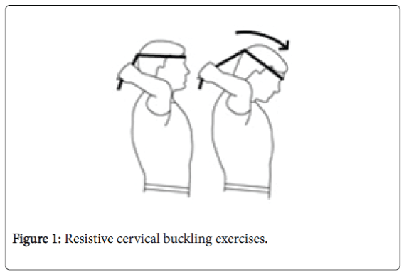
Sit or stand naturally and wrap elastic band around forehead. Hold two ends of the band and gently pull back. Look straight forward, and then bend neck down.
When maximum head movement is reached, hold for 10 seconds. Slowly return to start position and repeat for four times (Figure 1).
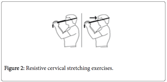
Sit or stand naturally and wrap elastic band around back of the head. Hold two ends of the band and gently pull forward. Look straight forward, and then tuck chin in. When maximum head movement is reached, hold for 10 seconds. Slowly return to start position and repeat for four times (Figure 2).
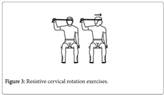
Sit or stand naturally and wrap elastic band around side of the head (above ear). Hold two ends of the band in right hand and gently pull right. Look straight forward, and then turn head to left. When maximum head movement is reached, hold for 10 seconds.
Slowly return to start position and repeat on the other side. Repeat the exercises on both sides for four times (Figure 3).
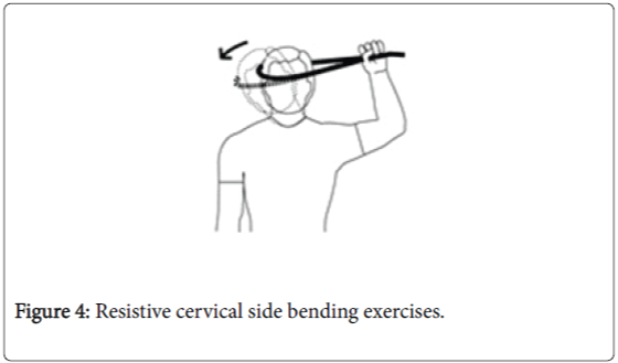
Sit or stand naturally and wrap elastic band around side the head (above ear). Hold two ends of the band in right hand and gently pull right. Look straight forward, and then bend neck towards left shoulder.
When maximum head movement is reached, hold for 10 seconds. Slowly return to start position and repeat on the other side. Repeat the exercises on both sides for four times (Figure 4).
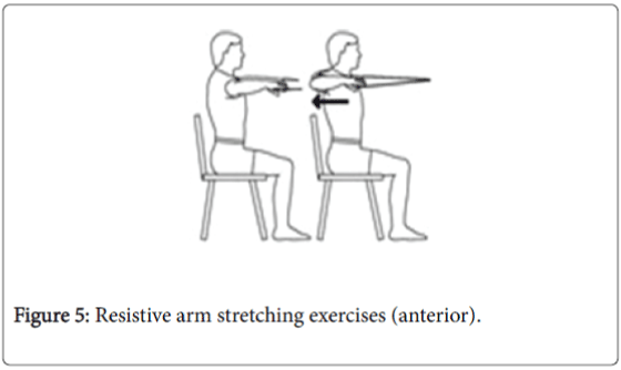
Sit naturally in chair with back unsupported. Hold elastic band in two hands and gently pull straight, raising arm to shoulder level. With one arm extending out straight, pull the other arm back.
When maximum hand movement is reached, hold for 5 seconds. Slowly return to start position and repeat on the other side. Repeat the exercises on both sides for twenty times (Figure 5).
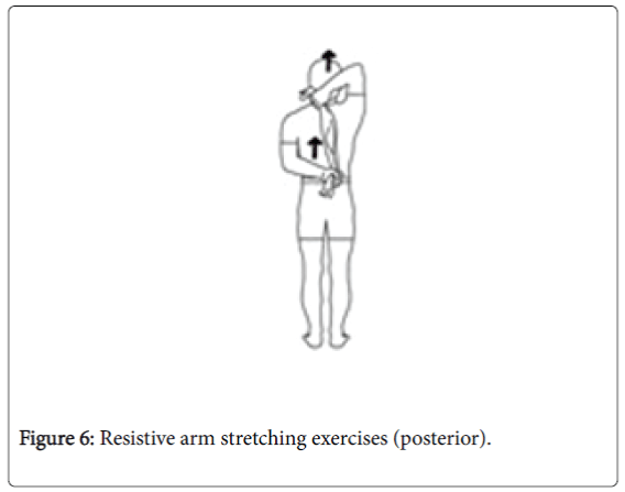
Stand naturally with both hands holding elastic band behind (Figure 6). Upper hand pulls up and lower hand pulls down. When maximum hand movement is reached, hold for 10 seconds. Return to start position and repeat with switched hands. Repeat the exercises for both hands for four times (Figure 6).
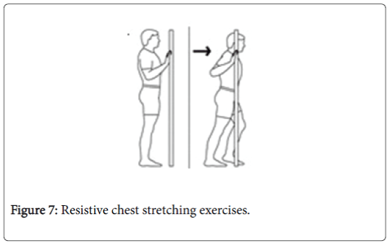
Stand naturally in front of an open door. Hold door frame with both hands and step up one foot. Keep the back straight, bend elbows and move chest forward until maximum (or until the chest is beyond the tip of toe of the front foot). Hold for 15 seconds. Return to start position and repeat for four times (Figure 7).
Data collection
Positive rake angle was measured by PWS mobility measurer (Jiangsu Qianjing Medical Equipment Co., China) [3].
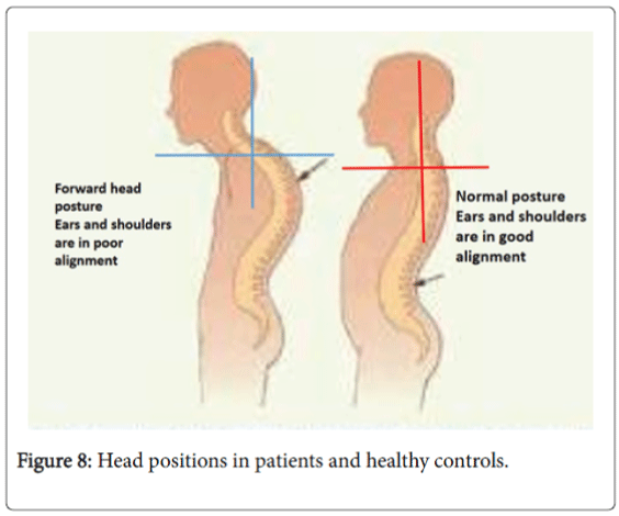
The angle between earlobe and acromion was measured as shown in Figure 8. Cervical vertebrae mobility was measured by PWS mobility measurer (Jiangsu Qianjing Medical Equipment Co., China).
The front-back (flection to extension), side binding (stretching from left to right) and rotation (from left to right) ranges were measured. Patients sat tight on a chair, with upper body straight up and back and shoulders clinging to the back of the chair (Figure 8).
The glenohumeral mobility (internal bending, external bending, external stretching, internal rotation and external rotation) was measured by PWS mobility measurer (Jiangsu Qianjing Medical Equipment Co., China) [3].
For internal bending, external bending and external stretching, the patient stood with back aligning on a straight column, with whole body stretching up, head, back and buttocks clinging to the column.
For internal rotation and external rotation, the patient lay up front. The measurement was described previously [3].
Statistical analysis
The data were analyzed by SPSS for Windows version 18.0 (SPSS Inc., Chicago, IL, USA). One-way ANOVA analysis and Newman– Keuls test were used when appropriate (p<0.05 was considered significant), and data were shown as Mean ± SD.
Results
Positive rake angle was decreased after rehabilitation treatment
The positive rake angle in 26 patients was decreased from 5.2 ± 2.3 (pre-treatment) to 2.2 ± 1.2 (post-treatment), and the difference is significant (p<0.05). By under loaded resistive sports medicine prescriptions and exercises, the strength of cervical extension muscle groups and upper back muscle groups was increased, especially for the and the endurance of muscles, which is essential for protection from excessive anterior positioning of head and cervical spine.
Our results indicate that the increase of positive rake angle may not be due to the change of cervical vertebrae itself, but more likely be the result of over-stretching of cervical vertebrae by anterior cervical muscle groups.
Cervical vertebra mobility was increased after rehabilitation exercises
In Table 1, significant improvement of cervical vertebra mobility was observed after resistive sports medicine exercises and stretching exercises on cervical vertebra, thoracic vertebra and arms (p<0.05).
| Movement | Flection-to-extension (80°) | Left-to-right bending (90°) | Left-to-right rotation (140°) | |||
|---|---|---|---|---|---|---|
| Before | After | Before | After | Before | After | |
| Angle | 56.8 ± 2.6 | 76.6 ± 2.2 | 46.1 ± 3.7 | 80.1 ± 3.4 | 80.5 ± 9.2 | 122.8 ± 9.6 |
Table 1: Cervical vertebra mobility in patient group (n=26, Mean ± SD).
When round shoulder, thoracic kyphosis, FHP or similar abnormal postures occur, the muscles around cervical vertebra become tense and spastic, thus limited the movement of cervical and thoracic vertebras. By resistive cervical exercises, the tense muscles could be effectively relaxed, which helps restoration of muscle elasticity, loosened intervertebral joints and re-activation of muscles.
Glenohumeral joint mobility was increased after rehabilitation exercises
In Table 2, post-treatment glenohumeral joint mobility was significantly increased comparing to that of pre-treatment (p<0.05).
| Movement | Internal bending (180°) | Extension (60°) | Internal rotation (90°) | External rotation (90°) | External stretching (180°) | |||||
|---|---|---|---|---|---|---|---|---|---|---|
| Before | After | Before | After | Before | After | Before | After | Before | After | |
| Angle | 144.7 ± 7.5 | 172.3 ± 6.8 | 43.4 ± 4.7 | 54.5 ± 4.1 | 77.3 ± 5.7 | 83.2 ± 6.6 | 69.8 ± 5.3 | 80.4 ± 7.3 | 146.1 ± 7.2 | 169.2 ± 8.9 |
Table 2: Glenohumeral joint mobility in patient group (n=26, Mean ± SD).
In patients with symptoms such as round shoulder, thoracic kyphosis, FHP, etc., round shoulder indicates that glenohumeral joint and scapula were far off their normal positions. The forward positioning of scapula caused excessive traction of muscles attached to scapula, leading to tension and spasm, which then limits the mobility of glenohumeral joints. The resistive exercises with arms could enhance the strength of muscles attached to scapula and increase the extensive capability of arms. The chest stretching exercises could effectively ease the tension and spasm of thoracic muscle groups and promote repositioning of scapula back to normal.
Discussion
Imbalanced muscle kinematics in round shoulder, thoracic kyphosis and FHP
Abnormal body postures (round shoulder, thoracic kyphosis and FHP) were mainly due to the imbalance among a few muscle groups, including the anterior and posterior cervical muscle groups, anterior thoracic muscle groups and upper back muscle groups.
First is the imbalance among anterior cervical flexor muscles and upper back extensor muscles. The anterior cervical flexor muscle group is consisted of sternocleidomastoid muscle, scalene muscle and hyoideus muscle. The main function of anterior cervical flexor muscles is to control the stabilization, rotation and bending of head and neck. Muscle contractions initiated by cervical vertebra allow people to lower their heads to read books, use mobile devices or operate computers. Sternocleidomastoid muscle and scalene muscle are major players during this process. The upper end of sternocleidomastoid muscle extends to outside of processus mastoideus and middle of superior nuchal line, and the lower end to presternum and preclavicular surface. Contraction of sternocleidomastoid muscle pulls the head to chest and makes the head moving forward [4]. Scalene muscle can be divided into three groups based on their location (anterior, medius and posterior). They attach to 2-7 Transverse processes of cervical vertebra and ends between 1-2 ribs. Scalene muscle controls the flection of cervical vertebra, and it serves as an assistant muscle during abnormal breathing. Injured scalene muscle could cause pressure on inferior aperture of thorax, which leads to arm pain and/or numbness. That is due to the fact that brachial arteries, axillary artery, subclavian artery and brachial plexus pass through anterior and medius scalene muscles, where the inferior aperture of thorax locates [5]. That may explain why quite a few patients with round shoulder, thoracic kyphosis and forward head posture also displayed symptoms of dysponea and pain and numbness of arms.
Upper back muscle groups competes against anterior cervical flexor muscles. Upper back muscles include suboccipital muscles (rectus capitis posterior major muscle, rectus capitis posterior minor muscle, obliquus capitis superior muscle and obliquus capitis inferior muscle), cucullaris muscle, levator muscle of scapula, splenius muscle of neck, rhomboideus muscle, etc., and their function is to extend and stabilize spine. When cervical vertebra undergoes internal bending activities, those muscles extend by centrifugal contractions. When a person lowers head to read a book or use a mobile device, it requires 30-40% energy of the extended spinal muscles, and ∼60% energy of head and neck muscles to maintain proper angle for reading, as the arm of force for neck muscles and tendons is smaller than that of the head movement [1]. Longer reading time is often accompanied with lower head position, due to continuously exhausted muscle energy from head, neck and spine area, and result in overtoil of upper back muscles and injuries of the microstructure of muscles. Suboccipital muscles and trapezius muscles would bear the most serious injuries. Suboccipital muscles locate within the triangle area of nuchal plane, transverse process of first cervical vertebra (atlas) and nuchal bone, and control the position of head. Suboccipital muscles work against sternocleidomastoid muscles, and both muscle groups display tension at the same time [5]. Trapezius muscle covers posterior cervical area, shoulder and upper back area with multi-functions. If trapezius muscle is under tension for a long time, the muscles harden and cause difficulties to raise head, and patients usually have limited mobility of cervical vertebra and obvious pain on neck and shoulder.
The second is the imbalance of thoracic muscles and middle back muscles. Among all the thoracic muscles, pectoralis major muscle is the key player related to round shoulder phenotype. Pectoralis major muscle fibers cling to the anterior thoracic area like an opening folding fan, and come together in front of humerus, covering sternoclavicular, acromioclavicular and glenohumeral joints. When under tension, pectoralis major muscle pulls shoulder bone forward, and causes dislocation of those three joints [6]. In this study, we found that in patients with round shoulder, thoracic kyphosis and FHP, the mobility of shoulder joints are much limited comparing to healthy people, especially for the backward extension of the arms. Malfunction of another muscle, pectoralis minor muscle, is responsible for thoracic kyphosis. Pectoralis minor muscle starts from the connection between ribs 2-5 and costal cartilage, and extends to the top of processus coracoideus. Tensed pectoralis minor muscle pulls scapula to thorax, and induces the antagonistic contraction of back muscles (including middle and lower trapezius muscles, rhomboid muscle and serratus posterior superior muscles). Most of the upper back muscles attach to and stabilize scapula between spine and shoulder bone, so as to complete the backward extension, outward rotation and movement towards spine. Tension and strong contraction of thoracic muscles could result in overfatigue and antagonistic injuries (such as injuries of rhomboideus muscle). Rhomboideus muscle pulls scapula away from thorax (and thus works against the upper back muscles to stabilize scapula). The tension of thoracic muscles could easily cause the tension and spasm, which lead to pain of upper back and shoulders, possible brachial plexus neuralgia and numbness in upper arm when raising the arm [7].
Rehabilitation of round shoulder, thoracic kyphosis and FHP
In this study, we designed rehabilitation exercises based on muscle kinematics analysis above, which focuses on extension exercises on strong and tensed thoracic muscles, ad antagonistic force exercises on comparably weak areas (neck and back muscles), so as to re-balance the force of body muscles. Usage of elastic bands could increase resistance to increase muscle force and restore elasticity of muscles. Lengthening the elastic band and slowly moving back to original position could make centrifugal contraction of muscles, and transmit the force to deeper layer of muscles, so as to improve endurance and elasticity of muscles [8,9]. Cervical flection, rotation and side bending in sports prescription could strengthen sternocleidomastoid muscle, scalene muscle and upper trapezius muscle. Cervical stretching could develop the muscle force of trapezius muscle, multifidi muscle, rotator muscle, splenius muscle of neck and longissimus capitis muscle, to prevent muscle force deficiency after long time cervical flection. Anterior and posterior arm stretching exercises could strengthen middle and lower trapezius muscle, rhomboideus muscle, supraspinatus muscle, bicipital muscle of arm, brachial triceps muscle and latissimus dorsi, stabilize scapula and remove the limitation of scapular upward rotation, outward rotation of shoulder bone and backward extension of arm, to restore the normal mobility of shoulder joints [10]. Chest stretching exercises could elongate the contracted pectoralis major and minor muscles, and restore normal contraction and extension of those muscles. During this exercise, to completely extend pectoralis major and minor muscles, it is recommended that the patient try to place palm at several positions (either equal to or above the shoulder), and hold for 15 seconds at each position, so that not only the muscle fiber, but also the tendons could be extended as well [11].
Conclusion
Long time lowering head and/or using mobile devices could induce round shoulder, thoracic kyphosis and FHP, which is due to imbalanced kinematics of cervical muscles, thoracic muscles and upper back muscles. Underloaded resistive sports prescription exercises and stretching of thoracic muscles could re-balance muscle force, restore muscle elasticity, re;move tension and spasm of muscles, and recover mobility of cervical vertebra and shoulder joints, and effectively improve the symptoms of round shoulder, thoracic kyphosis and FHP. We would also recommend patients displaying those symptoms to regularly keep normal posture (with chin tucked in, shoulders squared and opened naturally, abdomen drawn, etc.), so as to maintain the balance of muscle kinematics between thorax and shoulders and to prevent abnormal postures.
References
- Bolsterlee B, Veeger HEJ, van-der FCT (2014) Modelling clavicular and scapular kinematics: from measurement to simulation. Med Biol Eng Comput 52: 283-291.
- Richter P, Hebgen E (2008) Trigger Points and Muscle Chains in Osteopathy (1st edn.)Thieme Stuttgart, New York.
- Wang Y, Wang R, Chen P (2011) Sports Traumatology (Yun Dong Chuang Shang Xue). People's Militery Medical Press, Beijing.
- Clay JH, PoundsDM (2008) Basic Clinical Massage Therapy: Integrating Anatomy and Treatment(2nd edn.)Lippincott Williams & Wilkins.
- Hamilton N, Weimar W, Luttgens K (2011) Kinesiology: Scientific Basis of Human Motion(12th edn.) McGraw-Hill Education. 640.
- Cools AM, Dewitte V, Lanszweert F, Notebaert D, Roets A, et al.(2007) Rehabilitation of Scapular Muscle Balance: Which Exercises to Prescribe? Am J Sports Med 35: 1744-1751.
- Liao YP, JiaoW (2011) Effects of Different Exercise Modes on the MHC-?,COL-?and COL-? in Healing Process of Acute Skeletal Muscle Injury of Rats. Chinese Jouornal of Sports Medicine, 30: 1018-1022.
- Walker B (2007) The Anatomy of Stretching, Second Edition: Your Illustrated Guide to Flexibility and Injury Rehabilitation(2nd edn.)North Atlantic Books,California.
- Walker B (2005) The Anatomy of Sports Injuries (1st edn.)North Atlantic Books. California.
- Lv P (2016) A Study of Using Massage Therapy Accompanied with Stretching Exercise for Rehabilitation of Mammary Gland Hyperplasia. BioMed Res Int.
Citation: Chen X, Pin LV, Peng, Zhang Y, Ding K (2016) Kinematic Causes and Exercise Rehabilitations of Patients with Round Shoulder, Thoracic Kyphosis and Forward Head Posture (FHP). Epidemiology (Sunnyvale) 6:263. DOI: 10.4172/2161-1165.1000263
Copyright: © 2016 Chen X. This is an open-access article distributed under the terms of the Creative Commons Attribution License, which permits unrestricted use, distribution, and reproduction in any medium, provided the original author and source are credited.
Share This Article
Recommended Journals
Open Access Journals
Article Tools
Article Usage
- Total views: 20564
- [From(publication date): 10-2016 - Apr 02, 2025]
- Breakdown by view type
- HTML page views: 19258
- PDF downloads: 1306

