Isolated Fracture Dislocation of Navicular Bone: A Case Report
Received: 03-Feb-2018 / Accepted Date: 13-Feb-2018 / Published Date: 20-Feb-2018 DOI: 10.4172/2329-910X.1000262
Abstract
Isolated fracture dislocation of navicular bone is rare. It is due to a plantar flexion compressive injury, which crushes the bone. Sometimes it may displace a part of the fractured bone from the naviculocuneiform and the talonavicular joints. We present a case report of a 37 year old male who presented with isolated fracture dislocation of navicular. Open reduction and Internal Fixation was done on the dorsal surface using Navicular plate and screws. There is always a possibility of ischaemic necrosis and post-traumatic arthritis in such cases.
Keywords: Fracture dislocation navicular bone; Naviculocuneiform; Talonavicular joints; Navicular plate; Plantar flexion injury
Introduction
Navicular bone is a boat shaped bone, which is one of the seven tarsal bones of the human foot. It is situated midfoot towards the hallux side of the foot, maintaining the medial column of foot between talus and cuneiform bones [1]. Isolated fracture dislocation of this bone is rare because it is a very rigid bony and there are many ligamentous structures surrounding the navicular bone [1]. There are very few cases reported in literature to date [2,3]. Usually it is accompanied with the fracture of Navicular [4,5] itself or with multiple other bones of the foot [6-8]. It is most commonly seen in athletes [9-15]. Cause of injury is generally due to the severe abduction force when the foot is in plantar flexion [16,17].
The ligament attachments around the bone are very strong and due to the position of the navicular bone, fractures are much more common than dislocations. But, sometimes it may displace a part of the fractured bone from the naviculocuneiform and the talonavicular joints only, which is discussed in our case study [17]. Isolated fracture dislocation of navicular bone could have serious consequences because of the essential role of the talonavicular joint [18]. Disruption of talonavicular joint can result in loss of 90% or greater of complex hind foot motion.
Case Report
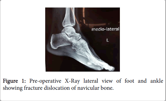
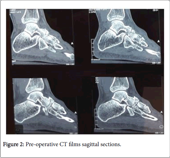
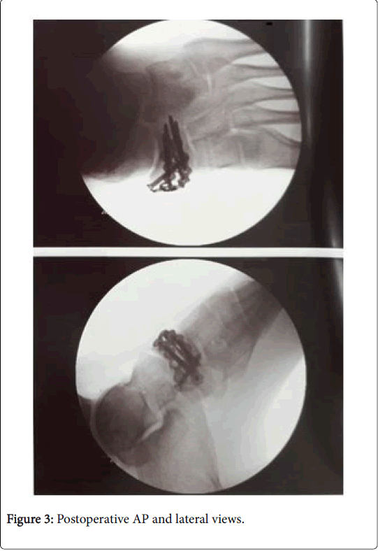
A 37 year old male presented himself with a 5 days old injury on the left foot. He was riding his motorcycle when he, at slow speed, put his left foot down to step off the bike. It was a jarring injury to his left foot and he bore a fracture dislocation of the talonavicular joint. He had swelling and pain and had been imaged with plain X-rays and a CT scan. Scans showed that the primary injury was really the navicular, which was fragmented and impacted by the talar head crushing into it. The dorsal lip of the talar head had been fractured off, and this represented an impaction fracture dislocation of the talonavicular joint. Patient was advised that the joint needed to be reduced and the fractured navicular needed to be bone-grafted and plated. He was also told possible risks of infection, DVT or non-union of the bone post the surgery. He was taken to the operation theatre where he was given spinal anaesthesia. This was 7 days post his injury. Under tourniquet, using manual traction, an antero-medial incision was given over foot and an open reduction was done. It was then confirmed under image intensifier. Navicular fracture reduction was temporarily maintained using K-wire fixation. Internal fixation with navicular plate and screws done with stryker navicular plate on the dorsal surface. K-wires were removed, and wound was closed in layers. Postoperatively patient was kept in below knee back slab.
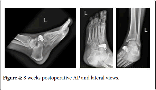
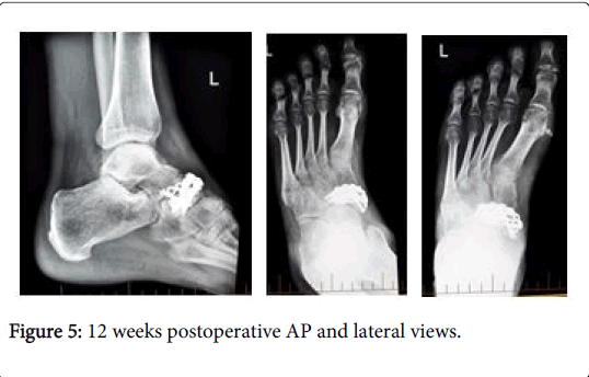
At 2 weeks postoperative follow-up, patient looked well, and wounds were clean, and the clips removed. He could then wash his foot and commence range of motion exercises. He was then allowed partial weight bearing with his boot on, touch weight bearing on the toes. He was advised to keep it elevated. He could remove the boot for washing, sleeping, and range of motion exercises. 8 weeks post-surgery, he was doing well. He still had marked swelling and some stiffness, but he was comfortable, he could take weight on his foot as his X-ray was satisfactory. He was allowed take weight and transition from his CAM boot to work boots over the next four weeks. He was also advised to swim, cycle and walk around the house. His follow-ups were scheduled for 4 weeks. At 12 weeks follow-up, the patient had returned to work and resumed his everyday activities. Till date, at 24 weeks post-surgery no complications have been noticed or recorded and patient is doing well and back to cycling (Figures 1-5).
Discussion
Fractures of the navicular are usually due to severe force to plantar flexed foot and most often occur without ligamentous injury. A vertical fracture dislocation of the navicular bone however is even more rare [19-22]. Till now, very few published case reports have been mentioned in literature and earliest published case was reported in 1924 [3]. A longitudinal force transmitted along the metatarsal ray compresses the navicular in a tight space between the cuneiforms and the head of the talus. This causes a force in line with the intercuneiform joints. The naviculocuneiform ligament tear, and the force along the lateral rays tend to crush the lateral fragment of the navicular while the remaining major fragment is extruded medially [21]. This type of injury disrupts the normal arch of the foot. When this happens, open reduction and anatomical fixation of this fracture-dislocation is mandatory accompanied by physiotherapy, since the tarsal navicular is the main reason for the medial longitudinal arch of the foot. Usually the injury occurs as fracture-dislocations of navicular itself or presents itself with multiple fractures, subluxation or includes fracture dislocations of talus, cuneiforms, cuboid and other tarsal bones [6-8]. Complex multidirectional forces are the root causes of such injuries [16,17].
According to Singh et al. [23] fracture dislocations of navicular itself are rare injuries and isolated navicular dislocations are even rarer. Exact mechanisms of such injuries are complex, and more studies are required for exact patho-mechanics. Management includes accurate reduction and fixation along with regular physiotherapy. Another, published case report by Yoshino et al. [24] found that fracturedislocation of the tarsal navicular is rare injury because of strong ligamentous support on both its plantar and dorsal sides. The authors reported a case of a twenty-five-year-old man who sustained bilateral isolated tarsal navicular fracture-dislocations, treated by open reduction and internal fixation combined with external fixation.
We present this case with isolated fracture dislocation of navicular bone in a young 37 year old male. Since the patient presented to us after 5 days, we did not find any tenderness on lateral column or anywhere else on the foot except navicular. Management includes close reduction or open reduction along with fixation (either internal or external) [19]. In our patient we planned open reduction and internal fixation because he was young and active, and navicular was reconstructable and there was only some damage to talus head articular cartilage present.
Since these patients are at a high risk of avascular necrosis of navicular, long term follow-up is a must to ensure that there are no late complications [2,25,26]. After fracture dislocation of the talonavicular joint, the only blood supply left is through the posterior tibial tendon attachment, since the Navicular bone has poor blood supply by itself [1]. Other complications may include flat foot deformity, stiffness of foot, secondary arthritis around navicular and residual subluxation of navicular [2,25,26].
Conclusion
We conclude that the isolated fracture dislocation of navicular bone is very rare injury. Being rare, these injuries remain poorly understood. Management includes accurate reduction and fixation along with regular physiotherapy. They are at increased risk of specific set of complication.
Level of Evidence
Level V.
Conflicts of Interest
None to declare
References
- Gray H (2005) Ankle and foot. Standring S Editon. Gray's Anatomy: The anatomical basis of clinical practice. 39th (Edn) New York: Elsevier churchill livingstone pp: 1443.
- Dhillon MS, Nagi ON (1999) Total dislocations of the navicular: Are they ever isolated injuries? J Bone Joint Surg 81: 881-885.
- Mathesul AA, Sonawane DV, Chouhan VK (2012) Isolated tarsal navicular fracture dislocation: A case report. Foot Ankle Spec 5: 185-187.
- Vaishya R, Patrick JH (1991) Isolated dorsal fracture-dislocation of the tarsal navicular. Injury 22: 47-48.
- Williams DP, Hanoun A, Hakimi M, Ali S, Khatri M (2009) Talonavicular dislocation with associated cuboid fracture following low-energy trauma. Foot Ankle Surg 15: 155-157.
- Meister K, Demos HA (1994) Fracture dislocation of the tarsal navicular with medial column disruption of the foot. J Foot Ankle Surg 33: 135-137.
- Rockwood CA, Green DP (2010) Fractures and dislocations of the midfoot and forefoot. Rockwood and Green's Fractures in Adults. 7th Edn Philadelphia: Lippincott Williams & Wilkins. pp: 2110-2120.
- Cohen M, Roman A, Lovins JE (1993) Totally implanted direct current stimulator as treatment for a nonunion in the foot. J Foot Ankle Surg 32: 375-381.
- Samoladas E, Fotiades H, Christoforides J, Pournaras J (2005) Talonavicular dislocation and nondisplaced fracture of the navicular. Arch Orthop Trauma Surg 125: 59-61.
- Pinney SJ, Sangeorzan BJ (2001) Fractures of the tarsal bones. Orthop Clin North Am 32: 21-33.
- Garland DE, Moses B, Salyer W (1991) Long-term follow-up of fracture nonunions treated with PEMFs. Contemp Orthop 22: 295-302.
- Fitch KD, Blackwell JB, Gilmour WN (1989) Operation for non-union of stress fracture of the tarsal navicular. J Bone Joint Surg Br 71: 105-110.
- Orava S, Hulkko A (1988) Delayed unions and nonunions of stress fractures in athletes. Am J Sports Med 16: 378-382.
- Coughlin L, Kwok D, Oliver J (1987) Fracture dislocation of the tarsal navicular. A case report. Am J Sports Med 15: 614-615.
- Sangeorzan BJ, Benirschke SK, Mosca V, Mayo KA, Hansen ST (1989) Displaced intra-articular fractures of the tarsal navicular. J Bone Jt Surg Am 71: 1504-1510.
- Rymaszewski LA, Robb JE (1988) Mechanism of fracture-dislocation of the navicular: Brief report. J Bone Jt Surg 70: 492.
- Cronier P, Frin JM, Steiger V, Bigorre N, Talha A (2013)Â Internal fixation of complex fractures of the tarsal navicular with locking plates. A report of 10 cases. Orthop Traumatol Surg Res 99: 241-249.
- Eftekhar NM, Lyddon DW, Stevens J (1969) An unusual fracture-dislocation of the tarsal navicular. J Bone Joint Surg 51: 577-581.
- Greenberg MJ, Sheehan JJ (1980) Vertical fracture-dislocation of the tarsal navicular. Orthopedics 3: 254-255.
- Main BJ, Jowett RL (1975) Injuries of the midtarsal joint. J Bone Jt Surg 57: 89-97.
- Nadeau P, Templeton J (1976) Vertical fracture-dislocation of the tarsal navicular. J Trauma 16: 669-671.
- Singh VK, Kashyap A, Vargaonkar G, Kumar R (2015) An isolated dorso-medial dislocation of navicular bone: A case report. J Clin Orthop Trauma 6: 36-38.
- Yoshino N, Noguchi M, Yamamura S (2001) Bilateral isolated tarsal navicular fracture dislocation: A case report. J Trauma 15: 77–80.
- Dhillon MS, Gupta R, Nagi ON (1999) Inferomedial (subsustentacular) dislocation of the navicular: a case report. Foot Ankle Int 20: 196-200.
- Pathria MN, Rosenstein A, Bjorkengren AG, Gershuni D, Resnick D (2016) Isolated dislocation of the tarsal navicular: A case report. Foot Ankle 9: 146-149.
Citation: Batra AV, O’Sullivan J (2018) Isolated Fracture Dislocation of Navicular Bone: A Case Report. Clin Res Foot Ankle 6: 262. DOI: 10.4172/2329-910X.1000262
Copyright: ©2018 Batra AV, et al. This is an open-access article distributed under the terms of the Creative Commons Attribution License, which permits unrestricted use, distribution, and reproduction in any medium, provided the original author and source are credited.
Share This Article
Recommended Journals
Open Access Journals
Article Tools
Article Usage
- Total views: 9349
- [From(publication date): 0-2018 - Apr 07, 2025]
- Breakdown by view type
- HTML page views: 8313
- PDF downloads: 1036

