Case Report Open Access
Isokinetic Cycling and Elliptical Stepping: A Kinematic and Muscle Activation Analysis
| Nur Azah Hamzaid1,2*Richard M Smith1Glen M Davis1 | ||
| 1Clinical Exercise and Rehabilitation Unit, Faculty of Health Science, The University of Sydney, 2141, NSW, Australia | ||
| 2Department of Biomedical Engineering, Faculty of Engineering, University of Malaya, 50603, Kuala Lumpur, Malaysia | ||
| Corresponding Author : | Nur Azah Hamzaid Department of Biomedical Engineering Faculty of Engineering, University of Malaya 50603, Kuala Lumpur, Malaysia Tel: +60379674487 Fax: +60379674579 E- mail: azah.hamzaid@um.edu.my |
|
| Received May 18, 2013; Accepted August 21, 2013; Published August 29, 2013 | ||
| Citation: Hamzaid NA, Smith RM, Davis GM (2013) Isokinetic Cycling and Elliptical Stepping: A Kinematic and Muscle Activation Analysis. Clin Res Foot Ankle 1:117. doi:10.4172/2329-910X.1000117 | ||
| Copyright: © 2013 Hamzaid NA, et al. This is an open-access article distributed under the terms of the Creative Commons Attribution License, which permits unrestricted use, distribution, and reproduction in any medium, provided the original author and source are credited. | ||
| Related article at |
||
Visit for more related articles at Clinical Research on Foot & Ankle
Abstract
Background: Semi-recumbent cycling exercise has been used as a strategy to complement gait retraining in individuals with disordered walking ability. However, seated elliptical stepping might be a more potent exercise modality for this purpose. Yet, there has not been a kinematic analysis of elliptical stepping exercise, whereby the movement path is produced by a slider-crank mechanism. This study compared the kinematic and leg muscle activation patterns of two isokinetic exercise modalities–cycling and elliptical stepping.
Methods: Electromyographic and kinematic signals were collected from twelve healthy able-bodied subjects who performed steady-state seated cycling and stepping exercise. Leg joint excursions of both exercise modes were analysed using 3-D motion analysis. Electromyographic analyses of 10 leg muscles were performed to analyse activation duration and volume (EMG amplitude by time).
Results: Kinematic analyses indicated that the elliptical stepping movement created significantly greater hip and knee extension compared to cycling. The ankle joint angles were significantly closer to neutral, with larger ranges of motion during elliptical stepping. EMG descriptors revealed that elliptical stepping elicited greater muscle activation than cycling (9% more volume, 54% longer duration), particularly for the vastii (94% more volume, 150% longer duration) and ankle dorsiflexor muscles (141% longer time) without affecting other muscles’ activation periods.
Conclusion: These findings support the efficacy of seated elliptical stepping exercise over cycling for lower-limb training. More potent gait rehabilitation for the neurologically-compromised population might be achieved via seated isokinetic elliptical stepping, since leg movements closer to walking can be executed in a safe environment.
| Keywords | |
| Isokinetic; Cycling; Elliptical stepping | |
| Introduction | |
| Leg pedalling exercise, such as cycling, is a widely accepted therapeutic modality for stroke, head injury and spinal cord-injured patients to increase their muscle strength and endurance. Numerous studies have been conducted to provide insight into the clinical efficacy of cycling exercise for the improvement of leg muscle function and coordination [1-5]. | |
| A physical rehabilitation modality for these populations, such as cycling, can be useful to combine multiple outcomes, such as stronger muscles, improved aerobic fitness or higher metabolic rate, and more effective coordination during walking. As the coordination of gait is a function of human motor control – itself a complex interrelationship between movement biomechanics and muscle timing [6] – any exercise mode, such as cycling, that entrains these two elements should also contribute towards motor re-learning during neurological rehabilitation. | |
| One of the key factors that affects muscle activation pattern during cycling is the pedalling rate, or cadence. Neptune et al. [7] demonstrated that gastrocnemius, hamstrings and vastus medialis muscles linearly increased their onset and offset activation timings as the pedalling rate increased, while gluteus maximus and soleus EMG burst timing increased in a quadratic trend with a rise of pedalling cadence. As pedal rate became faster during cycling, biarticulate muscles’ activation timing became more ‘in phase’ with the change in knee joint angle. The relationship between single joint muscle activation and changes in knee joint angle remained consistent within a single pedalling cadence [8]. | |
| Apart from pedalling cadence, the mechanical drive system, either a conventional crank cycle or elliptical slider-crank mechanism, may affect joint kinematics. Other factors that may affect joint kinematics include the effective number of mechanism linkages that connect all moving components of the lower limbs to the machine, the degrees of freedom around the joints and mechanical linkages, and the motion domain of the foot pedal [9]. In a study that investigated how neuromuscular and biomechanical coupling during voluntary cycling changed with alterations in the bicycle’s crank length, Mileva observed that a greater crank length increased knee joint angles and angular velocities during flexion and extension, and ankle joint angle during leg extension [6]. The authors also noted that when crank length was elongated, biceps femoris activation was spread over a longer duration with a concomitantly reduced EMG signal amplitude, while soleus and tibialis anterior’s activation was amplitude increased. Together, these studies demonstrated that muscle activation patterns are altered when the drive mechanism of a cycle ergometer is modified. | |
| We proposed that a new design of exercise ergometer – an elliptical stepping trainer based on a slider-crank mechanism – might shift the movement pattern closer to that of stepping and that this might produce differences in muscle activation and leg kinematics that might be more beneficial for leg rehabilitation. While there has been a kinematic study on recumbent cycling [10], there has not been a kinematic analysis of seated elliptical stepping exercise. A previous study utilized an elliptical chain ring to modify the angular speed throughout the crank cycle of a cycling movement [11]. Muscle activation amplitude changed significantly with the deployment of the elliptical chain ring, but the activation durations were not altered. Presumably, the muscle activity timing was not significantly affected because, even though the instantaneous angular velocity of the movement was changed by the elliptical chain ring, the overall resultant movement was still a circular movement. | |
| The current investigation compared leg movements and muscle activation patterns in able-bodied individuals performing two types of similar leg exercises; (i) seated circular cycling, and, (ii) seated elliptical stepping. In this study, the primary focus was to determine the extent of changes in leg muscle activation properties, such as EMG amplitude and timing, corresponding to alterations of movement patterns elicited via biomechanical adaptations to the ergometer. | |
| Methods | |
| This study was conducted in a movement analysis laboratory, within the Discipline of Exercise and Sport Science at the University of Sydney. Twelve healthy able-bodied volunteers (8 males, 4 females) aged 27.9 (SD 5.1) yr, of stature 1.72 (SD 0.099) m, and body mass 67.7 (SD 11.3) kg, were recruited. Written informed consent was obtained from each participant after the study had been approved by the University of Sydney Human Research Ethics Committee. | |
| The experiment was conducted on a single day for each subject. Subjects exercised on an isokinetic cycle ergometer (Motomed Viva 1, Reck Medizintechnik GmBH, Betzenweiller, Germany) to perform circular cycling movements. Elliptical stepping was facilitated by a semi-recumbent isokinetic elliptical exercise trainer (Biodex Medical System, Inc., New York, USA). Isokinetic (i.e. constant-velocity) exercise machines were selected to maintain the same movement velocity between exercise modes. Each subject’s anthropometric measurements of the leg segments were recorded along with their height and body mass. Subjects were then further prepared for the test by choosing a comfortable seat distance on each exercise device, and their feet were strapped (with comfortable shoes) onto the pedals with Velcro straps. Seat height and the subjects’ leg distance from seat to pedal were made equivalent between the two exercise devices, and the subjects’ maximum knee angle during furthest extension was approximately 170º. The order of leg exercise modes were randomized amongst subjects. | |
| Kinematic data acquisition | |
| Kinematic data was collected via a ten-camera 3-D motion analysis system (EVaRT, Motion Analysis Corporation, USA). Retroreflective markers were placed on 7 points of the body and right leg (C7, lateral side of lower rib, greater trochanter, lateral condyle of the knee, lateral malleolus, posterior calcaneus, hallux). The markers were attached on each body point to track the three dimensional motion of the trunk, thigh, shank and foot at sampling rate of 100Hz. EMG data were obtained using integrated analogue-to-digital electromyography synchronised with the motion analysis cameras. | |
| EMG signal acquisition | |
| Surface EMG recordings of the 10 superficial right leg muscles [12] comprising Rectus Femoris (RF), Vastus Lateralis (VL), Vastus Medialis (VM), Gluteus Maximus (GM), Biceps Femoris (BF), Adductor Longus (AL), Gracilis (G), Gastrocnemius (GS), Tibialis Anterior (TA), and Soleus (S) were collected using a wireless telemetric EMG system (Noraxon TeleMyo 2400, Noraxon MyoResearch U.S.A. Inc.) at a 1kHz sampling frequency. Disposable bipolar electrodes, (10mm disk Ag/AgCl adhesive pre-gelled, 20mm inter-electrode distance) were placed over each muscle belly, along a line parallel to the direction of the underlying muscle fibres, with a reference electrode placed at the ankle. Before attaching the electrodes, skin impedance below 2.5 kΩ was ensured by shaving, abrasion and cleaning the skin with isopropyl alcohol swab. | |
| Signals were preamplified (×500), digitised, and transmitted to the remote amplifier via telemetry (Noraxon Inc. USA). Offline, the series of digital EMG signals were corrected for offset, band pass-first order filtered at 30Hz to 400Hz cut-off frequency, then the signal amplitude was normalized against MVC values before being full-wave rectified [13]. | |
| EMG descriptors in this study referred to the muscle activation amplitude and time duration, which were measurable through surface EMG. It did not require needle biopsies as the muscles being investigated were all surface muscles, not deep muscles. | |
| Experimental procedure | |
| Maximal Voluntary Isometric Contractions (MVC) of each muscle group (Figure 1) was performed before each subject commenced the pedalling tests. Each muscle’s MVC was performed at a different posture and postural angles were held constant amongst subjects to elicit the highest possible EMG signal for each muscle group. Each muscle’s MVC values were then used to ‘normalize’ subsequent EMG data [13], and the dynamic movement of the muscles during exercise did not exceed the MVC achieved during isometric effort. | |
| Subsequently, all subjects pedalled both isokinetic exercise machines at 50 rev��?min-1 for 10-min at 30W and 45W power outputs, monitored from a user interface screen. The target power output was set low to match the maximum possible power output achievable by the intended user of the rehabilitation system [14], primarily SCI and stroke patients. During elliptical stepping and cycling, EMG signals were recorded for 15 s after at least 3-min of activity to ensure effective movement synergies by the primary functioning muscles and eliminate effects of warming up and familiarization for each exercise mode. Three sets of measurements from each power output were recorded for each subject within the 10-min duration of elliptical stepping or cycling. A short exercise bout was selected to minimise the effects of fatigue on muscle activation patterns, if any, by the duration of exercise was minimised. | |
| Data analysis | |
| Data analysis was performed separately for leg joint kinematics and muscle activation patterns. Within-subjects and between-subjects statistical comparisons revealed that there were no differences of kinematics or muscle activation patterns amongst the three trials and between power outputs (30 W and 45 W) for each subject (p>0.05). The main descriptors for muscle activation were each muscle’s EMG signal activation volume (the integration of burst amplitude by duration) and activation time. Leg joint kinematics analysis explained the movement of the leg segments by describing the joint angles and the Range of Motion (ROM). | |
| The EVaRT motion analysis system derived the hip, knee and ankle joint angles. The software-calculated angles were employed to obtain the average angle and the range of motion. Average angle was calculated for each subject as the mean of the maximum and the minimum angle produced by the two leg segments making up the angle. ROM was obtained by calculating the difference between the maximum and the minimum angle produced by every leg segment. Both parameters were used to illustrate the overall excursion of the legs throughout the cycling and stepping movement pattern. | |
| Between-subjects muscle activation patterns were analysed using linear envelope of the processed EMG signals by creating the interindividual ensemble averages over a complete cycling or stepping revolution. The linear envelope (cut off frequency=10 Hz) of all subjects’ muscle activities were plotted as mean ± 95% confidence interval against the crank rotational position, θ, of 360° with respect to top dead centre (TDC). Rotation started at the TDC, wherein the centre point was established as the average coordinate of the movement path trace. | |
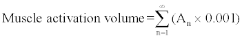
| For muscle activation analysis, the EMG signals were processed for activation time and integration. Onset and turn off times of EMG burst activity were established at the points at which muscle activity was greater than mean + 1.5 SD, or less than mean - 0.75 SD, respectively, of the running EMG signal, supplemented by repeated visual verification. Integral analysis was performed on the signals’ linear envelope by multiplying normalized amplitude value by time resolution of 0.001s, and then accumulated the total product of amplitude-by-time. | |
 (1) (1) |
|
| where An is the normalized amplitude value at each 0.001s duration. | |
| It was not known, however, whether more muscle mass were activated if volume is higher, as the measurement were derived through electrical signals produced by the working muscles instead of the muscles’ physical performance, which might be measured through other methods. | |
| The kinematic and EMG data of the independent variables were entered into a statistical software package (SPSS 15.0, SPSS Inc., Chicago, IL, USA). Paired t-test comparing stepping versus cycling were performed on all kinematic and muscle activation parameters (p<0.05). | |
| Results | |
| The average joint angles and ROM for hip, knee and ankle angles for both exercise modes are presented in Table 1. | |
| The kinematic analysis revealed that the hips and knees were significantly more extended during elliptical stepping compared to cycling. The ankle joint angles were significantly more neutral (closer to 90°), while ankles, knees and hips joint ranges of motion were significantly greater during elliptical stepping. | |
| To produce an elliptical stepping or a cycling movement, the leg muscles essentially function similarly – to extend and to flex the legs primarily using quadriceps and hamstrings. Figure 2 illustrates vastus lateralis (VL), vastus medialis (VM) and tibialis anterior (TA) muscles produced consistently longer activation periods and higher vastii EMG volume during elliptical stepping compared to cycling. The other muscles’ activation period was unchanged despite the change in movement pattern and the higher activation period of the mentioned muscles (VL, VM and TA). | |
| This was supported by statistically significant differences of activation duration per revolution of 1200ms – elliptical stepping vs cycling: VL 941 ± 216 ms vs 370 ± 380 ms, VM 997 ± 177 ms vs 419 ± 355 ms, TA 480 ± 584 ms vs 199 ± 273 ms and volume (%MVCâ�?�?second) – elliptical stepping vs cycling: VL 58.5 ± 24.6 vs 28.7 ± 16, VM 62.2 ± 27.4 vs 33.7 ± 17.3. | |
| Other muscles, such as rectus femoris, had less consistent activation patterns amongst subjects, with no significant differences observed between the two pedalling modalities. This reflected different synergistic activity of the muscles amongst subjects when the movements were modified. Muscle activation of the adductors; i.e. gracilis, G, and adductor longus, AL, (Figure 3) were not significantly different (G: p=0.457, AL: p=0.474) even though by visual inspection the activation volume appeared larger during cycling. | |
| Discussion | |
| This study revealed key differences in kinematics and EMGdetermined muscle activations of the lower limbs for closely-related pedalling movements. Cycling and elliptical stepping are produced by different mechanical joint and linkage systems, evoking adaptive responses in muscle lower-limb kinematic and EMG-activation patterns. While a cycling movement is mechanically facilitated by a simple rotating crank, the elliptical stepper used in our study had a secondary sliding link from the crank to the pedal that translated back-and-forth linearly at the pedal end. This created a greater linear component of the foot movement pattern, while a shorter revolving primary crank elicited a smaller angular component. The joint and linkage system of both movements allowed consistent and reproducible distal motion patterns. | |
| Muscle activation | |
| The activation patterns of the 10 muscle groups that we investigated were similar to a previous study into semirecumbent cycling [15], supporting the validity of the current EMG data. The earlier investigation also observed small EMG activity of the GM, confirming our premise that GM, being a deep-lying muscle covered by subcutaneous fat and skin, makes EMG signal detection more difficult during dynamic movements. During isokinetic exercise, when the angular velocity was held constant, a longer activation time translates into a greater joint angle over which the muscles were recruited. The current study revealed that elliptical stepping exercise evoked greater muscle activation times compared to cycling. This was accompanied by higher EMG activation amplitudes in the vastii muscles, thereby producing more muscle work. | |
| Ryan and Gregor performed a similar study that investigated cycling at constant workload and cadence. EMG signals were consistent within subjects and a single trial, but varied between subjects, a similar finding to the current investigation. Muscle activation variability amongst subjects was smallest in the single joint muscles (eg. vastus medialis, vastus lateralis and gluteus maximus) compared to two-joint muscles (eg. hamstrings and soleus) [13]. The muscle activation times in our study revealed that these were highest for the single joint quadriceps group (i.e. vastus lateralis and vastus medialis). | |
| Relationship of muscle activation to kinematic properties | |
| Different synergistic activities amongst subjects were demonstrated in our study as different muscle activation patterns between individuals when producing primarily reciprocating and repetitive movements. A single human joint is potentially actuated by many different combinations of muscle forces [16], thus a specific joint trajectory may be produced by involving different muscles with others being uninvolved or redundant. These variations of muscle combination amongst individuals explained the inconsistency and high dissimilarity of muscle activations for cycling and elliptical stepping, except for vastus lateralis and vastus medialis in the current study. | |
| Although in general muscle activation patterns were altered by varying the pedalling cadence, the primary function of all lower extremity muscles remains similar across all pedalling rates [7]. Other factors that may have affected leg muscle activation and coordination during cycling were the crank inertial load alteration and gear ratio [17], postural effects on biarticular and monoarticular muscles [18], and subject training background [19]. All these factors were not deemed relevant in the current study, as the load and gear ratio were constant, posture was carefully controlled, and subjects were introduced to both exercise systems only during the experimental session. | |
| Adductor muscle activation and their kinematic properties | |
| Each individual performed the cycling and elliptical stepping movements with slightly different muscle coordination to achieve essentially the same motion pattern. This slight disparity of neuromuscular coordination was observed especially for the lateral leg movements, affected by the adductor muscles, i.e. G and AL. | |
| The adductor muscles produced visibly larger, but not statistically significant (p>0.05), EMG activation amplitude and time during cycling compared to elliptical stepping. This suggested that during cycling, subjects tended to control their thigh abduction more than when they performed elliptical stepping. The author presumed that a mechanical advantage of the slider-crank mechanism that facilitated elliptical stepping was a second link that connected the foot pedal to the primary crank situated underneath the seat. This allowed the link to be virtually connected to the hip, as a closed-loop mechanical multibody system connected by joints and muscles as actuators [20], thereby increasing lateral stability of the leg during stepping and requiring lower leg adducting effort. | |
| In contrast, the cycling exercise machine was connected to the legs only at the foot pedals. In addition, the lateral knee movement is greater during cycling with greater variation in adductor muscles activities. These facts supported the author’s presumption that the lack of connecting linkages is the cause of lower lateral stability during cycling. Another factor of improved movement mechanics for elliptical stepping was illustrated by a series plot of joint angles over one pedal revolution, which demonstrated the better mechanical advantage of elliptical stepping linkages and the resulting dynamic stability of leg movement coordination (Figure 4). | |
| When the joint excursions were in phase with each other, the actions were simultaneous in a single stroke towards producing the desired movement. This may be translated into less effort of a single limb, than when each joint extended and flexed separately. | |
| Leg Joint Kinematic | |
| Kinematic properties of iFES-LST and iFES-LCE exercises were similar amongst able bodied people (Figure 4) and spinal injured individuals (Figure 5) [21], and were comparable to other published studies [22]. This was illustrated by comparable knee and hip joint angles throughout the cycle range and consistently greater plantarflexion when performing cycling as compared to stepping. The ankle joint was closer to normal with considerably more range of motion which included both dorsiflexion and plantarflexion during stepping exercise. This could greatly and safely improve ankle range of motion especially for people with stiff ankle joint. | |
| The kinematic properties, i.e. joint angles and range of motion, were similar irrespective of the exercisers’ conditions. This was because the movements were highly assisted and constrained by the machine linkages to the pedals, further proving that FES-evoked exercise using iFES-LST had valid kinematic properties. This allows the exercise device to have consistent and predictable kinematic property, which is useful for further research and training prescription. | |
| Do seated cycling and stepping have clinical efficacy for walking rehabilitation? | |
| The current investigation did not compare the two exercise modalities to a walking movement; neither did it compare the differences in a neurological population. However, it may be inferred from the established walking kinematics and EMG literature, that the movements during seated cycling or elliptical stepping possess similarities to walking with analogous biomechanics and muscle coordination components [23]. While a walking movement trace, (Figure 6), [24] was noted to be more similar to elliptical stepping movement than cycling, the muscle activation patterns from our study indicated that there were broad similarities in the leg muscle activation sequences of all three movements [25,26,27]. In particular, muscle timing and activation similarities were identified in rectus femoris, vastus lateralis, and biceps femoris in Winter’s study [28] compared to the elliptical stepping muscle activation pattern of the current investigation. | |
| The differences between the two activation patterns may be due to the nature of walking itself compared to seated stepping or cycling, such as the lack of hip extension during seated movement, and the absence of body weight support, upper body and leg swing momentum control and propulsion that is present during walking but not during a seated stepping or cycling exercise [29]. The main reason of the choice of the hip position in this thesis was the priority of seated exercise amongst disabled individuals who might not have the ability and strength to perform any exercise while in a standing position. | |
| Conclusion | |
| This study compared lower limb kinematic and leg muscle EMGderived activation during seated cycling and elliptical stepping movements. This study revealed significantly consistent differences in vastus lateralis and vastus medialis muscles’ activation patterns when producing elliptical stepping movement. Tibialis anterior produced greater activation time, but not activation amplitude, while other muscles’ activation was unaffected. Kinematic analysis of the leg movement pattern demonstrated that elliptical stepping and cycling were elicited by altered leg joint excursions and movement pattern. | |
| This investigation also inferred some potential advantages of utilizing an elliptical stepping trainer over cycling exercise to train the legs for upright stepping during gait. This finding might potentially contribute to a new exercise modality and therapeutic approach during walking rehabilitation by positing that leg movements, which are closer to walking, could be executed in a safe environment, especially for populations with pathologically disordered gait biomechanics. | |
| Acknowledgements | |
| The authors acknowledge the contributions of Dr Peter Sinclair, Dr Gustavo Braz and Dr Che Fornusek for their technical help. This project was funded by a Program Grant from the NSW Office for Science and Medical Research. | |
| References | |
References
- value="1" id="Reference_Titile_Link">Chen K, Chen SC, Tsai KH, Chen JJ, et al.(2004) An improved design of home cycling system via functional electrical stimulation for paraplegics. International Journal of Industrial Ergonomics 34: 223-235.
- value="2" id="Reference_Titile_Link">Gföhler M, Angeli T, Eberharter T, Lugner P, Mayr W, et al. (2001) Test bed with force-measuring crank for static and dynamic investigations on cycling by means of functional electrical stimulation. IEEE Trans Neural Syst Rehabil Eng 9: 169-180.
- value="3" id="Reference_Titile_Link">Gfohler M, Lugner P (2000) Cycling by means of functional electrical stimulation. IEEE Transactions on Rehabilitation Engineering 8: 233-243.
- value="4" id="Reference_Titile_Link">Gföhler M, Lugner P (2004) Dynamic simulation of FES-cycling: influence of individual parameters. IEEE Trans Neural Syst Rehabil Eng 12: 398-405.
- value="5" id="Reference_Titile_Link">van Soest AJ, Gföhler M, Casius LJ (2005) Consequences of ankle joint fixation on FES cycling power output: a simulation study. Med Sci Sports Exerc 37: 797-806.
- value="6" id="Reference_Titile_Link">Mileva K, Turner D (2003) Neuromuscular and biomechanical coupling in human cycling: adaptations to changes in crank length. Exp Brain Res 152: 393-403.
- value="7" id="Reference_Titile_Link">Neptune RR, Kautz SA, Hull ML (1997) The effect of pedaling rate on coordination in cycling. J Biomech 30: 1051-1058.
- value="8" id="Reference_Titile_Link">Suzuki S, Watanabe S, Homma S (1982) EMG activity and kinematics of human cycling movements at different constant velocities. Brain Res 240: 245-258.
- value="9" id="Reference_Titile_Link">Yoshizawa Y,Watanabe K, Kyuichi N (2006) Kinematic and Static Analyses of the Pedaling by Means of New Slider-Crank Mechanism. Paper presented at: 15th International Conference on Mechanics in Medicine and Biology, Singapore.
- value="10" id="Reference_Titile_Link">Gregor SM, Perell KL, Rushatakankovit S, Miyamoto E, Muffoletto R, et al. (2002) Lower extremity general muscle moment patterns in healthy individuals during recumbent cycling. Clin Biomech (Bristol, Avon) 17: 123-129.
- value="11" id="Reference_Titile_Link">Neptune RR, Herzog W (2000) Adaptation of muscle coordination to altered task mechanics during steady-state cycling. J Biomech 33: 165-172.
- value="12" id="Reference_Titile_Link">Shewman T, Konrad P (NA) Clinical SEMG Electrode Sites. In: SEMG-Muscle-Chart. AZ, USA.
- value="13" id="Reference_Titile_Link">Ryan MM, Gregor RJ (1992) EMG profiles of lower extremity muscles during cycling at constant workload and cadence. J Electromyogr Kinesiol 2: 69-80.
- value="14" id="Reference_Titile_Link">.Hamzaid N A, Fornusek C, Ruys A J, Davis GM. (2009) Development of an Isokinetic FES Leg Stepping Trainer (iFES-LST) for Individuals with Neurological Disability. Paper presented at: IEEE 11th International Conference on Rehabilitation Robotics, Kyoto, Japan.
- value="15" id="Reference_Titile_Link">Trumbower RD, Faghri PD (2004) Improving pedal power during semireclined leg cycling. IEEE Eng Med Biol Mag 23: 62-71.
- value="16" id="Reference_Titile_Link">Stelzer M, von Stryk O (2006) Efficient Forward Dynamics Simulation and Optimization of Human Body Dynamics. J. Appl. Math. Mech. 86:828-840.
- value="17" id="Reference_Titile_Link">Duc S, Villerius V, Bertucci W, Pernin JN, Grappe F (2005) Muscular activity level during pedalling is not affected by crank inertial load. Eur J Appl Physiol 95: 260-264.
- value="18" id="Reference_Titile_Link">Li L, Caldwell GE (1998) Muscle coordination in cycling: effect of surface incline and posture. J Appl Physiol 85: 927-934.
- value="19" id="Reference_Titile_Link">Jammes Y, Arbogast S, Faucher M, Montmayeur A, Tagliarini F, et al. (2001) Interindividual variability of surface EMG changes during cycling exercise in healthy humans. Clin Physiol 21: 556-560.
- value="20" id="Reference_Titile_Link">Rasmussen J, Christensen ST, Gföhler M, Damsgaard M, Angeli T (2004) Design optimization of a pedaling mechanism for paraplegics. Structural and Multidisciplinary Optimization 26:132-138
- value="21" id="Reference_Titile_Link">Hamzaid NA, Pithon K, Baek I (2009) Metabolic Cost and Mechanical Efficiency of FES-Evoked Leg Cycling and Elliptical Stepping. 14th Annual International Functional Electrical Stimulation Society Conference. Seoul, Korea.
- value="22" id="Reference_Titile_Link">Franco JC, Perell KL, Gregor RJ, Scremin AM (1999) Knee kinetics during functional electrical stimulation induced cycling in subjects with spinal cord injury: a preliminary study. J Rehabil Res Dev 36: 207-216.
- value="23" id="Reference_Titile_Link">Zajac FE, Neptune RR, Kautz SA (2002) Biomechanics and muscle coordination of human walking. Part I: introduction to concepts, power transfer, dynamics and simulations. Gait Posture 16: 215-232.
- value="24" id="Reference_Titile_Link">Whittle MW (1996) Gait Analysis an Introduction: Butterworth-Heinemann.
- value="25" id="Reference_Titile_Link">Courtine G, Schieppati M (2003) Human walking along a curved path. II. Gait features and EMG patterns. Eur J Neurosci 18: 191-205.
- value="26" id="Reference_Titile_Link">Hof AL, Elzinga H, Grimmius W, Halbertsma JP (2005) Detection of non-standard EMG profiles in walking. Gait Posture 21: 171-177.
- value="27" id="Reference_Titile_Link">Yang JF, Winter DA (1985) Surface EMG profiles during different walking cadences in humans. Electroencephalogr Clin Neurophysiol 60: 485-491.
- value="28" id="Reference_Titile_Link">Winter DA, Yack HJ (1987) EMG profiles during normal human walking: stride-to-stride and inter-subject variability. Electroencephalogr Clin Neurophysiol 67: 402-411.
- value="29" id="Reference_Titile_Link">Durward BR, Baer GD, Rowe PJ (1999) Functional Human Movement: Measurement and Analysis: Butterworth-Heinemann.
Relevant Topics
Recommended Journals
Article Tools
Article Usage
- Total views: 16938
- [From(publication date):
November-2013 - Apr 03, 2025] - Breakdown by view type
- HTML page views : 12258
- PDF downloads : 4680
Peer Reviewed Journals
Make the best use of Scientific Research and information from our 700 + peer reviewed, Open Access Journals