Intussusception of the Colon: Rare Case of Bowel Obstruction
Received: 04-May-2023 / Manuscript No. roa-23-99642 / Editor assigned: 06-May-2023 / PreQC No. roa-23-99642 (PQ) / Reviewed: 20-May-2023 / QC No. roa-23-99642 / Revised: 23-May-2023 / Manuscript No. roa-23-99642 (R) / Published Date: 30-May-2023
Abstract
An intussusception happens when a portion of the intestine folds over like a glove and enters the intestinal segment immediately downstream. As a result of this “telescoping”, the digestive tunics that form the wall of the gastrointestinal tract become nested, forming an intussusception loop that has a head and a neck. Its diagnosis is not simple and should be suspected in the absence of a particular background or with recurrent spontaneously resolving occlusive episodes.
Case history
A 77-year-old male patient consulted the emergency department with a cessation of matter and gas for 3 days in association with abdominal pain, melena, vomiting as well as abdominal distension.
The clinical examination showed an important abdominal distension as well as signs of dehydration however, the patient was stable on respiratory and hemodynamic plans and there was no fever. His biological workup didn’t show any abnormalities.
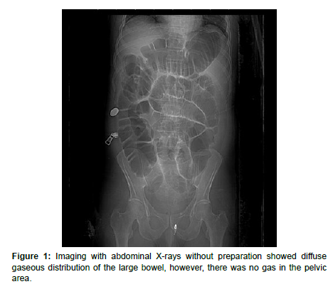
Imaging with abdominal X-rays without preparation showed diffuse gaseous distribution of the large bowel, however, there was no gas in the pelvic area (Figure 1).
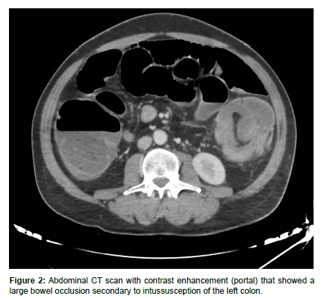
Then the patient underwent an abdominal CT with contrast enhancement (portal) that showed a large bowel occlusion secondary to intussusception of the left colon (Figure 2).
The patient went immediately to the operating room, where the diagnosis was confirmed.
Discussion
Intussusception affects children 20 times more frequently than adults.
Adult intussusceptions represent 5% of all intussusceptions [1] and 1–5% of bowl occlusions in adults [2]. It happens when one segment invaginate into the immediately adjacent segment; it affects more frequently the small bowel and less frequently the large bowel.
An intussusception loop is defined by its head (lead point) and neck. As opposed to children, this lead point in adults contain more often a pathological lesion, any abnormality the disrupt the bowel peristalsis can cause intussusception, most of it cause by a lead point -1-, which can be neoplasm, diverticula, intestinal ulcer, this lead point is initially caught by adjacent segment, the pulled further by peristalsis. Primary intussusception (without a pathological lead point) in adults is present in only 8–20% of cases [3].
Pain is the most frequent symptom, which can occur up to 80% of the time, as described by several studies [4,5].
Other symptoms include abdominal tenderness. Usually, fever is an indicator of intestinal necrosis. In addition to symptoms of parietal peritoneal irritation, failure to pass gas, abdominal lumps, and diarrhea, even with bloody stool, decreased or missing bowel sounds may also be present.
CT scan is considered the gold standard for diagnosing intussusception, as it can highlight the location, lead point, as well as signs of complications (necrosis, perforation).
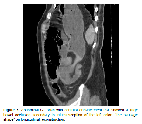
Pathognomonic radiological aspects of intussusception are represented by “the target sign” or “concentric ring sign” on axial reconstruction (Figure 1) or “the sausage shape” on longitudinal reconstruction (Figure 3).
As was previously mentioned, a pathologic lead point frequently contributes to adult intussusception. For those reasons, as opposed to the pediatric population, surgery has traditionally been used to treat intestinal intussusception, which results in obstruction. The attempt of hydrostatic reduction in the adult population is not recommended; on the other hand, in the pediatric population, this is the preferred course of therapy in most circumstances; in fact, the proportion of surgical treatment in this later age group is so much lower than 10% of the reported instances [6].
Conflict of Interest
The authors declare no competing interest.
Contributions
All authors contributed equally in this work.
Funding statement
The authors received no specific funding for this work.
Patient consent statement
Informed consent for patient information to be published in this article was obtained.
References
- Panzera F, Di Venere B, Rizzi M, Biscaglia A, Praticò CA, et al. (2021) Bowel intussusception in adult: prevalence, diagnostic tools and therapy. World J Methodol 11: 81-87.
- Marinis A, Yiallourou A, Samanides L, Dafnios N, Anastasopoulos G, et al. (2009) Intussusception of the bowel in adults: a review. World J Gastroenterol. 15: 407-411.
- Ghaderi H, Jafarian A, Aminian A, Daryasari SAM (2010) Clinical presentations, diagnosis and treatment of adult intussusception, a 20 years survey. Int J Surg 8: 318-320.
- Hong KD, Kim J, Ji W, Wexner SD (2019) Adult intussusception: a systematic review and meta-analysis. Tech Coloproctol 23: 315-324.
- Mrak K (2014) Uncommon conditions in surgical oncology: acute abdomen caused by ileocolic intussusception. J Gastrointest Oncol 5: E75-E79.
- Lee EH, Yang HR (2020) Nationwide Population-Based Epidemiologic Study on Childhood Intussusception in South Korea: Emphasis on Treatment and Outcomes. Pediatr Gastroenterol Hepatol Nutr 23: 329-345.
Indexed atGoogle Scholar Crossref
Indexed atGoogle Scholar Crossref
Indexed atGoogle Scholar Crossref
Indexed atGoogle Scholar Crossref
Indexed atGoogle Scholar Crossref
Citation: Rostoum S, Fadil M, Zhim M, Naggar A, Laamrani FZ, et al. (2023)Intussusception of the Colon: Rare Case of Bowel Obstruction. OMICS J Radiol12: 446.
Copyright: © 2023 Rostoum S, et al. This is an open-access article distributed under the terms of the Creative Commons Attribution License, which permits unrestricted use, distribution, and reproduction in any medium, provided the original author and source are credited.
Share This Article
Open Access Journals
Article Usage
- Total views: 1101
- [From(publication date): 0-2023 - Mar 29, 2025]
- Breakdown by view type
- HTML page views: 836
- PDF downloads: 265


