Intraosseous Dermoid Cyst of the Proximal Phalanx of a Little Toe: A Case Report
Received: 07-Feb-2022 / Manuscript No. CRFA-22-53752 / Editor assigned: 01-Jan-1970 / PreQC No. CRFA-22-53752 (PQ) / Reviewed: 23-Feb-2022 / QC No. CRFA-22-53752 / Revised: 28-Feb-2022 / Manuscript No. CRFA-22-53752 (R) / Accepted Date: 04-Mar-2022 / Published Date: 07-Mar-2022 DOI: 10.4172/2329-910X.1000331
Abstract
Dermoid cysts are rare benign lesions which are seldom found intraosseous. We describe the case of a 56-y.o. male diabetic with an abscess formation on the dorsum of the foot, between MT4 and MT5 with suspected osteomyelitis of the adjacent phalanx. Surgery for excision of the abscess and amputation of the little toe was performed. Intraoperatively the proximal Phalanx showed an opened intraosseous cyst with squamous cells and specialized skin appendage.
Histopathological the dermoid cyst was confirmed. We would like to showcase this rare incident of an intraosseous dermoid cyst of the proximal phalanx of the little toe.
Keywords: Cyst; Phalanx; Neurulation
Introduction
Dermoid cysts are rare benign lesions predominantly of soft tissues, but can occur in every part of the human body [1]. The pathogenesis is not completely understood, there are two primary theories the congenital/ dysontogenetic and traumatic/ acquired [2].
The more frequently mentioned theory is the dysontogenetic [3]. Abnormal embryological development e.g. a fault in the folding process during neurulation (3-5week of gestation) leads to aberrant ectodermal inclusion within the underlying mesoderm [4,5]. Also an ectodermal enclavement of parts of ectodermal tissue during the time of calescence at epithelial fusion lines can lead to an inclusion of such tissues and furthermore to the formation of a dermoid cyst.
On the other hand, there is the traumatic or acquired theory of development of dermoid cysts. A local penetrating trauma or iatrogenic measure causes an implantation of fragments of epithelial tissues or nail bed tissues in underlying soft tissues even bone [5,6]. This type of pathogenesis is more often encountered in the hands at sites of penetrating trauma [6].
Given the rare occurrence of intraosseous dermoid cysts on the extremities data for incidence is hard to come by. Also, the fact that in some cases there is no accurate differentiation between dermoid cysts and epidermoid cysts complicates to find reliable data.
The difference between the two is found histologically, so for diagnostic confirmation a histologic examination is necessary [5,7]. According to Meyer et al. there are three dysontogenetic cysts that can be differentiated [2,8].
Epidermoid cyst
A cystic lesion composed of ectodermal structures, lined by simple stratified squamous epithelium.
Teratoid Cyst
A complex cyst, also epithelial-lined but they incorporate also meso- and endodermal elements, like connective tissue or parenchyma [9].
Dermoid Cyst
It has an epithelial lined cavity consisting of keratinizing squamous cells and also shows evidence of specialized skin appendages, like hair follicles, sebaceous and sweat glands, which all derive from the ectoderm [10,11]. The lumen can include keratin, cholesterol, sebaceous glands excretion, hair and cellular debris [12]. The growth process of the cyst is slowly because it goes along with the proliferation of the squamous cells and the subsequent accumulation of keratindebris in the lumen of the cyst [13]. In the case of the dermoid cyst the lumen can additionally contain excretion of sebaceous and sweat glands, cholesterol, hair and cellular debris [12]. Depending on the location the slowly increasing dermoid cysts can cause problems (for instance neurological, pain, or osteolysis), due to local exertion of pressure or endogenous inflammatory response in the event of rupture of the cyst [4]. An intraosseous localization of a dermoid cyst can lead to resorptive alterations in the bone structure due to the growth of the cyst [13].
The occurrence of dermoid cysts is predominantly in soft tissues. The intraosseous localization is rather rare [3]. Intraosseous occurrences were described for the cranio-facial region [10,14,15 ]. The only dermoid cyst of the lower extremity we found in the literature was a case report of Behr et al. who described a dermoid cyst of the lower leg with an underlying case of foot duplication [16]. On the other hand however interosseous epidermoid cyst are more common at the extremities, especially on the distal phalanges of the hand and we found seven cases of interosseous epidermoid cases of the toes. 15 With the epidermoid cyst the traumatic etiology is most commonly hypothesized [17,18].
We did not find a single case of an intraosseous dermoid cyst of the lower extremity or foot in our online research through pub med [12,19].
We present a rare case of an intraosseous dermoid cyst of the proximal phalanx of the little toe.
Case Presentation
We report the case of a 56-year old patient who presented at our emergency department with chronic-recurrent infection of the 4th webb space of the left foot. The patient reports a confined swelling and redness as well as pus-like excretion between the 4th and 5th metatarsal heads of the dorsum of the foot over the last 6 months. Last secretion was about 10 days ago. No reported systemic inflammatory symptoms, e.g.fever and no local pain. The patient who was diagnosed with diabetes mellitus type 2 in 2009 was already treated with i.v. antibiotics in our hospital because of erysipelas of the left lower leg one year prior to his current presentation. Alongside diabetes this patient has diagnosis of cardiomyopathy of unknown origin, a severe case of respiratory partial-insufficiency, active smoker and severe sleep-apnea. Four months prior he underwent surgery for a Y-roux- gastric bypass because of obesity WHO grade 3 (BMI 40.4 kg/m2).
Clinically the patient presents with a small (about 1 cm in diameter), dry, round induration on the dorsum of the left lateral foot with localized erysipelas, with no current pus-secretion. Pulses of the foot were palpable.
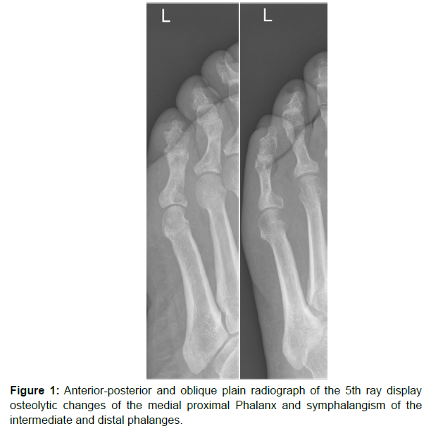
The laboratory workup revealed normal white blood cell (WBC) count, but elevated C-reactive protein (CRP) of 68 mg/l (normal <5). The X-ray showed an osteolytic lesion on the medial side of the base of the proximal phalanx (Figure 1).
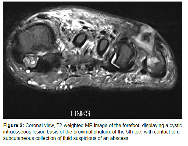
Sonography revealed a collection of heterogenous slightly hyperechoic content between 4th and 5th Metatarsal head, with dimensions of 9 x 8 x 16 mm. In the MRI findings the radiologist described a destructive process of the metaphysis of the proximal phalanx compatible with osteomyelitis as well as an abscess in the adjacent soft tissue with a fistula to the dorsum of the foot (Figure 2).
We assumed an intermetatarsal abscess with a fistula and osteomyelitis of the base of the proximal Phalanx of the 5th toe. Consequently, we decided to excise the abscess and amputate the little toe to cope with the infection.
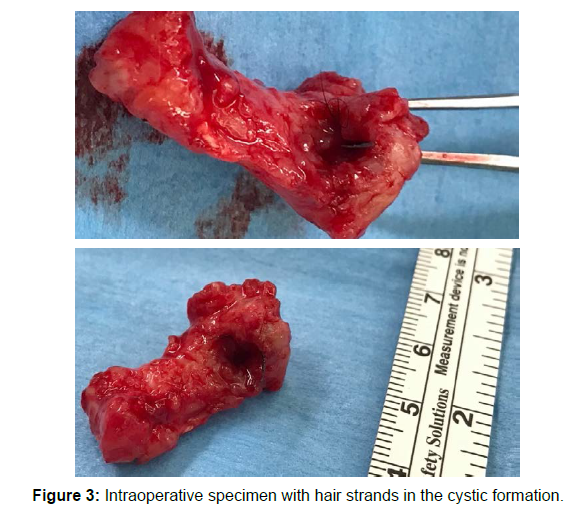
During surgery we made a fusiform excision of the fistula and the Abscess. We took soft tissue samples for microbiological examination. During exarticulation of the proximal Phalanx of the metatarsophalangeal- joint 5 (MTP-5) the osteolytic-cystic lesion of the proximal phalanx presented itself spontaneously opened. During inspection there where bunches of black hair inside the lumen (Figure 3). The whole proximal Phalanx was sent in for histopathological examination.
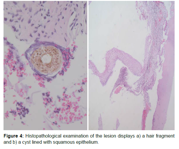
In microscopic examination the paraffin embedded specimen showed a chronic granulating and fibrosing Osteomyelitis with a Destruction of the bone and inclusion of squamous epithelium plus hair fragments in the bone. The histopathological examination confirmed the suspected dermatoid cyst plus osteomyelitis (Figure 4).
Because of detected Enterococcus faecalis in the soft tissue samples the established antibiotic treatment was continued for 20 days. Wound healing was successful and the patient is free of complaints.
Discussion
The occurrence of dermoid cysts in the lower extremities is rather seldom and rarely described in the literature respectively. A pubmed research did not reveal a single case describing an intraosseos dermoid cyst in a foot.
As a differential diagnosis for osteolytic lesions osteomyelitis, as well as benign and malign tumors like enchondromas, intraosseous ganglion, osteoid osteoma, simple bone cysts, aneurysmatic bone cyst, giant cell tumors and metastasis should be considered [4].
In the presented case we initially did not assume the presence of a dermoid cyst. Clinically an infectious situation was present, the radiological findings let us lead to the conclusion of osteomyelitis. It’s reasonable to believe that the intraosseous cyst opened up to release its content. A draining sinus tract could have formed over time to release the cell-debris content, which the patient has seen excreted from time to time. These way bacteria could have infiltrated the soft tissues and also the bone. It’s agreed on that intact dermoid cysts do not lead to inflammatory reactions [11]. Otherwise, an abscess formation as commonly found among diabetics could have randomly formed near the intraosseous dermoid cyst and therefore the cyst has been accidentally found. Either way with the little abscess formation and the external draining tract Osteomyelitis had to be suspected or so the amputation was vindicated and necessary given that the histopathological examination showed infection of the cyst as well.
Retrospectively also in synopsis with our radiology department and our present state of knowledge we could not have known of the dermoid cyst pre-operatively because differentiation between an intraosseous dermoid cyst and osteomyelitis on plain radiographs and MRI is difficult [7, 20]. Histopathological differentiation between dermoid cyst and epidermoid cyst is also difficult, because the entire cyst wall has to be adequately sampled and examined to confirm dermal adnexal structures and consequently the diagnosis. Some Authors even argue not to claim dermoid cysts and epidermoid cysts as separate entities given their similarities and lack of clinical importance [7].
According to the present literature a complete extraction of the cyst is treatment of choice. Incomplete Excision can lead to recurrence [18 ].
Conclusion
Dermoid Cysts are rare benign, epithelial lined malformations which contain dermal adnexal structures in the cyst wall. The inclusion of epithelial rests in embryogenesis or traumatic inclusion leads to their formation. The intraosseous location in extremities is very seldom or at least not often diagnosed and therefore not often encountered by orthopedic surgeons, whereas in other disciplines dermoid cysts seem to occur more frequently. Even with MRI-Examination preoperative Diagnosis of a dermoid cyst is difficult. Histopathological examination will show dermal adnexal structures and therefore differentiate between other cystic malformations. Complete extraction of the cyst is treatment of choice.
Consent
Informed consent was obtained from the patient for publication of this case report and accompanying images.
Financial Support / Conflict of interest
None
Acknowledgement
We would like to thank Dr. med. Ruggero Biral for the histopathological examination.
References
- Rubin E, Reisner HM (2014) Essentials of Rubin’s Pathology. Wolters Kluwer Health/Lippincott Williams & Wilkins (6th edn).
- Rapidis AD, Angelopoulos AP, Scouteris C (1981) Dermoid cyst of the floor of the mouth. Report of a Case. Br J Oral Surg 19:43-51.
- Freyschmidt J, Ostertag H, Jundt G (2010) Tumorahnliche Knochenlasionen („tumor-like lesions“). Knochentumoren Mit Kiefertumoren. Springer Berlin Heidelberg pp: 739-944.
- Reissis D, Pfaff MJ, Patel A, Steinbacher DM (2014) Craniofacial dermoid cysts: Histological analysis and inter-site comparison. Yale J Biol Med 87:349-357.
- Wang BY, Eisler J, Springfield D, Klein MJ (2003) Intraosseous epidermoid inclusion cyst in a great toe. A case report and review of the literature. Arch Pathol Lab Med 127: e298-e300.
- Reid J, Baker J, Davidson D (2019) Dermoid Inclusion Cyst Following Percutaneous Needle Fasciotomy: A Novel Complication. J Hand Surg Asian Pac 24:116-117.
- Balasundaram P, Garg A, Prabhakar A, Joseph Devarajan LS, Gaikwad SB, et al. (2019) Evolution of epidermoid cyst into dermoid cyst: Embryological explanation and radiological-pathological correlation. Neuroradiology 32:92-97.
- Meyer I (1955) Dermoid cysts (dermoids) of the floor of the mouth. Oral Surgery, Oral Medicine, Oral Pathology 8:1149-1164.
- http://www.pathologyoutlines.com/topic/mandiblemaxilladermoid.html
- Patel K, Bhuiya T, Chen S, Kenan S, Kahn L (2006) Epidermal inclusion cyst of phalanx: a case report and review of the literature. Skeletal Radiol 35:861-863.
- Choi JS, Bae YC, Lee JW, Kang GB (2018) Dermoid cysts: Epidemiology and diagnostic approach based on clinical experiences. Arch Plast Surg 45:512-516.
- Fanous AA, Gupta P, Li V (2014) Analysis of the growth pattern of a dermoid cyst. J Neurosurg Pediatr 14:621-625.
- Pryor SG, Lewis JE, Weaver AL, Orvidas LJ (2005) Pediatric dermoid cysts of the head and neck. Otolaryngol - Head Neck Surg 132:938-942.
- Richardson MP, Foster JR, Logan DB (2017) Intraosseous Epidermal Inclusion Cyst of the Proximal Phalanx of the Fifth Toe and Review of the Literature: A Case Study. Foot Ankle Spec 10:470-472.
- Byers P, Mantle J, Salm R (1966) Epidermal cysts of phalanges. British volume J Bone Surg 48: 577-581.
- Behr JT, Kohli KK, Millar EA (1986) Giant dermoid associated with foot duplication. J Pediatr Orthop 6:486-488.
- Parman LM, Murphey MD (2000) Alphabet soup: Cystic lesions of bone. Semin Musculoskelet Radiol 4:89-101.
- Reda B (2018) Cystic bone tumors of the foot and ankle. J Surg Oncol 17:1786-1798.
- Menditti D, Laino L, Ferrara N, Baldi A (2008) Dermoid cyst of the mandibula: a case report. Cases J 1:260.
- Hamad AT, Kumar A, Anand Kumar C (2006) Intraosseous epidermoid cyst of the finger phalanx: a case report. J Orthop Surg (Hong Kong) 14:340-342.
Google Scholar Crossref Indexed at
Google Scholar Crossref Indexed at
Google Scholar Crossref Indexed at
Google Scholar Crossref Indexed at
Google Scholar Crossref Indexed at
Google Scholar Crossref Indexed at
Google Scholar Crossref Indexed at
Google Scholar Crossref Indexed at
Google Scholar Crossref Indexed at
Google Scholar Crossref Indexed at
Google Scholar Crossref Indexed at
Google Scholar Crossref Indexed at
Citation: Kreuzer S, Schober M, Hess R, Gebel JP (2022) Intraosseous Dermoid Cyst of the Proximal Phalanx of a Little Toe: A Case Report. Clin Res Foot Ankle, 10: 331. DOI: 10.4172/2329-910X.1000331
Copyright: © 2022 Kreuzer S, et al. This is an open-access article distributed under the terms of the Creative Commons Attribution License, which permits unrestricted use, distribution, and reproduction in any medium, provided the original author and source are credited.
Share This Article
Recommended Journals
Open Access Journals
Article Tools
Article Usage
- Total views: 3254
- [From(publication date): 0-2022 - Apr 26, 2025]
- Breakdown by view type
- HTML page views: 2687
- PDF downloads: 567


