Case Report Open Access
Intramuscular Lipoma of Cheek: A Rarity
| S Jayachandran1 and Koijam Sashikumar Singh2* | |
| 1Department of Oral Medicine and Radiology, Tamilnadu Government Dental College and Hospital, Chennai, Tamil Nadu, India | |
| 2Directorate of Medical & Health Services, Government of Manipur, Imphal, Manipur, India | |
| Corresponding Author : | Dr. Koijam Sashikumar Singh MDS, Dental Surgeon Directorate of Medical & Health Services Government of Manipur, Imphal Manipur, India Tel: 8974067254 E-mail: koijamsas@gmail.com |
| Received July 10, 2012; Accepted August 27, 2012; Published August 30, 2012 | |
| Citation: Jayachandran S, Singh KS (2012) Intramuscular Lipoma of Cheek: A Rarity. OMICS J Radiology. 1:108. doi: 10.4172/2167-7964.1000108 | |
| Copyright: © 2012 Jayachandran S. This is an open-access article distributed under the terms of the Creative Commons Attribution License, which permits unrestricted use, distribution, and reproduction in any medium, provided the original author and source are credited. | |
Visit for more related articles at Journal of Radiology
Abstract
Intramuscular lipomas are uncommon benign mesenchymal tumors which infiltrate skeletal muscle. It is very rarely encountered in head and neck region and often located in limbs. A consistent feature is infiltration with dissociation of the surrounding muscle fibers which makes it difficult to distinguish from well-differentiated liposarcomas. Its recurrence is very high. Imaging such as CT, MRI and ultrasonography gives valuable diagnostic information which seldom requires histopathology for diagnosing these lesions. Here, we report a case of an intramuscular lipoma of the masseter muscle in a 50-year-old male patient who presented with a growing mass in the left cheek. The radiographic imaging features are also discussed.
| Keywords |
| Intramuscular lipoma; Computed tomography; Ultrasonography |
| Introduction |
| Lipoma is a benign tumor of mature fat cells [1]. The incidence of oral lipoma (OL) is thought to be 1% to 4% of all benign oral lesions [2]. Approximately 220 cases of intraoral lipoma have been reported in the literature [3]. Lipomas may be either superficial or deep, with the former being more common [4]. Roberts et al. [5] further classified the superficial palpable fatty masses into encapsulated and unencapsulated. The deep lipoma is usually larger and deforms the surrounding tissue as compared with superficial lipomas, which are generally more circumscribed. The subfascial or deep lipomas can be classified as parosteal, interosseous, or visceral and intermuscular or intramuscular. Lipomas are typically asymptomatic unless they compress neurovascular structures [4]. |
| Infiltrating lipomas are usually located in the limbs [3]. They are rarely encountered in the oral cavity and an extensive search of literature found 4 cases in tongue [6-9], 3 cases in cheek [3,10,11], 2 cases in infraorbital region [11], 1 case in submandibular area [6], 1 case in infratemporal fossa [7], and 1 case in mental region [11]. Here, we highlight a case of intramuscular lipoma involving masseter muscle. |
| Report of a Case |
| A 50-years-old male patient was reported to the Department of Oral Medicine and Radiology, Tamilnadu Govt. Dental College and Hospital, Chennai with the chief complain of pain and swelling in the left cheek region. Patient gave history of a small swelling over left cheek 6 months back and gradually increased in size to attend the present size. There was association of pain for the past 1 month. |
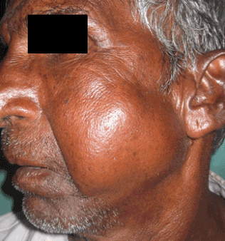
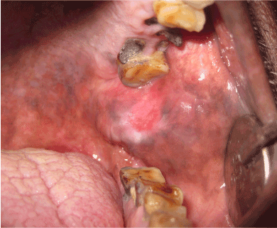
| Extra-orally, a single swelling over left side of face measuring approximately 10 cm × 8 cm was present. Superiorly, it extended upto the left zygomatic arch and zygoma, inferiorly upto the left body of mandible and left angle region, anteriorly upto naso-labial fold and posteriorly upto left angle region (Figure 1). The swelling was soft in consistency. On palpation, firm mass deep into the lesion was felt. On clinching the teeth, the swelling became firm and prominent; and soft and diffused on relaxation. There was no skin ulceration present. A single tender left submandibular lymph node measuring 1 cm was palpable. Intra-orally, a mild swelling was present over the left buccal mucosa in 28, 38 regions and anterior to left pterygomandibular raphe. There was mild erythema surrounded by whitish area over the swelling due to traumatic irritation from 27 (Figure 2). |
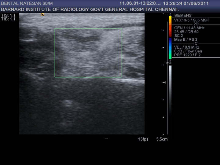
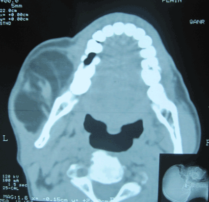
| Ultrasonography revealed hypoechoic appearance with evidence of disorderly echo within the lesion (Figure 3). On color Doppler ultrasonography, no vascularity was evident (Figure 3). CT revealed hypodense lesion separated by hyperdense muscle fibers into multiple compartments (Figure 4). Hounsfield unit of the hypodense area was measured on CT and it was found to be -118HU. The lesion involved left masseter muscle. Continuous fibers of buccinator muscle lined the lateral aspect of the lesion. Aspiration was attempted and it revealed no aspirate. Fine needle aspiration was done and it showed moderately cellular smears showing mature adipocytes clusters and fibroblasts. |
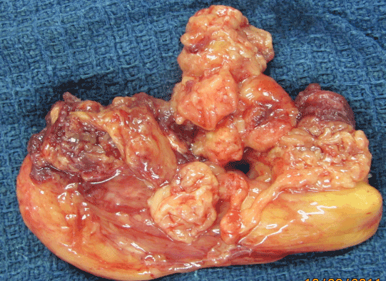
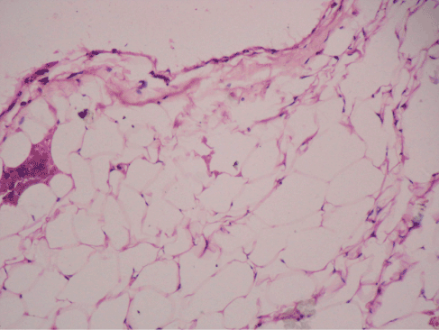
| Surgery was done under general anesthesia. An extra-oral submandibular incision was given on the left side. The lesion was explored with blunt dissection of tissues and muscles overlying it. Once exposed, the lesion was removed along with the adjoining muscle mass. The gross appearance of the lesion was an irregular, lobulated, firm, fibrofatty lesion (Figure 5). The cut surface was pale yellow. The post-operative histopathological report confirmed to be a lipoma (Figure 6). |
| A post-operative CT was taken and it revealed complete removal of the lipomatous tissue. Normal mouth opening and masticatory function was achieved within 3 weeks post-operatively. 1 year followup was done. There was no significant morbidity or signs of recurrence. |
| Discussion |
| Lipomas are common, benign, slow-growing, soft tissue neoplasms of mature adipocytes. Lipoma is different from normal body fat in that its lipids are not available for metabolism, and it has been suggested that this fact, together with its autonomous growth, may warrant its classification as a true benign neoplasm [3]. |
| Conventional radiography is of no use in providing a clue for lipoma. Of late, prior to biopsy, the diagnosis of lipomatous lesions is enhanced by the availability of CT. On CT, fat has low attenuation, that is, less than -20 Hounsfield units and typically between -65 and -120 Hounsfield units [4]. Plain CT scans of lipoma shows homogeneous hypodensity. Large infiltrating intramuscular lipomas may show numerous hyperdense band-like structures representing muscle fibers running across the background of homogeneous hypodense area representing lipoma. Contrast CT scans can help in distinguishing intramuscular lipoma from angiolipoma [12] as it shows enhancement in angiolipoma. It remains controversial whether lipomas are hypoechoic or hyperechoic on ultrasonograms. But many have agreed that the ultrasonographic appearance of most lipomas is hypoechoic, while the distribution of the internal echogenic patterns might be influenced by the number of tissue interfaces [13]. Infiltrating intramuscular lipoma may show a mixed pattern of hypo- and hyper-ecogenicity due to the intermingling nature of the lipoma within the muscle fibers. |
| In our case, as the measurement of Hounsfield unit on CT was -118 and was almost confirmatory of a fatty tissue lesion, MRI was not taken. The classic MRI signal intensity for lipomas is similar to subcutaneous fat [4]. On T1-weighted image, lipomas show high signal intensity. On T2-weighted MR images, the fat shows relatively lower signals. However, on fast spin echo (FSE), T2-weighted images, the signal intensity lipomas remain high. With fat suppression, lipoma reveals a low signal intensity patterns. Lipomas show no significant enhancement following the injection of contrast. |
| In cases of lipomas, differential diagnosis of liposarcoma can be made. The presence of lipoblasts or atypical adipocytes would raise the concern for liposarcoma [4]. Histologically, lipomas resemble normal fat, whereas well-differentiated liposarcomas, although similarly resembling fat histologically, also have dense bands of collagen traversing the mass with associated gelatinous areas [4]. |
| Because of the infiltrative nature and potentially high rate of recurrence of intramuscular lipoma after inadequate surgery, complete surgical excision is mandatory. Recurrence is always attributable to technical error [3]. |
| Conclusion |
| Although intramuscular lipomas are rare, they must be kept in mind while working a differential diagnosis of mass involving muscles. Computed tomography with measurement of Hounsfield Unit is almost diagnostic. MRI provides better soft tissue details than CT but cost limits its use. |
References |
|
Figures at a glance
 |
 |
 |
| Figure 1 | Figure 2 | Figure 3 |
 |
 |
 |
| Figure 4 | Figure 5 | Figure 6 |
Relevant Topics
- Abdominal Radiology
- AI in Radiology
- Breast Imaging
- Cardiovascular Radiology
- Chest Radiology
- Clinical Radiology
- CT Imaging
- Diagnostic Radiology
- Emergency Radiology
- Fluoroscopy Radiology
- General Radiology
- Genitourinary Radiology
- Interventional Radiology Techniques
- Mammography
- Minimal Invasive surgery
- Musculoskeletal Radiology
- Neuroradiology
- Neuroradiology Advances
- Oral and Maxillofacial Radiology
- Radiography
- Radiology Imaging
- Surgical Radiology
- Tele Radiology
- Therapeutic Radiology
Recommended Journals
Article Tools
Article Usage
- Total views: 9112
- [From(publication date):
October-2012 - Jul 02, 2025] - Breakdown by view type
- HTML page views : 4433
- PDF downloads : 4679
