Make the best use of Scientific Research and information from our 700+ peer reviewed, Open Access Journals that operates with the help of 50,000+ Editorial Board Members and esteemed reviewers and 1000+ Scientific associations in Medical, Clinical, Pharmaceutical, Engineering, Technology and Management Fields.
Meet Inspiring Speakers and Experts at our 3000+ Global Conferenceseries Events with over 600+ Conferences, 1200+ Symposiums and 1200+ Workshops on Medical, Pharma, Engineering, Science, Technology and Business
Case Report Open Access
Intestinal Obstruction Due to Adhesions around VP Shunt Tip in a Case of Pyogenic Meningitis
| Priyanka Arora*, Gayatri Madhukar Autkar, Shephali Shrikant Pawar, Ajay Kumar and Avinash A Gutte | |
| Grant Government Medical College and Sir J.J Group of Hospitals Mumbai, Maharashtra, India | |
| Corresponding Author : | Priyanka Arora Grant Government Medical College and Sir J.J group of Hospitals Mumbai Maharashtra, India Tel: +919167302587 E-mail: prankie18feb@gmail.com |
| Received October 07, 2014; Accepted November 18, 2014; Published November 21, 2014 | |
| Citation:Priyanka Arora, Gayatri Madhukar Autkar, Ajay Kumar, Shephali Shrikant Pawar, Avinash A Gutte (2014) Intestinal Obstruction Due to Adhesions around VP Shunt Tip in a Case of Pyogenic Meningitis. OMICS J Radiol 3:172. doi: 10.4172/2167-7964.1000172 | |
| Copyright: © 2014 Arora P, et al. This is an open-access article distributed under the terms of the Creative Commons Attribution License, which permits unrestricted use, distribution, and reproduction in any medium, provided the original author and source are credited. | |
Visit for more related articles at Journal of Radiology
Abstract
Small bowel obstruction caused by adhesions around the VP shunt is a rare and severe complication of the shunt which may require laparotomy and resection of bowel. We report a case in which small bowel obstruction was caused by adhesions around the VP shunt.
| Keywords |
| Intestinal obstruction; Adhesions; VP shunt |
| Case Report |
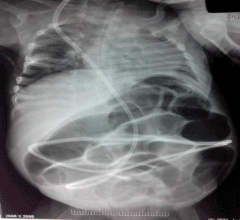
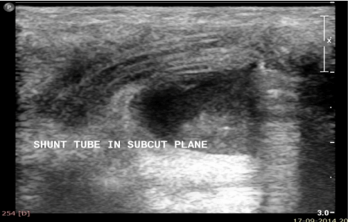
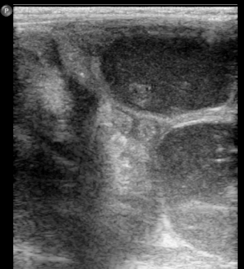
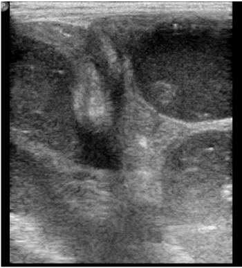
| An infant was admitted to the emergency department with abdominal distension and repeated vomiting. She had been operated for meningomyelocoele with non communicating hydrocephalus in the newborn period. Ventriculo-peritoneal shunt was inserted 4 weeks after birth. A base line abdominal radiograph showed dilated small bowel loops. An ultrasound was requested which revealed tip of the VP shunt in the pelvis with dilated small bowels loops and absent peristalsis .Transition point between the dilated and collapsed loops was seen in close proximity to the VP shunt tip. Diagnosis of small bowel obstruction was made. Operative findings revealed that the small bowel obstruction was due to adhesions at the VP shunt tube tip for which adhesiolysis was done. |
| Discussion |
| Ventriculoperitoneal shunt surgery is the most commonly used technique for the management of hydrocephalus in paediatric cases [1]. Distal end of the shunt catheter is usually placed in the peritoneum. This procedure is associated with various complications. The most common complications are shunt infection and obstruction due to the ventriculoperitoneal shunt device [2,3]. Others complications are intra-abdominal ascites, regional cyst formation, hydrocele, intestinal injury, intestinal obstruction, mechanical blockage of the distal end by omentum causing shunt failure [2], formation of abdominal pseudocyst. Shunt catheters can migrate to a number of locations like anus, scrotum, urethra umbilicus, or mouth or it may migrate to internal organs. Spontaneous bowel perforation is a rare complication of VPS surgery (0.01-0.07%) [4], although some studies have reported higher rates (2.51%) [5]. The sharp tip at the distal end of the catheter is blamed for higher complication rates. It should be kept in mind that this is a rare but a highly mortal complication (15%) [6]. Diagnosis of the intra-abdominal complications of VP shunt needs thorough physical neurologic and careful abdominal examination. Imaging modalities like plain radiograph, ultrasound and computed tomography are of immense help. Plain radiographs help to locate intraperitoneal position of catheter and status of bowel loops. Ultrasound and CT give a better and exact idea of the etiology (Figures 1-4). A high index of suspicion along with optimum clinical assessment and imaging will improve the management of shunt complications and decrease morbidity. |
References |
|
Tables and Figures at a glance
 |
 |
 |
 |
| Figure 1 | Figure 2 | Figure 3 | Figure 4 |
Post your comment
Relevant Topics
- Abdominal Radiology
- AI in Radiology
- Breast Imaging
- Cardiovascular Radiology
- Chest Radiology
- Clinical Radiology
- CT Imaging
- Diagnostic Radiology
- Emergency Radiology
- Fluoroscopy Radiology
- General Radiology
- Genitourinary Radiology
- Interventional Radiology Techniques
- Mammography
- Minimal Invasive surgery
- Musculoskeletal Radiology
- Neuroradiology
- Neuroradiology Advances
- Oral and Maxillofacial Radiology
- Radiography
- Radiology Imaging
- Surgical Radiology
- Tele Radiology
- Therapeutic Radiology
Recommended Journals
Article Tools
Article Usage
- Total views: 16156
- [From(publication date):
December-2014 - Jul 06, 2025] - Breakdown by view type
- HTML page views : 11557
- PDF downloads : 4599
