Inflammation Risks of Diabetic Foot with Toe Deformation are Reflected by Plantar Loading
Received: 03-May-2023 / Manuscript No. crfa-23-98410 / Editor assigned: 05-May-2023 / PreQC No. crfa-23-98410 / Reviewed: 19-May-2023 / QC No. crfa-23-98410 / Revised: 23-May-2023 / Manuscript No. crfa-23-98410 / Published Date: 31-May-2023
Abstract
Diabetes is one of the most common chronic diseases in the world. The aim of this study was to quantitatively evaluate the foot load-bearing characteristics of diabetic patients with fifth toe deformity through comparative analysis with diabetic patients with normal foot and healthy. Six female diabetic neuropathic subjects with fifth toe deformity and six age-matched diabetic neuropathic subjects without any foot deformity participated in the test. walk test. The dynamic pressure of bare feet is measured with Novel's EMED force plate. Peak pressure and pressure-time integration for all 7 forefoot, midfoot, lateral forefoot, central forefoot, medial forefoot, forefoot, and other toes were be collected. Peak pressure was significantly higher in patients with toe deformity in the posterior forefoot, midfoot, and big toe region than in the control group. Meanwhile, the force retention time was longer in the big toe region of the deformed group compared with the control group, and the center of pressure was almost located in the big toe region during the toe deformity phase. Diabetics with fifth toe deformity may experience decreased contact area on the sole of the foot in the rest of the toe and increased load on the big toe. Results showed that 5th toe deformity was associated with potential ulcer risk, especially in the big toe region.
Keywords
Diabetes; Foot; Ulcer risk
Introduction
Diabetes is one of the most common chronic diseases affecting people's daily lives. It is estimated that the number of people with diabetes worldwide will exceed 365 million by 2030. Diabetic feet suffer from foot ulcers or foot deformities that impair normal mobility. It is the result of long-term load on the soleus surface during walking that has changed or shifted to specific regions in patients with diabetes mellitus and peripheral neuropathy. Excessive pressure in the unprotected foot is considered a major risk factor for plantar ulceration, which is the most common precursor to lower extremity amputation in diabetics [1]. Peripheral neuropathy can cause lower extremity damage and even disability in people with diabetes. Due to impaired sensation in the foot nerves, foot injuries can be easily missed, increasing the risk of ulcers or skin damage. At least 15% of these ulcers result in some form of foot amputation. A previous study found that the feet in early-stage diabetics tend to deform in the toes and imbalance in the muscles in the feet or lower extremities, which then causes abnormal pressure in the lumen foot. When human foot structure and human mobility are damaged or dysfunctional, foot pressure and foot load distribution will change accordingly. It is clear that the occurrence of diabetic foot ulcers is largely due to changes in the bearing properties of the foot. And diabetic feet can change the distribution of pressure in the legs, leading to uneven distribution of blood flow in the legs, thereby destroying the blood supply to the feet, eventually leading to foot ulcers and even amputation. However, there are rare reports of kinetic changes in toe deformity, which is an early sign of diabetic foot deformity and a major cause of foot load changes [2-5]. Therefore, this study aimed to measure the soleus pressure of diabetic patients with fifth toe deformity. Analysis of the pressure distribution in the soles of the feet in normal gait was conducted, and the foot load characteristics in the fifth toe deformed foot were illustrated by comparison with patients with diabetes mellitus. line with normal and healthy feet, aims to provide useful suggestions for the design of footwear for diabetics, relieve pressure, relieve pain in the patient's deformed foot, and reduce the incidence of diabetes rates of ulcers or even amputation of the feet due to diabetes.
Participants
A total of 12 people with diabetes of the same age participated in the experiment, 6 of them had damaged toes and 6 others had healthy feet. Subjects in the experimental group were elected based on the deformity manifesting in the fifth toe under no-load conditions, considering hammer toes or crooked toes leading to deformity unlikely to show a significantly different biomechanics [6]. All had no history of foot surgery or rehabilitation treatment. Although the deformed fifth toe was claw- or hammer-shaped without any signs of swelling in the foot, the deformed participants and the control group were able to walk normally. This study was approved by the Ethics Committee of Ningbo University. Prior to testing, subjects were informed of the walking test requirements and procedures. All gave their written consent to participate in the study.
Statistical analysis
Novel EMED pressure plate is used to measure plant pressure data. Before the test, the participants had to adjust their walking speed by placing their right foot on a pressure pad, which was mounted in the middle of the aisle. During testing, subjects walked along an aisle in a straight line with comfortable equipment and chose to exercise their normal gait characteristics. Each participant walked six consecutive trials to demonstrate their normal gait. In order to accurately and thoroughly illustrate the characteristics of plantar load, the foot has been divided into seven anatomical regions, namely, posterior forefoot, midfoot, lateral forefoot, central part of the forefoot the middle of the forefoot, the big toe, and the other toes [7-9]. For each zone, peak pressure, pressure-time integral, and pressure center trajectories were collected, and the mean of the six walk tests was used to analyze the data to minimize errors. All statistical tests were performed using SPSS and statistical test data from both groups of subjects, that is, mean, standard deviation and significance statistics. A significance level of p = 0.5 was used for all analyses.
For the trajectories of the center of pressure, the deformed group and the control group did not show significant differences in CoP scores in the posterior and midfoot regions, but showed significant differences in the forefoot region. front and toes. Especially, for the CoP line in the forefoot area, the deformed group tended to slightly shift to the midline, while the displacement amplitude of the control group increased [10]. The present results confirm recent reports of the forefoot pressure group with predominantly mid-forefoot lesions, and the control group pressures with a consistent concentration in the central and medial regions of the forefoot. front foot. From the standing interval of the total pressure away from the midline of the forefoot area, it can be easily concluded that the time ratio of the deformity group was significantly lower than that of the control group, while in the toe, the time rate of the deformity group was markedly higher than that of the control group [11-15]. It should be noted that in the toe area, the pressure center of the deformed group shifted completely to the phantom area, while the control group remained between the first and second toes.
Discussion
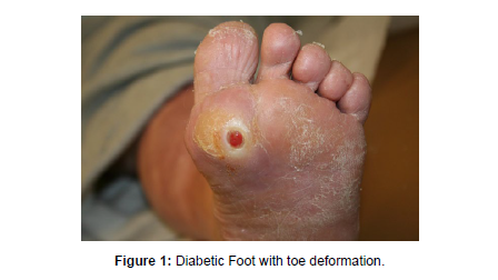
The clinical importance of studying the biomechanics of toe deformity in neuropathic diabetic patients has been demonstrated by several prospective population-based studies showing abnormality of the toe. This structure is a significant independent predictor of plantar ulceration in diabetic patients. The results of this study clearly show that fifth toe deformity is associated with significantly altered foot load parameters, such as peak pressure, pressure-time integral and fundus. pressure center at the plantar surface of the foot. This result shows that the 5th toe deformed foot of the diabetic patient causes a greater impact load in the patient's gait. From the vegetation pressure point of view, the deformed group's CoP flow shifted to the corridor part during walking. As the fifth toe was significantly deformed and could not withstand the higher pressures, it showed a marked decrease in peak pressure in the lateral forefoot and other toe regions. The contact area in the other toes of the deformed group was significantly lower than that of the control group, which is similar to previous studies. An important function of the toes in gait is to touch the surface and apply enough pressure to achieve a fixed point from which the body can be pushed. Due to the deformity of the toes and reduced contact area, the pressure on the soles of the toes shifts to the forefoot. Patients with fifth toe deformity showed a decreased area of contact with the sole of their foot and a significant increase in the pressure-time integral. Unlike other distortions, the reduced peak functions can be compensated by increasing the load on other regions. As toe function declines, the pressure that the metatarsal part of the forefoot is resisting during the push-up phase increases dramatically and the load the toes carry decreases. The results of these findings were inconsistent with the results of previous studies, and the toe area resisted impact and pressure was significantly reduced in patients with diabetes. This will cause the pressure to shift to a nearby area with an increase in the pressure peak. However, expansion of peak pressure leads to necrosis of muscle tissue and atrophy of muscle fibers and exacerbates the risk of injury in these regions of increased peak pressure. The cause of increased pressure peak in the deformed 5th toe group may be that the toes are deformed, leading to the destruction of fatty tissue leading to unable to withstand extreme pressure, so the pressure in the ankle area increases suddenly variable to maintain body stability(Figure 1).
Conclusion
This study found that the big toe and forefoot are the load-bearing parts of the foot in the 5th toe deformity group and are the most susceptible to foot ulceration due to the long-term effects of the collapse. Changes in pressure distribution in the soles and center of pressure trajectories should be considered when analyzing and treating plantar ulcers associated with diabetic deformity. Quantitative illustration of the foot load characteristics of fifth toe deformity can be of great benefit to diabetics to understand their foot load status and to physicians or rehabilitative therapists function to develop protocols that prevent or even restore foot ulcers associated with fifth toe deformity and other injuries. Some functional shoes are needed to reduce the risk of ulcers in the affected areas, normalizing the distribution of diabetics with fifth toe deformity.
References
- Flint JH, Conley AP, Rubin ML, Feng L, Lin PP, et al. (2020) Clear Cell Chondrosarcoma: Clinical Characteristics and Outcomes in 15 Patients. Sarcoma 2386191.
- Chondrosarcoma.
- Thorkildsen J, Taksdal I, Bjerkehagen B, Haugland HK, Borge Johannesen T, et al. (2019) Chondrosarcoma in Norway 1990-2013; an epidemiological and prognostic observational study of a complete national cohort. Acta Oncol Stockh Swed 58:273–282.
- Collins MS, Koyama T, Swee RG, Inwards CY (2003) Clear cell chondrosarcoma: radiographic, computed tomographic and magnetic resonance findings in 34 patients with pathologic correlation. Skeletal Radiol 32:687–694.
- Unni KK, Dahlin DC, Beabout JW, Sim FH (1976) Chondro sarcoma: clear-cell variant. A report of sixteen cases. J Bone Joint Surg Am 58:676–683.
- Bjornsson J, Unni KK, Dahlin DC, Beabout JW, Sim FH (1984) Clear cell chondrosarcoma of bone. Observations in 47 cases. Am J Surg Pathol 8:223-230.
- Kaim AH, Hugli R, Bonél HM, Jundt G (2002) Chondroblastoma and clear cell chondrosarcoma: radiological and MRI characteristics with histopathological correlation. Skeletal Radiol 31:88–95.
- Kumar R, David R, Cierney G (1985) Clear cell chondrosarcoma. Radiology 154:45-48.
- Laitinen M, Nieminen J, Pakarinen TK (2014) An Unusual Case of Clear Cell Chondrosarcoma with Very Late Recurrence and Lung Metastases, 29 Years after Primary Surgery. Case Rep Orthop e109569.
- Ogose A, Hotta T, Kawashima H, Hatano H, Umezu H, et al. (2001) Elevation of serum alkaline phosphatase in clear cell chondrosarcoma of bone. Anticancer Res 21:649-655.
- McLoughlin GS, Sciubba DM, Wolinsky JP (2008) Chondroma/Chondrosarcoma of the spine. Neurosurg Clin N Am 19:57-63.
- Chick JF, Chauhan NR, Madan R (2013) Solitary fibrous tumors of the thorax: nomenclature, epidemiology, radiologic and pathologic findings, differential diagnoses, and management. AJR Am J Roentgenol 200: 238-248.
- Flint A, Weiss SW (1995) CD-34 and keratin expression distinguishes solitary fibrous tumor (fibrous mesothelioma) of the pleura from desmoplastic mesothelioma. Hum Pathol 26: 428-431.
- Dalton WT, Zolliker AS, McCaughey WT (1979) Localized primary tumors of the pleura: an analysis of 40 cases. Cancer 44: 1465-1475.
- Witkin GB, Rosai J (1989) Solitary fibrous tumor of the mediastinum: a report of 14 cases. Am J Surg Pathol 13: 547-557.
Indexed at, Google Scholar, Crossref
Indexed at, Google Scholar, Crossref
Indexed at, Google Scholar, Crossref
Indexed at, Google Scholar, Crossref
Indexed at, Google Scholar, Crossref
Indexed at, Google Scholar, Crossref
Indexed at, Google Scholar, Crossref
Indexed at, Google Scholar, Crossref
Indexed at, Google Scholar, Crossref
Citation: Li X (2023) Inflammation Risks of Diabetic Foot with Toe Deformation are Reflected by Plantar Loading. Clin Res Foot Ankle, 11: 413.
Copyright: © 2023 Li X. This is an open-access article distributed under the terms of the Creative Commons Attribution License, which permits unrestricted use, distribution, and reproduction in any medium, provided the original author and source are credited.
Share This Article
Recommended Journals
Open Access Journals
Article Usage
- Total views: 956
- [From(publication date): 0-2023 - Apr 07, 2025]
- Breakdown by view type
- HTML page views: 728
- PDF downloads: 228

