Infiltrating Syringomatous Adenoma of the Nipple
Received: 02-Apr-2022 / Manuscript No. JCD-22-59352 / Editor assigned: 05-Apr-2022 / PreQC No. JCD-22-59352(PQ) / Reviewed: 19-Apr-2022 / QC No. JCD-22-59352 / Revised: 25-Apr-2022 / Manuscript No. JCD-22-59352(R) / Accepted Date: 27-Apr-2022 / Published Date: 02-May-2022 DOI: 10.4172/2476-2253.1000146
Abstract
Background: Infiltrating Syringomatous adenoma of the nipple is a very rare benign tumour of the breast. It often presents with a lump in areolar/ nipple complex and can occur in women of all ages. The clinical and radiology finding are suggestive of breast cancer. It is known to cause local infiltration, but according to the literature review, there has been no reported case of metastasis. Surgical excision of the lump is the curative management.
Case Presentation: We present a case report of a 43 years old woman with no family history of breast cancer or other cancer types and no significant risk factors for breast pathology, who presented with history of left breast pain and mass. Clinical examination showed thickening of the nipple in an otherwise normal breast and axilla. Ultrasound of the affected breast showed several scattered cysts in the area of the lesion and Mammogram showed micro calcification in the nipple. Biopsy was done and histopathology showed branching cords of cells forming a glandular structure infiltrating the surrounding stoma which was in keeping with infiltrating Syringomatous adenoma of the nipple. She underwent lump excision successfully and was discharged.
Conclusion: We present a rare case of infiltrating Syringomatous adenoma of the nipple highlighting it as a possible differential diagnosis of areolar and nipple lumps to avoid misdiagnosis and unnecessary management.
Keywords
Introduction
Syringomatous Adenoma of the Nipple (SAN) is one of the rare benign breast tumors that was first described by Rosen in 1983 [1] known to show local infiltrative proliferation but does not metastasize [2]. Only about 40 cases have been reported so far [3]. The tumor usually occurs in women between 11 to 76 years with a mean age of presentation of 40 years [4]. This is case report of 43-year-old female who underwent local excision of left nipple areola complex after a confirmed diagnosis of Syringomatous adenoma.
Case Report
This is a case of 43 years old female who presented to the breast clinic with two months history of left breast pain. She was initially seen in a private clinic where they found a large mass in the left upper quadrant of the breast. However, the mass resolved following treatment with oral medication which was prescribed from the same clinic. She is not known to have any medical conditions. In history, the patient denied having risk factors for breast pathology. Age of Menarche was at 13 years. She is married with 6 children; all were by spontaneous vaginal delivery and breastfed. Her age of first pregnancy was18 years and she denied taking contraceptive hormone, she had no family history for a breast, ovarian or other malignancies. Physical examination showed thickening of the left nipple however the rest of her examination was unremarkable.
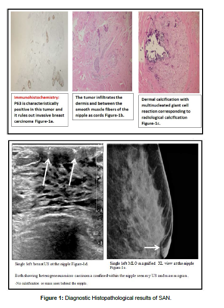
X-ray mammography was done and was reported as a heterogeneously dense fibro glandular tissue seen in both breasts with the right breast showed 3 foci of punctate macro calcifications in the outer lower quadrant and having a benign morphology. While in left breast, there was an irregular dense mass seen in the inner upper breast. It is the posterior fat/fibro glandular interface. It persisted on the compression views but became denser to the surrounding. It measures 1 cm x 0.9 cm. Also, there were heterogeneous macro calcifications seen in the left nipple at the lower aspect.
Ultrasound findings in the left breast showed several scattered cysts corresponding to the site of palpable abnormality there was a cyst at 1 o’clock and an oval shaped macro lobulated lesion seen deep at 11:30. It measured 9 mm x 3.8 mm x 9.3 mm. No internal vascularity was detected. The micro calcifications were seen in the nipple inferiorly and no retro areolar mass were seen. In the right breast, Small scattered cysts were detected. Axillary lymph nodes were looking benign in both breasts. Surgical biopsy (Wedge biopsy) was done: The pathology of the wedge biopsy taken from the left nipple revealed infiltrating adnexal tumor, favouring infiltrating Syringomatous adenoma of the nipple. The deep margins of biopsy where involved. Ultrasound guided core biopsy of left breast was taken at 11 o’clock lesion and reported as a fibro adenoma. Patient was admitted for left nipple areolar complex resection and left fibro adenoma excision. Hook wire was deployed across the lesion under ultrasound guidance and the operation was uneventful. She was discharged on the second day.
Discussion
SAN is easily confused with carcinomas of the breast because of their expansive as well as infiltrating pattern of growth often noted on histology. They have a tendency to recur locally especially following incomplete excision however they are not known to metastasize.
Clinical features
The typical clinical presentation is a unilateral single firm breast mass that causes itching, pain, nipple discharge, inversion or ulceration [1]. Because of the clinical presentation of nipple ulceration and crusting, SAN might be mistaken with Paget disease, prompting unnecessary mastectomy [5]. The tumor usually occurs in women between 11 to 76 years with a mean age of presentation of 40 years [4].
The diagnosis of SAN can be difficult with nipple adenomas and low-grade adenosquamous carcinomas as top differential [1, 2] however; nipple duct adenomas are well circumscribed usually ulcerates and does not invade the underlying tissue [5].
Low-grade adenosquamous carcinomas usually show an adenomalike structure derived from salivary gland duct. Thus, these carcinomas can be differentiated from their sites of origin [6].
Imaging
On imaging SAN appears as ill-defined with heterogeneous internal echoes on ultrasonography [2], SAN usually appears as a high-density mass in the sub areolar region with an irregular outline, spicule formation, and micro calcification foci on mammogram. SAN is more obvious in MRI than MMG. In fact, the imaging findings often resemble those of malignant tumors, thus tissue study is needed to distinguish between SAN and carcinoma [4].
Pathological features
Grossly, SAN is a firm tumor from 1 to 3 cm in diameter size. Cut section shows an ill-defined tumor with small cystic spaces around the nipple. The SAN tumor microscopically, has an infiltrating pattern characterized by branching cords of cells forming glandular structures. The tumor cells infiltrate the stoma and usually invade the per neural region and smooth muscle bundle [7, 8]. The surrounding breast tissue might appear normal or show hyperplastic changes. It does not involve the overlying skin or nipple epidermis. On histological study SAN appears similar to nipple adenoma; a benign variant of intraductal papilloma associated with serous or bloody nipple discharge and squamous metaplasia may be present in both SAN and nipple adenoma. Microscopically, the nipple adenoma shows epithelial hyperplasia arising from a lactiferous duct displacing the nipple stoma. On the other hand, nipple Syringomatous adenoma displays stromal infiltration [6]. The followings are the diagnostic histopathological criteria of SAN (1) location in dermis and sub cutis of nipple or areola; (2) irregular, compressed, or comma-shaped tubules infiltrating into smooth muscle bundles and/or nerves; (3) presence of my epithelial cells around the tubules; (4) presence of cysts lined by stratified squamous epithelium and filled with keratinous material; and (5) absence of mitotic activity and necrosis [4] (Figure 1).
Management
The management of SAN is complete local excision of the nipple areola complex to achieve histologically negative margins with the risk of recurrence high if not totally excised [6]. Studies found that with complete local excision no did not show evidence of recurrence as reported on a follow-up period of 1 to 6 years. Hence, close follow up to detect local recurrence is considered necessary [6,9]. Fortunately, most of the recurrences were managed with local re-excision. Although SAN shows local infiltration and recurrence, it is not known to metastasize [7,10]. If a patient wishes to undergo Nipple-sparing resection this option can be considered for its excellent cosmetic results. However, careful postoperative follow up is necessary as recurrence period range from 1.5 months to 4 years [7].
Conclusion
We reported the case of 43 years old woman with SAN. Patient underwent uneventful left NAC EXCISION.
Conflict of Interest
The Authors declare no conflict of interest in this study.
References
- Rosen PP (1983) Syringomatous adenoma of the nipple. Am J Surg Pathol 7:739-745.
- Carter E, Dyess DL (2004) Infiltrating syringomatous adenoma of the nipple: a case report and 20-year retrospective review. Breast J 10:443-447.
- Montgomery ND, Bianchi GD, Klauber-Demore N, Budwit DA (2014) Bilateral Syringomatous Adenomas of the Nipple: Case Report With Immunohistochemical Characterization of a Rare Tumor Mimicking Malignancy. Am J Clin Pathol 141:727-731.
- Oo KZ, Xiao PQ (2009) Infiltrating syringomatous adenoma of the nipple: Clinical presentation and literature review. Arch Pathol Lab Med 133:1487-1489.
- Ishikawa S, Sako H, Masuda K, Tanaka T, Akioka K, et al. (2015) Syringomatous adenoma of the nipple: a case report. J Med Case Rep 9:256.
- Rosen PP (2001) Syringomatous adenoma of the nipple. In Rosen's Breast Pathology 111–114. (Lippincott, Williams & Wilkins, Philadelphia).
- Jones MW, Norris HJ, Snyder RC (1989) Infiltrating syringomatous adenoma of the nipple: a clinical and pathological study of 11 cases. Am J Surg Pathol 13:197-201.
- Slaughter MS, Pomerantz RA, Murad T, Hines JR (1992) Infiltrating syringomatous adenoma of the nipple. Surgery 111:711-713.
- Eusebi V, Mai K, Taranger-Charpin A (2003) Tumours of the nipple. In: Tavassoli FA, Deville P, eds Pathology & Genetics of Tumours of the Breast and Female Genital Organs WHO 104–105.
- Suster S, Moran CA, Hurt MA (1991) Syringomatous squamous tumors of the breast. Cancer 67:2350-2355.
Google Scholar, Crossref, Indexed at
Google Scholar, Crossref, Indexed at
Google Scholar, Crossref, Indexed at
Google Scholar, Crossref, Indexed at
Google Scholar, Crossref, Indexed at
Google Scholar, Crossref, Indexed at
Citation: Aljarrah A, Harrasi NA, Alazri M, Aboje A, Ameri UA, et al. (2022) Infiltrating Syringomatous Adenoma of the Nipple. J Cancer Diagn 6: 146. DOI: 10.4172/2476-2253.1000146
Copyright: © 2022 Aljarrah A. This is an open-access article distributed under the terms of the Creative Commons Attribution License, which permits unrestricted use, distribution, and reproduction in any medium, provided the original author and source are credited.
Share This Article
Recommended Journals
Open Access Journals
Article Tools
Article Usage
- Total views: 1855
- [From(publication date): 0-2022 - Apr 04, 2025]
- Breakdown by view type
- HTML page views: 1393
- PDF downloads: 462

