Make the best use of Scientific Research and information from our 700+ peer reviewed, Open Access Journals that operates with the help of 50,000+ Editorial Board Members and esteemed reviewers and 1000+ Scientific associations in Medical, Clinical, Pharmaceutical, Engineering, Technology and Management Fields.
Meet Inspiring Speakers and Experts at our 3000+ Global Conferenceseries Events with over 600+ Conferences, 1200+ Symposiums and 1200+ Workshops on Medical, Pharma, Engineering, Science, Technology and Business
Case Report Open Access
Incidental Left Ventricular Pseudoaneurysm Discovered 5 Years after Myocardial Infarction
| Richard AP Takx1*, Christian Fink2 and Thomas Henzler2 | |
| 1Department of Radiology, University Medical Center Utrecht, Utrecht, the Netherlands, The Netherlands | |
| 2Institute of Clinical Radiology and Nuclear Medicine, University Medical Center Mannheim, Medical Faculty Mannheim, Heidelberg University, Germany | |
| Corresponding Author : | Richard Takx Department of Radiology UMC Utrecht, Heidelberg laan 100 3508 GA, Utrecht, The Netherlands Tel: +31-887559529 Fax: +31-302581098 E-mail: rtakx@umcutrecht.nl |
| Received April 19, 2013; Accepted May 08, 2013; Published May 13, 2013 | |
| Citation: Takx RAP, Fink C, Henzler T (2013) Incidental Left Ventricular Pseudoaneurysm Discovered 5 Years after Myocardial Infarction. OMICS J Radiology 2:126. doi: 10.4172/2167-7964.1000126 | |
| Copyright: © 2013 Takx RAP, et al. This is an open-access article distributed under the terms of the Creative Commons Attribution License, which permits unrestricted use, distribution, and reproduction in any medium, provided the original author and source are credited. | |
Visit for more related articles at Journal of Radiology
| Case Report |
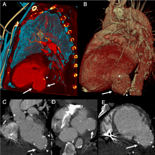
| GA 65 year-old man was admitted to the intensive care unit with candida sepsis. A suspected pulmonary etiology prompted a contrast enhanced Computed Tomography (CT) scan of the chest. CT revealed dilated cardiomyopathy, lateral and apical myocardial wall thinning, diffuse coronary artery calcifications, and a large, calcified left ventricular pseudoaneurysm arising from the basal lateral myocardial wall. Five years prior, the patient was diagnosed with Myocardial Infarction (MI) of the circumflex and first diagonal coronary arteries, which was managed medically. There was no record of subsequent free wall rupture or pseudoaneurysm formation. |
| Left ventricular (LV) pseudoaneurysms are rare, frequently asymptomatic and often fatal when left untreated. Unlike a true LV aneurysm, a LV pseudoaneurysm contains no endocardium or myocardium. LV pseudoaneurysm is most commonly the result of a transmural acute MI when myocardial rupture is contained by pericardial adhesions or scar tissue. Diagnosis can be complicated because patients usually present with symptoms similar to coronary artery disease [1]. CT accurately delineates the extent of a LV pseudoaneurysm and allows for assessment of both intra-cardiac and extra-cardiac anatomy. Owing to the risk of subsequent fatal rupture, regardless of chronicity [2], surgery is often recommended [3]. Surgical repair provides good symptomatic relief and long-term survival in case of dyskinetic or akinetic aneurysms [4]. However, taking into account the surgical risk, conservative treatment should be considered in asymptomatic individuals. Moreover, long-term survival of patients treated conservatively appears to be relatively benign [5]. This represents an unusual case of a stable post-MI LV pseudoaneurysm discovered years after the original insult (Figure 1). |
References |
|
Figures at a glance
 |
| Figure 1 |
Post your comment
Relevant Topics
- Abdominal Radiology
- AI in Radiology
- Breast Imaging
- Cardiovascular Radiology
- Chest Radiology
- Clinical Radiology
- CT Imaging
- Diagnostic Radiology
- Emergency Radiology
- Fluoroscopy Radiology
- General Radiology
- Genitourinary Radiology
- Interventional Radiology Techniques
- Mammography
- Minimal Invasive surgery
- Musculoskeletal Radiology
- Neuroradiology
- Neuroradiology Advances
- Oral and Maxillofacial Radiology
- Radiography
- Radiology Imaging
- Surgical Radiology
- Tele Radiology
- Therapeutic Radiology
Recommended Journals
Article Tools
Article Usage
- Total views: 13274
- [From(publication date):
July-2013 - Mar 29, 2025] - Breakdown by view type
- HTML page views : 8797
- PDF downloads : 4477
