Incidental Gallbladder Adenosquamous Carcinoma: Case Report and Literature Review
Received: 25-Sep-2020 / Accepted Date: 14-Oct-2020 / Published Date: 21-Oct-2020
Abstract
Gallbladder Cancer (GBC) is a rare but aggressive malignancy with marked ethnic and geographic variations. About 90% OF gallbladder cancers are Adenocarcinomas (AC) which are usually well to moderately differentiated. Adenosquamous/Squamous Cell Carcinoma (AS/SCC) of the gallbladder are a rare histopathologic subtype and account for nearly 1% to 12% of all GBC cases. The few cases in the literature suggest that these rare GBC tumors are more aggressive in comparison to adenocarcinoma and often infiltrate the adjacent viscera. Adenosquamous cell carcinoma shows a mixture of glandular and squamous elements with the squamous component between 25% and 99%. If the squamous component is less than 25%, the tumors are described as adenocarcinoma with focal squamous change. Pure squamous cell carcinoma is even rarer and is described in literature mainly in the form of case reports. These tumors exhibit exclusively squamous differentiation without any recognizable invasive glandular component.
Keywords: Gallbladder; Adenosquamous carcinoma; Cholelithiasis
Patient Presentation
63 years old female with PMH of DM, HTN presented with abdominal pain that started the previous night during dinner. This was a severe sharp constant RUQ abdominal pain radiating to her right chest and back, which she has never experienced before. It continued as several episodes of non-bile non-bloody vomiting, and pain with deep breathing in the RUQ. She denied fevers, diarrhea, dysuria, focal numbness, weakness, weight loss, dysphagia, or reflux, and no history of gallbladder stones or surgery.
Abdominal ultrasound revealed
1. Cholelithiasis and biliary sludge with gallbladder wall thickening and a positive sonographic Murphy’s sign, concerning for acute cholecystitis.
2. Borderline hepatomegaly with mildly echogenic liver. Patient underwent laparoscopic cholecystectomy the next day after admission and recovered well post-operatively, stable for discharge after two days.
However, pathologic examination of the surgical specimen gallbladder revealed moderately differentiated mural adenosquamous carcinoma.
The Gross and Morphological Features of Gallbladder Carcinoma
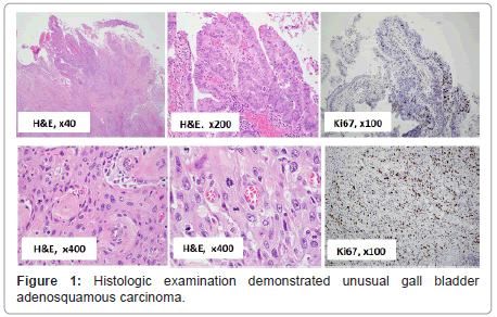
The gross evaluation of the gallbladder revealed a saccular 9.0 × 4.5 cm gallbladder with multiple gallstones and red to tan velvety mucosa with a firm mass measuring 5.0 × 3.0 × 1.5 cm on the lateral fundus, 5.0 cm away from the cystic duct margin [1]. The external surface serosa is smooth and glistening. Within the gallbladder, there were approximately 20 hard black faceted gallstones, 4 × 4 × 3 cm in aggregate. No gallstone was lodged within the cystic duct. Histologic examination demonstrated unusual gall bladder adenosquamous carcinoma (Figure 1). Submitted entirely, the carcinoma infiltrated the fundus wall locally but was confined within the serosa. The liver margin was negative for carcinoma cells, while tumor invaded peri muscular connective tissue on the peritoneal side without involvement of the serosa (visceral peritoneum). The remainder of the gallbladder mucosa showed changes of chronic cholecystitis and multiple gallbladder cystic diverticula (Rokitansky Aschoff sinuses).
Immunohistochemistry Stains
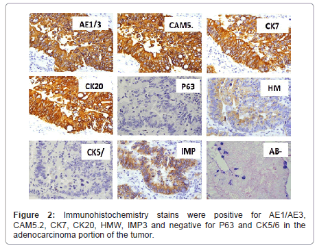
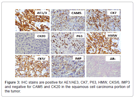
Immunohistochemistry stains were positive for AE1/AE3, CAM5.2, CK7, CK20, HMW, IMP3 and negative for P63 and CK5/6 in the adenocarcinoma portion of the tumor (Figure 2), and IHC stains are positive for AE1/AE3, CK7, P63, HMW, CK5/6, IMP3 and negative for CAM5. 2 and CK20 in the squamous cell carcinoma portion of the tumor (Figure 3). Special stain AB-PAS was positive for the mucin in adenocarcinoma portion of the tumor (Figure 2). The findings were consistent with an adenosquamous carcinoma.
Multiphase CT Evaluation
The pathological findings mandated a follow-up workup for possible metastatic tumor. Multiphase CT on liver revealed a 2.5 cm low density hepatic lesion on arterial phase images. Abdominal MRI showed multiple low-density hepatic lesions, concerning for metastatic gallbladder carcinoma. CT Guided biopsy of the low-density hepatic lesion demonstrated metastatic poorly differentiated adenosquamous carcinoma. The patient was started on appropriate chemotherapy immediately.
The Prevalence of the GB Carcinoma
Gallbladder cancer (GBC) is a rare but aggressive malignancy with marked ethnic and geographic variations. About 90% of gallbladder cancers are Adenocarcinomas (AC) which are usually well to moderately differentiated. Adenosquamous/Squamous Cell Carcinoma (AS/SCC) of the gallbladder are a rare histopathologic subtype and account for nearly 1% to 12% of all GBC cases [2-4]. The few cases in the literature suggest that these rare GBC tumors are more aggressive in comparison to adenocarcinoma and often infiltrate the adjacent viscera [5]. Adenosquamous cell carcinoma shows a mixture of glandular and squamous elements with the squamous component between 25% and 99%. If the squamous component is less than 25%, the tumors are described as adenocarcinoma with focal squamous change. Pure squamous cell carcinoma is even rarer and is described in literature mainly in the form of case reports [6]. These tumors exhibit exclusively squamous differentiation without any recognizable invasive glandular component [7].
According to Surveillance, Epidemiology, and End Results (SEER) Program of the National Cancer Institute, out of 3038 patients with gallbladder cancer, only 45 (1.7%) were squamous cell carcinoma [8,9]. In one study, the incidence of adenosquamous carcinoma (defined as 25%-99% of the tumor being squamous) was 6%, and that of pure squamous cell carcinoma (without any documented invasive glandular component) was 2%. Other reports are ranging from 1.4% to 10.6% of all cases of gallbladder carcinoma [5,6,10].
The Clinicopathologic Features of the GB Carcinoma
Given the rarity, limited data is available in medical literature regarding the clinicopathologic features of these tumors. As reported, clinical presentation of adenosquamous carcinoma/squamous cell carcinoma is vague and non-specific [11,12]. In the present case, the cancer was found incidentally with cholelithiasis and chronic cholecystitis expected. Mean age of AS/SCC was 58 to 60 years whereas it was 56 years in gallbladder adenocarcinoma cases [10,13,14]. Most common site of these tumors was the fundus and association with gallstones was identified in 50% of cases 14. The average tumor size in literature was 4-5 cm at the time of presentation. Tumor thickness was 1.4-3.1 cm. The histologic differentiation of adenocarcinoma element is typically well to moderate differentiated while squamous carcinoma element is poorly differentiated with no keratin pearls. But mucin may be present in the neoplastic glands. Adenosquamous carcinoma of the gallbladder with human chorionic gonadotropin (hCG) production, foci of spindle cells, and neuroendocrine and gastric foveolar type differentiation have been described in the literature [15-17].
These tumors are usually silent in initial stages and are detected in advanced stages. In one study, 23/29 patients were categorized with T3 or T4 tumors that invaded adjacent organs [18]. In another study, 6/8 patients (75%) were in higher stage (T3 or T4). The early stage of the AS/SCC of gallbladder presents with nonspecific symptoms or is asymptomatic, as in the present AS/SCC case which was diagnosed incidentally after laparoscopic cholecystectomy clinically due to cholecystitis.
Tumorigenesis about the GB Carcinoma
Tumorigenesis of these tumors remains unclear and may be due to response to irritation resulting in mucosal squamous metaplasia or originates in pre-existing adenocarcinoma [19]. Pure squamous cell carcinoma often arises in association with squamous metaplasia in other organ systems such as lung.
Patients with adenosquamous/squamous cell carcinoma of the GB are frequently diagnosed at an advanced stage with a bulky tumor and adjacent organ involvement. This has been attributed to a high proliferative index of the squamous component in these tumors [5]. In 20 cases of adenosquamous carcinoma of gall bladder study, the positive rate of immunostaining for proliferating cell nuclear antigen in the squamous areas (mean 20.55%) was higher than that of adenocarcinoma areas (mean 11.40%; p=0.0029) indicating that the squamoid areas had a greater proliferative capacity than that of adenocarcinoma [20]. In the present case, the Ki-67 proliferation index was much higher in the squamoid areas than the adenocarcinoma areas (35% versus 16%). The liver metastasis of the gallbladder carcinoma showed squamoid differentiation in 70% of cells which suggests the squamoid part of adenosquamous cell carcinoma is more aggressive than the adenocarcinoma component in this case.
The Metastatic Features of the GB Carcinoma
Mechanism of spread of these tumors includes early metastasis as well as extension from the primary, as patients with adenosquamous/ squamous cell carcinoma typically present at an advanced stage. Lymph node metastases of purely squamous carcinoma were observed in only one case [21]. Other authors have described an aggressive disease in 85.7% cases of adenosquamous cell/squamous cell carcinoma [21] and 57.1% cases [5]. The present case is negative for tumor involvement of the gallbladder liver-bed, but multiple metastases found in the liver lobes which confirmed by CT-guided biopsy. This suggests that lymphocytic or blood borne metastases could happen before the tumor direct invasion to the surrounding organs.
Declaration of Conflicting Interests
The author(s) declare no potential conflicts of interest with respect to the research, authorship, and/or publication of this article.
Funding
The author(s) received no financial support for the research, authorship, and/or publication of this article.
References
- Hopper KD, Landis JR, Meilstrup JW, McCauslin MA, Sechtin AG (1991) The prevalence of asymptomatic gallstones in the general population. Invest Radiol 26: 939-945.
- Ishikawa Y, Yoshida H, Mamada Y, Taniai N, Kawano Y (2004) Squamous cell carcinoma of the gall bladder. J Nippon Med Sch 71: 417-420.
- Duffy A, Capanu M, Abou-Alfa GK, Huitzil D, Jarnagin W (2008) Gall bladder cancer: 10 year experience at Memorial Sloan-Kettering Cancer Centre (MSKCC) J Surg Oncol 98: 485-489.
- Henson DE, Albores-Saavedra J, Corle D (1992) Carcinoma of the gall bladder. Histologic types, stage of disease, grade and survival rates. Cancer 70: 1493-1497.
- Kalayarasan R, Javed A, Sakhuja P, Agarwal AK (2013) Squamous variant of gall bladder cancer: Is it different from adenocarcinoma? Am J Surg 206: 380-385.
- Rao S, Arya A, Aggarwal S, Gupta K, Arora R (2007) Pure squamous cell carcinoma of the gall bladder. Indian J Pathol Microbiol 50: 599-600.
- Roa JC, Tapia O, Cakir A, Basturk O, Dursun N (2011) Squamous cell and adenosquamous carcinomas of the gallbladder: Clinicopathological analysis of 34 cases identified in 606 carcinomas. Mod Pathol 24:1069-1078.
- Henson DE, Albores-Saavedra J, Corle D (1992) Carcinoma of the gall bladder. Histologic types, stage of disease, grade and survival rates. Cancer 70: 1493-1497.
- Waisberg J, Bromberg SH, Franco MIF, Yamagushi N, dos Santos PA (2011) Squamous cell carcinoma of the gallbladder. Sao Paulo Med J 119: 43.
- Gulwani HV, Gupta S, Kaur S (2017) Squamous cell and adenosquamous carcinoma of gall bladder: A clinicopathological study of 8 cases isolated in 94 cancers. Indian J Surg Oncol 8: 560-566.
- Kondo M, Dono K, Sakon M, Shimizu J, Nagano H (2002) Adenosquamous carcinoma of the gall bladder. Hepato-Gastroenterology 49: 1230-1234.
- Lada PE, Taborda B, Sanchez M, Tommasino J, Rosso FF (2007) Adenosquamous and squamous carcinoma of the gall bladder. Cir Esp 81: 202-206.
- Kim WS, Jang KT, Choi DW, Choi SH, Heo JS (2011) Clinicopathologic analysis of adenosquamous/squamous cell carcinoma of the gallbladder. J Surg Oncol 103: 239-242.
- Song HW, Chen C, Shen HX, Ma L, Zhao YL (2015) Squamous/adenosquamous carcinoma of the gall bladder: Analysis of 34 cases and comparison of clinicopathologic features and surgical outcomes with adenocarcinoma. J Surg Oncol 112: 677-680.
- Nishihara K, Takashima M, Furuta T, Haraguchi M, Tsuneyoshi M (1995) Adenosquamous carcinoma of the gall-bladder with gastric foveolar-type epithelium. Pathol Int 45: 250-256.
- Fukuda T, Ohnishi Y (1990) Gallbladder carcinoma producing human chorionic gonadotropin. Am J Gastroenterol 85: 1403-1406.
- Oohashi Y, Shirai Y, Wakai T, Nagakura S, Watanabe H (2002) Adenosquamous carcinoma of the gallbladder warrants resection only if curative resection is feasible. Cancer 94: 3000-3005.
- Roppangi T, Takeyoshi I, Ohwada S, Sato Y, Fujii T (2000) Minute squamous cell carcinoma of the gall bladder: A case report. Jpn J Clinc Oncol 30:43-45.
- Nishihara K, Nagai E, Izumi Y, Yamaguchi K, Tsuneyoshi M (1994) Adenosquamous carcinoma of the gall bladder: A clinicopathological, immunohistochemical and flow-cytometric study of twenty cases. Jpn J Cancer Res 85: 389-399.
- Mingoli A, Brachini G, Petroni R, Antoniozzi A, Cavaliere F (2005) Squamous and adenosquamous cell carcinomas of the gall bladder. J Exp Clin Cancer Res 24: 143-150.
- Chan KM, Yu M, Lee WC, Jan YY, Chen MF (2007) Adenosquamous/squamous cell carcinoma of the gall bladder. J Surg Oncol 95: 129-134.
Citation: Jia Y, Samadzadeh S, Cornford M, Ji P, French SW (2020) Incidental Gallbladder Adenosquamous Carcinoma: Case Report and Literature Review. J Clin Exp Pathol 10: 383.
Copyright: © 2020 Jia Y et al. This is an open-access article distributed under the terms of the Creative Commons Attribution License, which permits unrestricted use, distribution, and reproduction in any medium, provided the original author and source are credited.
Share This Article
Recommended Journals
Open Access Journals
Article Usage
- Total views: 2697
- [From(publication date): 0-2020 - Apr 29, 2025]
- Breakdown by view type
- HTML page views: 1881
- PDF downloads: 816


