Research Article Open Access
In Vitro Comparison of Three Desensitizing Prophylaxis Pastes: A Morphological Analysis
Anna Maria Genovesi1, Simone Marconcini1,2*, Marco Lelli3, Enrica Giammarinaro1, Antonio Barone1,2 and Ugo Covani1,21Versilia General Hospital, Lido di Camaiore, Italy
- *Corresponding Author:
- Simone Marconcini
Versilia General Hospital
Lido di Camaiore, Lucca, Italy
Tel: + 39 0584 9853
E-mail: s.marconcini1977@libero.it
Received Date: August 08, 2015; Accepted Date: September 07, 2015; Published Date: September 12, 2015
Citation: Genovesi AM, Marconcini S, Lelli M, Giammarinaro E, Barone A, et al. (2015) In Vitro Comparison of Three Desensitizing Prophylaxis Pastes: A Morphological Analysis. J Oral Hyg Health 3:186. doi: 10.4172/2332-0702.1000186
Copyright: © 2015 Genovesi AM,et al. This is an open-access article distributed under the terms of the Creative Commons Attribution License, which permits unrestricted use, distribution, and reproduction in any medium, provided the original author and source are credited.
Visit for more related articles at Journal of Oral Hygiene & Health
Abstract
Background and Objective: Dentine hypersensitivity (DH) characterizes for short sharp pain arising from exposed dentine. Nowadays, there is a vast choice of products to overcome it. The aim of the present observational study was to compare the efficacy of three prophylaxis pastes in occluding dentinal tubules from in an in vitro setting.
Methods: Longitudinal mesio-distal sections were obtained from a sample of ten extracted teeth. Morphological analysis with scanning electron microscopy (SEM) assessed the penetration of three different prophylaxis pastes in dentinal tubules: Nupro by GSK, Stomyprox by Biorepair, Colgate Pro-Sollievo. The use of the Energy Dispersive X-ray Analysis (EDX probe) allowed assessing the exact composition of each product.
Results: The SEM analysis revealed that only Stomyprox by Biorepair could penetrate the dentinal tubule due to its low crystallinity. Its small sized particles (˜1.60 μ) fit better the tubules lumen if compared to the other two pastes with higher sized granules. The small sample size and the lacking of a quantitative evaluation of tubules filling limit this result.
Conclusion: The occlusion of dentinal tubules is one of the possible ways to reduce dentine hypersensitivity in a professional setting. Small sized particles fit better the lumen of tubules. In conclusion, there is a biological rationale in preferring prophylaxis pastes characterized by a low crystallinity. It is worthy to investigate furthermore, and in a clinical setting, the efficacy of the prophylaxis paste made by small sized particles of Hydroxyapatite.
Keywords
Dentine hypersensitivity; Prophylaxis paste; Hydroxyapatite
Introduction
Dentine hypersensitivity (DH), clinically, is as an exaggerated response to a non-noxious sensory stimulus (osmotic, thermal or mechanical changes). The prevalence of DH in general population ranges between 4-57% and females are more affected [1]. DH derives from the underlying exposed dentine, after the enamel or cementum at the root surface has been eroded away [2]. DH is most commonly reported at the buccal-cervical zone of permanent teeth [3] and this is due to its peculiar etiology that is multifactorial. The most common clinical cause of DH is gingival recession, which exposes the root surface, due to periodontal treatment, surgical/dental operative procedures, gum diseases, aging and incorrect tooth brushing or association of two or more of these factors. Other factors include patients’ deleterious habits, poor hygiene and diet, exposure of teeth to chemical products, excessive occlusal forces and premature occlusal contacts [4].
Histologically, sensitive dentine is characteristic for the patency of dentinal tubules, and it has been reported that there is a positive correlation between the density of open dentinal tubules and the intensity of pain responses [5]. Therefore, plugging tubules should overcome DH symptoms, as well as preventing fluid flow, or dulling the nerves.
An accurate diagnosis is the key in preventing the occurring or the recurring of DH. In fact, the treatment should be appropriate to the specific etiology of DH in each patient. Individualization of treatment protocols is the best way to obtain both hygienist and patient satisfaction. This is true not only for timings and maneuvers, but for specific product usage also.
Nowadays, there is a vast choice of products that both the doctor and the patient could use at the end of an oral hygiene session to reduce dentine hypersensitivity. They come in different formulas such as solutions, gels and prophylaxis pastes containing various components. There could be fluorides, calcium hydroxide, strontium chloride, potassium nitrate, sodium citrate, glutaraldehyde and hydroxyethyl methacrylate, potassium, or ferric oxalate [6].
The aim of the present observational study was to compare the efficacy of three prophylaxis pastes (Nupro by GSK, Colgate Pro-sollievo, Stomyprox by Biorepair) from a histological point of view. Our null hypothesis was that there was no significant difference between the three pastes in occluding dentinal tubules.
Materials and Methods
The study took place at the Stomatologic Institute of the Versilia hospital (Lido di Camaiore). Ten extracted permanent teeth constituted the sample of the present in vitro observational study. The small sample size is due to the nature of this in vitro research, which is a pilot observational study. Three molars, three premolars and four incisors composed the sample in analysis. Inclusion criteria for the extracted teeth were absence of decays, absence of brushing abrasion, absence of root canal therapy, and absence of prosthetic restorations. The reason for extraction was severe periodontal disease for every tooth in the sample.
The teeth have been washed out with a 5.25% sodium hypochlorite solution to dissolve any residual proteic material that could interfere with the morphological analysis. The operator let the teeth rest in the sodium hypochlorite solution for a night after a few shake of the test tube containing them. This procedure led to the standardization of the sample: the rinsed teeth resulted in a uniform surface with patent tubules ready to receive the product.
One of the ten teeth (one of the four incisors) was left untreated, in order to perform an initial testing of the Scanning Electron Microscopy (SEM), in order to visualize correctly tooth surface and its tubules. Once cleaned with the sodium hypochlorite solution, the remaining nine teeth were divided in three groups: each group consisted of a molar, a premolar, and an incisor. The three different prophylaxis pastes were randomly assigned to one of the three groups. The operator distributed uniformly one of the three products on the entire tooth surface with a small rotatory brush for one minute.
The laboratory-trained technician cut the teeth in clean longitudinal sections in a mesio-distal direction. The thickness of each section was ˜1 mm. The sections obtained at the mid-point of each tooth were analyzed at the SEM to visualize the penetration of products in dentinal tubules. The probe for the Energy Dispersive X-ray Analysis (EDX probe) helped to visualize the exact composition of the materials in study.
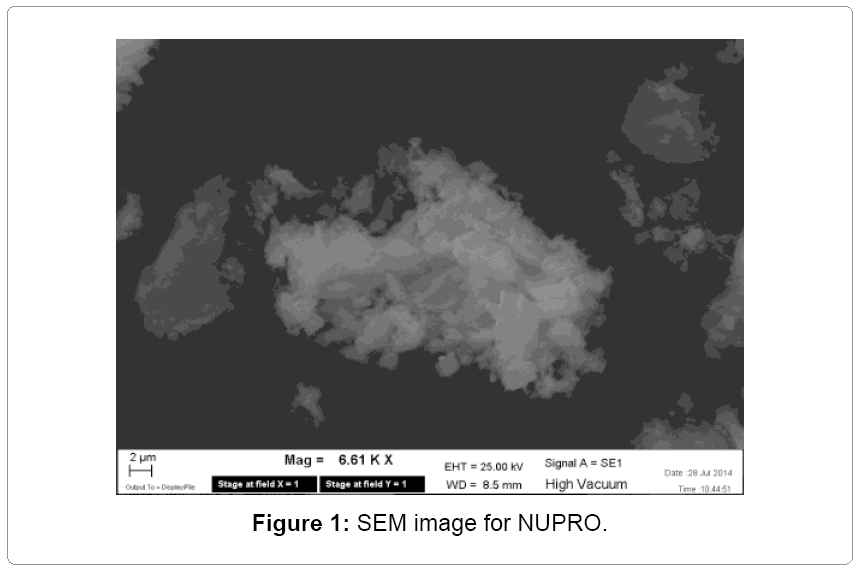
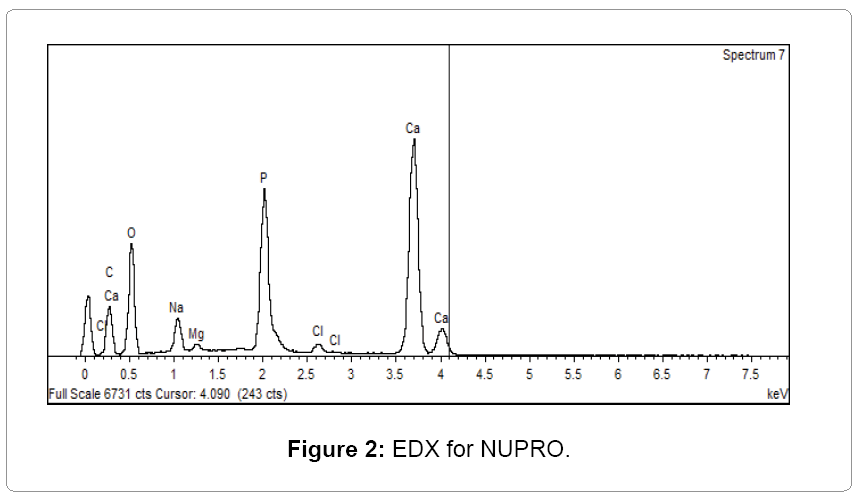
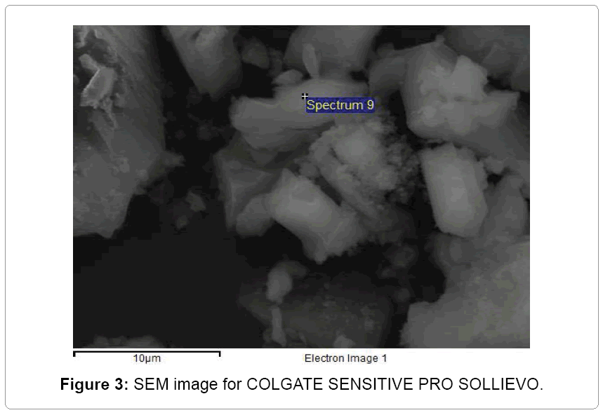
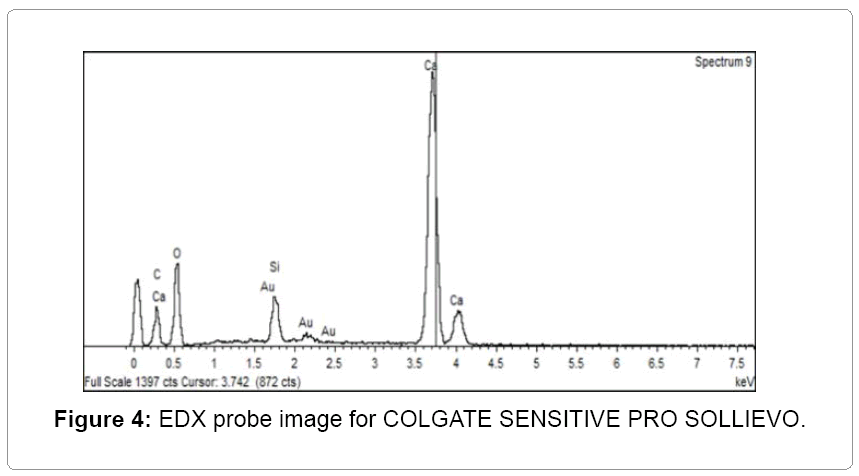
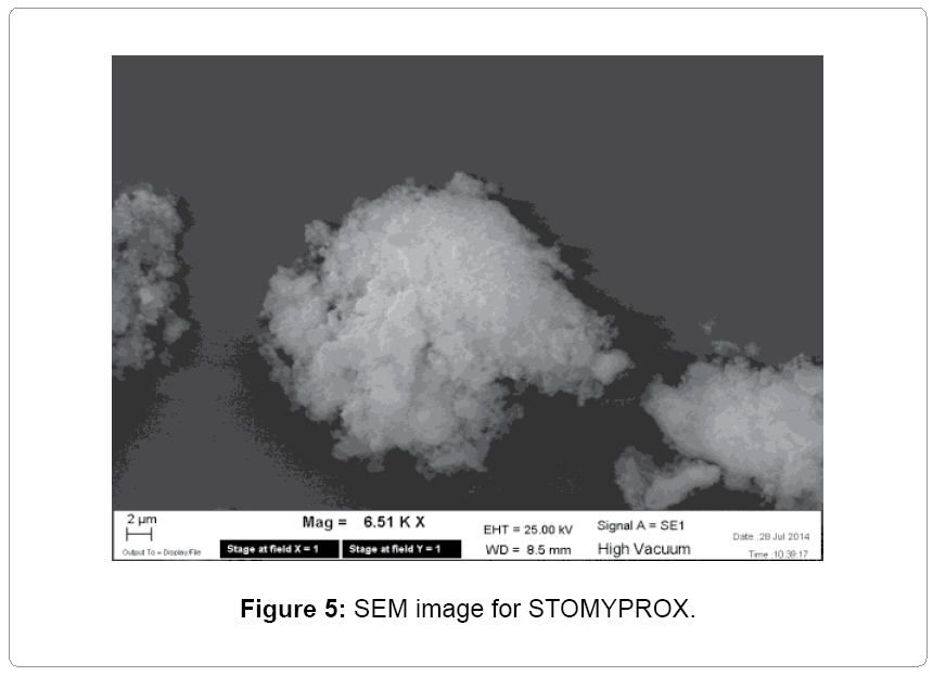
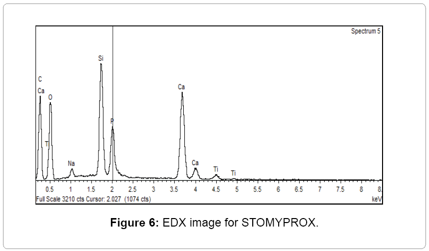
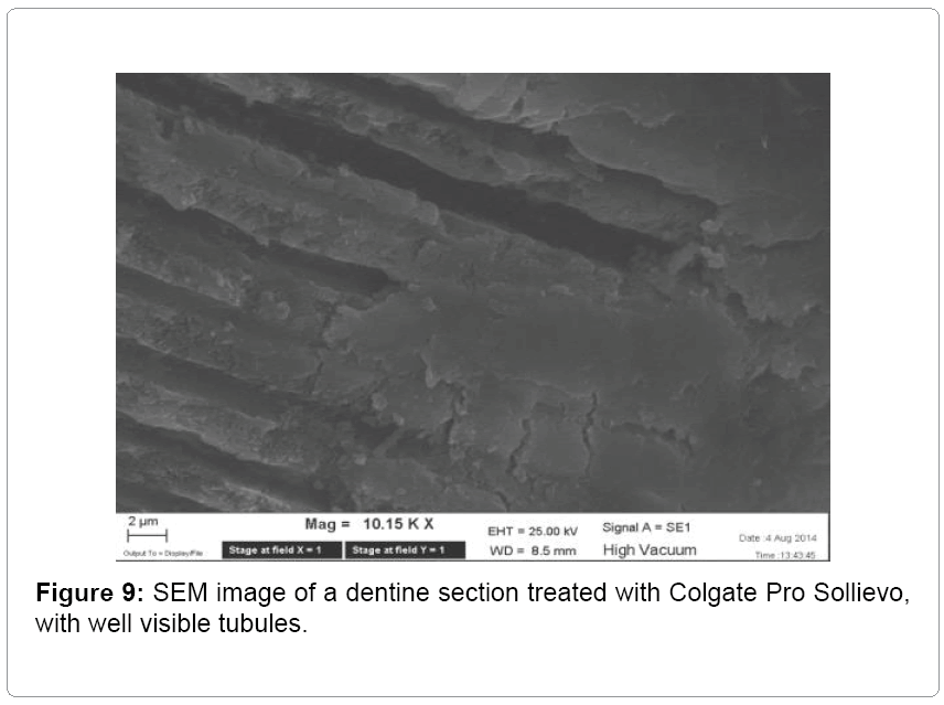
Nupro by GSK is a prophylaxis paste containing fluoride and a part of calcium sodium phosphosilicate (Figures 1 and 2). The formula of Colgate Pro-Sollievo contains arginine, which is an amino acid naturally present in the saliva, and to an insoluble calcium composite in the form of calcium carbonate (Figures 3 and 4). Stomyprox by Biorepair is a prophylaxis paste based on hydroxyapatite with low crystallinity, with dimensions and morphology able to fit the tubules (Figures 5 and 6).
Results
The SEM analysis, conducted on the sections treated with Nupro by GSK, presents a product consisting of particles sized few microns (˜4.80 μ). The EDX probe image (Figure 2) shows that the composition is mainly calcium and phosphate.
Figure three shows that Colgate Sensitive Pro Sollievo is made of irregular micrometric aggregates (˜12.0 μ). In Figure 4 (EDX probe), we can appreciate how Colgate Sensitive Pro Sollievo is mainly constituted by calcium, as described in the data sheet.
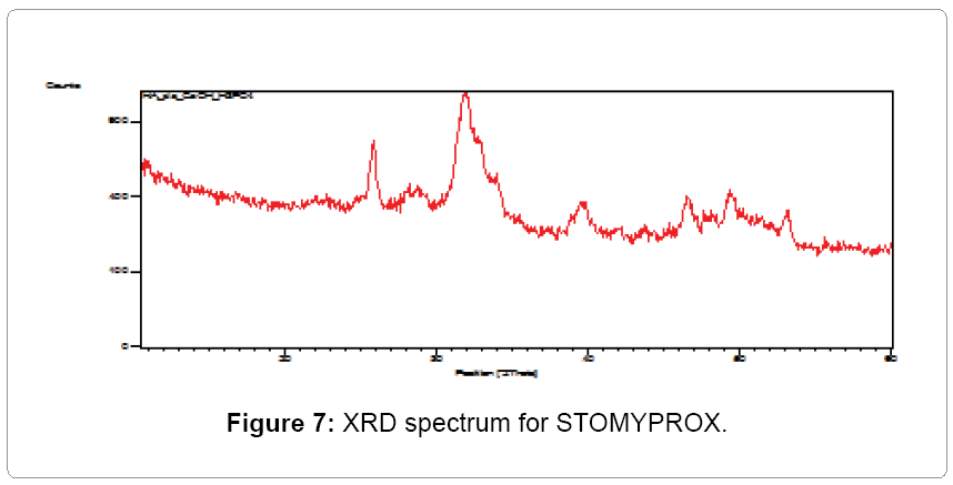
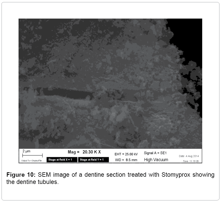
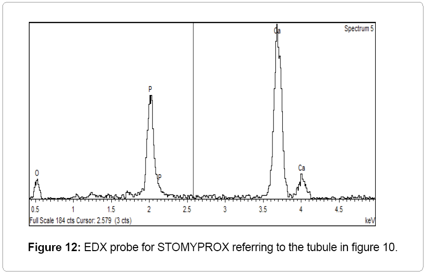
Figures five and six (EDX probe) show that Stomyprox by Biorepair is made of calcium-phosphate irregular micro aggregates with smaller sizes (˜1.60 μ) than those found in the previous pastes. The ratio calciumphosphorus identified through the EDX probe is different from that found in Nupro by GSK for the same elements. The image shows the presence of silica as well. An extra analysis carried out through X-Ray diffraction (XRD) revealed that the inorganic phase in Stomyprox was made of micrometric hydroxyapatite; unlike Nupro, which contains both calcium and phosphorus, but in salt and not in apatite form.
The mid-point 1 mm section, obtained from each tooth type, was analyzed with the SEM. For simplification, the possible result was dichotomous: absence of tubules filling, presence of tubules filling. There were no differences in tubules filling, based on the type of tooth treated. Instead, the filling was consistently different between treatment groups, as described below.
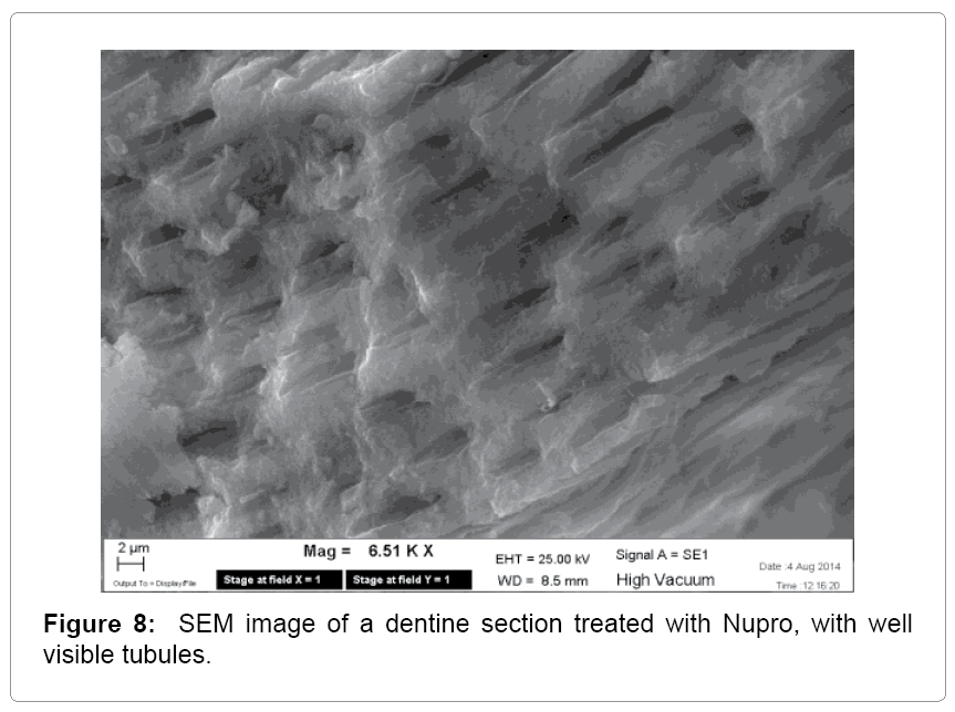
Figure 8 reports a dentine section treated with Nupro. This image shows that the dentine tubules appear perfectly clear and with no filling. Figure 9 reports the dentine tubules treated at the surface with Colgate Sensitive Pro Sollievo: even in this case the tubules appear to be empty. Figure 10 shows the SEM analysis of a tooth section treated with Stomyprox. The tubule visible at the center of Figure 10 is full of tiny micrometric granules.
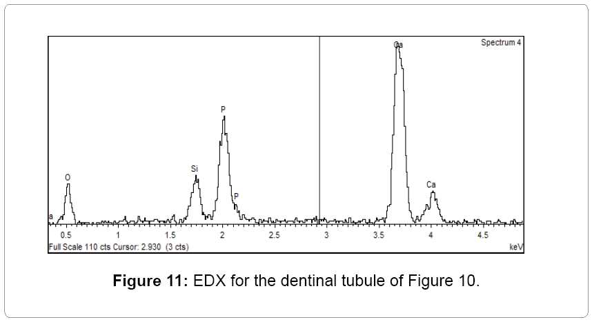
A deeper investigation through the EDX probe (Figure 11) shows that there are only calcium and phosphorus in a precise ratio where the filling is not present.
The EDX probe investigation (Figure 12) of the tubule visible in Figure 10 shows the presence of calcium and phosphorus (in a ratio similar to that of Figure 11) and silica, which is obviously not present on the dentine, but it comes from the formula of the product Stomyprox, as previously evidenced in EDX analysis (Figure 6).
Discussion
The main aim of the present study was to evaluate the in vitro efficacy of three prophylaxis pastes in occluding dentinal tubules. Our results disproved the hypothesis that there were no differences in tubule filling between the three pastes tested. The prophylaxis paste containing hydroxyapatite in small granules (˜1.60 μ) resulted in the occlusion of the lumen of dental tubules despite the type of tooth treated as evaluated on the morphological analysis with the SEM. The other two prophylaxis pastes, characterized by bigger particles (˜4.80 μ and ˜12.0 μ), resulted in no filling of dental tubules, in spite of the site and the tooth-type treated.
One of the limitations of the present observational study is the small sample size. That was due to the preliminary nature of the research, which was originally designed as a pilot study. The next step would be the identification of a standardized unit measure to describe tubules filling, despite their orientation, and despite the tooth site. Of course, the study lacks also in the statistic aspect, since there was no quantitative evaluation of the entity of filling, or counting the number of tubules occluded by the pastes granules. At last, a clinical evaluation would be useful, in order to match the histological evidence with the eventual symptoms relief.
The etiology of DH is multifactorial and the most widely accepted explanatory theory is the hydrodynamic one, proposed by Brannstrom [7], who suggested that exposed dentine with patent tubules, allows the movement of tubule fluid, which leads to dentine sensitivity. The centrifugal fluid movement activates the nerve endings at the end of dentinal tubules or at the pulp-dentinal complex.
The pathogenesis of DH includes two stages: lesion localization and lesion initiation. The localization stage refers to the moment when loss of enamel occurs; the lesion initiation is secondary to the removing of the protective covering of smear layer, leading to exposure and opening of dentinal tubules [5]. Scanning electron microscopy (SEM), in fact, has shown that tubules of teeth clinically characterized as ‘sensitive’ are eight times more numerous, two times wider in diameter and more penetrable [8].
The branching pattern of dentinal tubules reveals an intricate anastomosing system, criss-crossing the intertubular dentine. Major branches, 0.5-1.0 micron diameter, are typically at the periphery of the toothy. Fine branches, of 300-700 nm in diameter, forked off at 45 degrees, are abundant where the density of the tubules is relatively low. Micro-branches, 25-200 nm in diameter, extend at right angles from the tubules and are the most common [9].
Free liquids account for 75% of the dentinal tubule volume. A slight reduction of the dentinal tubule diameter can greatly reduce the flow of liquid within the dentinal tubules, subsequently alleviating the sensitivity of the nerve [10].
The results of the present study on the SEM reveals that, by comparing three prophylaxis pastes, with different major components, only the Stomyprox, containing hydroxyapatite, succeeded in filling dentinal tubules. Hydroxyapatite is the major component of human bones and teeth. It is a mineral form of calcium apatite with the formula Ca5(PO4)3(OH).
Research into the use of the mineral components of the inorganic portion of the tooth structure has inspired the preparation of hydroxyapatite (HA), for occlusion of dentinal tubules [11]. The rationale of using HA as a desensitizer is that it would permanently occlude the open dentinal tubules and blend with them. Synthetic HA comes in various forms and size of its particles. The recent biomimetic approach in biochemical research is leading to the development of nanomaterials for inclusion in a variety of dental products. Examples are pastes containing nano-apatites for biofilm management at the tooth surface, and products that contain nanomaterials for the remineralization of early enamel lesions [12]. There are clinical studies investigating the desensitizing property of formulas containing HA [13]. SEM images showed that ordinary dentifrice with added HA leads to a significantly increased effect of dentinal tubule occlusion after only 7 days of brushing: the measured rates of dentinal tubule occlusion by the HA dentifrice were all above 90% [14-19].
Conclusion
In the present pilot study, we analyzed the efficacy of three prophylaxispastes (Stomyprox by Biorepair, Colgate Pro-Sollievo and Nupro by GSK) in occluding dentinal tubules. The SEM analysis evidenced that only Stomyprox is able to enter the dentinal tubules occluding them physically, while this is not visible for the other products, Nupro and Colgate Pro-Sollievo. This effect is due to the small sized particles of hydroxyapatite constituting the product. Even if this is only a pilot observational study, we can conclude that there isa histologic rationale in preferring prophylaxis pastes made by micro or nano-aggregates, capable of fitting the tubules lumen. A larger study, with bigger sample and eventual clinical randomized trial, is necessary to match the morphological efficacy of Stomyprox in occluding tubules with the eventual efficacy of treating dentine hypersensitivity symptoms better.
Conflict of Interest
The authors report no conflicts of interest related to this study.
References
- Rees JS, Addy M (2002) A cross-sectional study of dentine hypersensitivity. J ClinPeriodontol 29: 997-1003.
- Curro FA (1990) Tooth hypersensitivity in the spectrum of pain. Dent Clin North Am 34: 429-437.
- Dababneh RH, Khouri AT, Addy M (1999) Dentine hypersensitivity - an enigma? A review of terminology, mechanisms, aetiology and management. Br Dent J 187: 606-611.
- Ladalardo TC, Pinheiro A, Campos RA, BrugneraJúnior A, Zanin F, et al. (2004) Laser therapy in the treatment of dentine hypersensitivity. Braz Dent J 15: 144-150.
- Miglani S, Aggarwal V, Ahuja B (2010) Dentin hypersensitivity: Recent trends in management. See comment in PubMed Commons below J Conserv Dent 13: 218-224.
- Gillam DG, Orchardson R, Närhi MVO, and Kontturi-Närhi V. (2000) Present and future methods for the evaluation of pain associated with dentine hypersensitivity. Tooth wear and sensitivity. Martin Dunitz, London, 283-297.
- Brännström M. (1963) A hydrodynamic mechanism in the transmission of pain producing stimuli through the dentine: Sensory mechanisms in dentine, ed. D. J. Anderson. Oxford: Pergamon Press, 73-79.
- Absi EG, Addy M, and Adams D. (1987) Dentine hypersensitivity. J Clinical Periodontol 14: 280-284.
- Mjör IA, Nordahl I (1996) The density and branching of dentinal tubules in human teeth. Arch Oral Biol 41: 401-412.
- Yuan P, Shen X, Liu J, Hou Y, Zhu M, et al. (2012) Effects of dentifrice containing hydroxyapatite on dentinal tubule occlusion and aqueous hexavalent chromium cations sorption: a preliminary study. PLoS One 7: e45283.
- Shreya S, Kohad R, and Yeltiwar R. (2010) Hydroxyapatite as an in-office agent for tooth hypersensitivity: a clinical and scanning electron microscopic study. JPeriodontol 81: 1781-1789.
- Hannig M, Hannig C (2010) Nanomaterials in preventive dentistry. Nat Nanotechnol 5: 565-569.
- Vano M, Derchi G, Barone A, andCovani U. (2013) Effectiveness of nano-hydroxyapatite toothpaste in reducing dentine hypersensitivity: A double-blind randomized controlled trial. Quintessence Int 45: 703-711.
- Lavender SA, Petrou I, Heu R, Stranick MA, Cummins D, et al. (2010) Mode of action studies on a new desensitizing dentifrice containing 8.0% arginine, a high cleaning calcium carbonate system and 1450 ppm fluoride. Am J Dent 23 Spec No A: 14A-19A.
- Surabhi J, Gowda AS, and Joshi C. (2013) Comparative evaluation of NovaMin desensitizer and Gluma desensitizer on dentinal tubule occlusion: a scanning electron microscopic study.J Periodontal Implant Sci43: 269-275.
- Holland GR, Narhi MN, Addy M, Gangarosa L, Orchardson R (1997) Guidelines for the design and conduct of clinical trials on dentine hypersensitivity. J ClinPeriodontol 24: 808-813.
- Chen CL, Parolia A, Pau A, Celerino de Moraes Porto IC. (2015) Comparative evaluation of the effectiveness of desensitizing agents in dentine tubule occlusion using scanning electron microscopy. Aust dent J60: 65-72.
- Orchardson R, Gillam DG (2006) Managing dentin hypersensitivity. J Am Dent Assoc 137: 990-998.
- Umberto R, Claudia R, Gaspare P, Gianluca T, Alessandro del V (2012) Treatment of dentine hypersensitivity by diode laser: a clinical study. Int J Dent 2012: 858950.
Relevant Topics
- Advanced Bleeding Gums
- Advanced Receeding Gums
- Bleeding Gums
- Children’s Oral Health
- Coronal Fracture
- Dental Anestheia and Sedation
- Dental Plaque
- Dental Radiology
- Dentistry and Diabetes
- Fluoride Treatments
- Gum Cancer
- Gum Infection
- Occlusal Splint
- Oral and Maxillofacial Pathology
- Oral Hygiene
- Oral Hygiene Blogs
- Oral Hygiene Case Reports
- Oral Hygiene Practice
- Oral Leukoplakia
- Oral Microbiome
- Oral Rehydration
- Oral Surgery Special Issue
- Orthodontistry
- Periodontal Disease Management
- Periodontistry
- Root Canal Treatment
- Tele-Dentistry
Recommended Journals
Article Tools
Article Usage
- Total views: 15786
- [From(publication date):
November-2015 - Apr 24, 2025] - Breakdown by view type
- HTML page views : 11112
- PDF downloads : 4674

