Imaging on: Infection of the Skeletal System
Received: 01-Nov-2022 / Manuscript No. roa-22-82183 / Editor assigned: 03-Nov-2022 / PreQC No. roa-22-82183 (PQ) / Reviewed: 17-Nov-2022 / QC No. roa-22-82183 / Revised: 23-Nov-2022 / Manuscript No. roa-22-82183 (R) / Published Date: 30-Nov-2022 DOI: 10.4172/2167-7964.1000414
Image Article
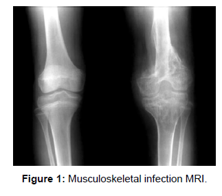
Diagnostic imaging plays a crucial role in confirming the presence of contamination in the outer musculoskeletal system (MSK) and determining the severity and extent of the infection. Due to the hazy display of agony and enlarging, the clinical conclusion may be challenging [1]. Certain highlights on diagnostic imaging are pathognomonic for contamination. However, there are a lot of instances in which the imaging highlights are less clear and cross over with noninfectious etiologies, making imaging diagnosis of disease challenging. Prior conditions, such as explosive joint inflammation, can make it difficult to diagnose infection because irritation from any source, such as autoimmunity or a septic organism can produce similar abnormalities on imaging [2] (Figure 1).
The tissues immediately surrounding cutting-edge imaging modalities like computed tomography (CT) and magnetic resonance imaging (MRI) can be clouded by antiquities from previous procedures, particularly those involving joint replacements or the use of metal. Doctors’ ability to identify MSK infection has improved as a result of ongoing innovative advancements, resulting in greater responsiveness, specificity, and accuracy.
References
- Hogan JI, Hurtado RM, Nelson SB (2017) Mycobacterial Musculoskeletal Infections. Infect Dis Clin North Am 31: 369-382.
- Ranson M (2009) Imaging of pediatric musculoskeletal infection. Semin Musculoskelet Radiol 13: 277-99.
Indexed at, Google Scholar, Crossref
Citation: Gupta S (2022) Imaging on: Infection of the Skeletal System. OMICS J Radiol 11: 414. DOI: 10.4172/2167-7964.1000414
Copyright: © 2022 Gupta S. This is an open-access article distributed under the terms of the Creative Commons Attribution License, which permits unrestricted use, distribution, and reproduction in any medium, provided the original author and source are credited.
Share This Article
Open Access Journals
Article Tools
Article Usage
- Total views: 1486
- [From(publication date): 0-2022 - Mar 29, 2025]
- Breakdown by view type
- HTML page views: 1165
- PDF downloads: 321

