Imaging of Abdominal Lymphoma: A Comprehensive Overview
Received: 02-May-2023 / Manuscript No. roa-23-100630 / Editor assigned: 04-May-2023 / PreQC No. roa-23-100630 (PQ) / Reviewed: 17-May-2023 / QC No. roa-23-100630 / Revised: 22-May-2023 / Manuscript No. roa-23-100630 (R) / Published Date: 29-May-2023 DOI: 10.4172/2167-7964.1000449
Introduction
Abdominal lymphoma refers to the presence of lymphoma, a type of cancer that originates in the lymphatic system, within the abdominal region. It is a diverse and complex disease with various subtypes, including Hodgkin’s lymphoma and non-Hodgkin’s lymphoma. Accurate imaging plays a crucial role in the diagnosis, staging, and treatment planning of abdominal lymphoma. In this article, we will provide a comprehensive overview of the different imaging modalities and their significance in evaluating abdominal lymphoma [1].
Imaging modalities for abdominal lymphoma
Computed tomography (CT): CT scan is the primary imaging modality used for evaluating abdominal lymphoma. It provides detailed cross-sectional images of the abdomen, allowing for the detection and characterization of lymphoma lesions. CT scans can accurately identify lymphadenopathy, which is a hallmark of lymphoma. Additionally, CT helps evaluate organ involvement, detect complications such as bowel obstruction, and assess the overall disease burden. CT-guided biopsies can also be performed to obtain tissue samples for histopathological confirmation [2].
Positron emission tomography (PET): PET scan, often performed in conjunction with CT (PET-CT), is a valuable tool for staging and restaging abdominal lymphoma. PET imaging utilizes a radioactive tracer, typically fluorodeoxyglucose (FDG), which is taken up by metabolically active cells. Malignant lymphoma cells have a higher metabolic rate and thus show increased FDG uptake. PET-CT can accurately determine the extent of disease involvement, identify sites of active lymphoma and differentiate between active disease and scar tissue or residual masses after treatment.
Magnetic resonance imaging (MRI): MRI provides excellent soft tissue contrast and is particularly useful for assessing organ involvement and evaluating complications associated with abdominal lymphoma. It is especially valuable in evaluating the liver, spleen, and bone marrow involvement, as well as detecting lymphoma infiltrations in solid organs. Diffusion-weighted imaging (DWI) and magnetic resonance spectroscopy (MRS) are advanced MRI techniques that can further aid in characterizing lymphoma lesions.
Ultrasonography: Ultrasonography is a non-invasive and widely available imaging modality used to evaluate abdominal lymphoma. It is particularly useful for assessing lymph nodes close to the surface, such as those in the neck, axilla, and inguinal regions. Ultrasonography can help identify abnormal lymph nodes, assess their size, shape, and internal characteristics, and guide needle biopsies for tissue sampling. However, its utility may be limited in cases where deeper lymph nodes or intra-abdominal organs are involved.
Interventional radiology procedures
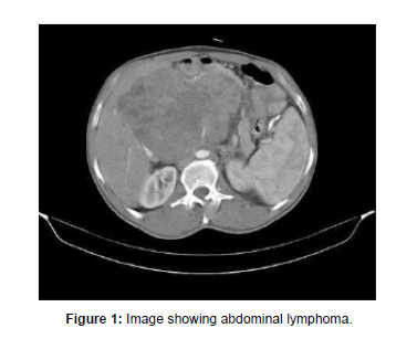
Various interventional radiology procedures play a crucial role in the management of abdominal lymphoma. These include imageguided biopsies, percutaneous drainage of abscesses or ascites, and radiofrequency or microwave ablation of localized lymphoma masses. These procedures are minimally invasive and provide valuable diagnostic and therapeutic options for patients with abdominal lymphoma (Figure 1).
Imaging plays a vital role in the diagnosis, staging, and management of abdominal lymphoma. Computed tomography (CT) remains the cornerstone of imaging evaluation, providing detailed anatomical information. Positron emission tomography (PET) is valuable for assessing disease activity and response to treatment. Magnetic resonance imaging (MRI) offers excellent soft tissue characterization, and ultrasonography aids in evaluating superficial lymph nodes. Interventional radiology procedures further contribute to the management of abdominal lymphoma. A multimodal approach, integrating different imaging techniques, helps clinicians accurately stage the disease, guide treatment decisions and monitor response to therapy, ultimately improving patient.
Acknowledgement
None
Conflict of Interest
None
References
- Radan L, Fischer D, Bar-Shalom R, Dann JE, Epelbaum R, et al. (2008) FDG avidity and PET/CT patterns in primary gastric lymphoma. Eur J Nucl Med Mol Imaging 35: 1424-1430.
- Anis M, Irshad A (2008) Imaging of abdominal lymphoma. Radiol Clin North Am 46: 265-285.
Indexed at, Google Scholar, Crossref
Citation: Joel J (2023) Imaging of Abdominal Lymphoma: A Comprehensive Overview. OMICS J Radiol 12: 449. DOI: 10.4172/2167-7964.1000449
Copyright: © 2023 Joel J. This is an open-access article distributed under the terms of the Creative Commons Attribution License, which permits unrestricted use, distribution, and reproduction in any medium, provided the original author and source are credited.
Share This Article
Open Access Journals
Article Tools
Article Usage
- Total views: 927
- [From(publication date): 0-2023 - Mar 10, 2025]
- Breakdown by view type
- HTML page views: 839
- PDF downloads: 88

