Imaging for osteoarthritis of the knee
Received: 05-May-2022 / Manuscript No. roa-22-64829 / Editor assigned: 07-May-2022 / PreQC No. roa-22-64829 (PQ) / Reviewed: 21-May-2022 / QC No. roa-22-64829 / Revised: 23-May-2022 / Manuscript No. roa-22-64829 (R) / Published Date: 30-May-2022 DOI: 10.4172/2167-7964.1000383
Image Article
Osteoarthritis (OA) otherwise called degenerative joint disease (DJD), is the most well-known type of arthritis, being broadly pervasive with high morbidity and social cost.
Epidemiology
Osteoarthritis is normal, influencing 25% of adults. The prevalence increments with age in the age group below 50 years, men are all the more frequently affected, while in the more established populace the disease is more normal in women. It is assessed that more than 300 million individuals on the planet suffered from OA in 2017 [1].
Knee OA is exceptionally normal and is the most common joint disease in the elderly. Locally, it is assessed to influence 12.5% of patients>45 years.
The average femorotibial joint compartment is more commonly affected and often more severe compared to the lateral.
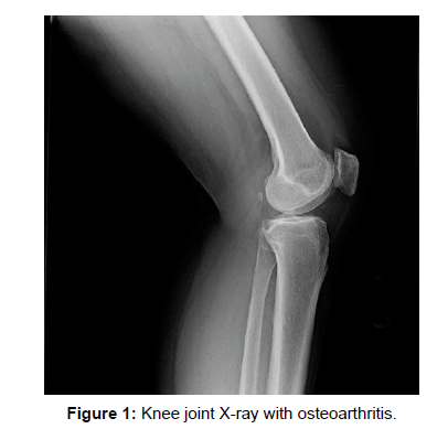
Osteoarthritis (OA) is a broadly prevalent disease overall and, with a rising ageing society, is a test for the field of physical and rehabilitation medicine. Technologic advances and implementation of sophisticated post-processing instruments and scientific procedures have brought about imaging playing an increasingly more significant part in understanding the disease process of OA [2]. Radiography is still the most generally involved imaging modality for establishing an imaging-based diagnosis of OA. The requirement for a successful non- surgical OA treatment is profoundly desired, however in spite of on-going examination endeavors no disease adjusting OA drugs have been discovered or approved to date. MR imaging-based investigations have revealed some of the limitations of radiography. The capacity of MR to image all pertinent joint tissues within the knee and to visualize cartilage morphology and arrangement has brought about MRI playing a vital part in grasping the natural history of the disease and in the quest for new treatments (Figure 1).
Treatment and prognosis
Non-operative management includes simple analgesia and weight reduction. In any case, patients will frequently in the long run require joint replacement. Total knee joint replacement is successful. Unicompartmental joint replacement might be viewed as in some institutions for situations where the disease is transcendently isolated to a single joint compartment.
References
- Guermazi A, Hayashi D, Eckstein F, Hunter DJ, Duryea J, et al. (2013) Imaging of osteoarthritis. Rheum Dis Clin North Am 39: 67-105.
- Hayashi D, Roemer FW, Guermazi A (2016) Imaging for osteoarthritis. Ann Phys Rehabil Med 59: 161-169.
Indexed at, Google Scholar, Crossref
Citation: Sharma P (2022) Imaging for osteoarthritis of the knee. OMICS J Radiol 11: 383. DOI: 10.4172/2167-7964.1000383
Copyright: © 2022 Sharma P. This is an open-access article distributed under the terms of the Creative Commons Attribution License, which permits unrestricted use, distribution, and reproduction in any medium, provided the original author and source are credited.
Share This Article
Open Access Journals
Article Tools
Article Usage
- Total views: 1221
- [From(publication date): 0-2022 - Mar 29, 2025]
- Breakdown by view type
- HTML page views: 799
- PDF downloads: 422

