Image Quality of Low-Ultra Low Dose: Computed Tomography
Received: 04-Jul-2022 / Manuscript No. roa-22-70960 / Editor assigned: 06-Jul-2022 / PreQC No. roa-22-70960 (PQ) / Reviewed: 20-Jul-2022 / QC No. roa-22-70960 / Revised: 22-Jul-2022 / Manuscript No. roa-22-70960 (R) / Published Date: 29-Jul-2022 DOI: 10.4172/2167-7964.1000394
Image Article
The radiological local area is trying to raise the awareness about the radiation induced cancer. Computed Tomography (CT) is the fundamental wellspring of medical irradiation. Makers gave productive mechanical devices on CT to accomplish a significant radiation portion decrease while keeping a symptomatic quality of the image. However, the execution of this multitude of enhancements permits the Low-Dose (LD) and Ultra-Low-Dose (ULD) CT imaging experiences issues to grab hold. Radiologists don’t effortlessly acknowledge reading images with a debased image quality although diagnostic. As each cultural change, even in a radiological division the usual meaning of the LD/ ULD-CT imaging demands time. Steady gatherings with significant exempla and valuable conversations among radiologists, without unexpected adjustments to the CT conventions in clinical practice, are the way in to the achievement.
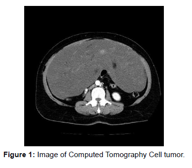
The decision-making process for patients’ consideration is progressively subject to Computed Tomography (CT). CT upset medication with an unmistakable decrease of morbidity and mortality [1]. Regardless of a considerable rundown of benefits there is a serious disadvantage addressed by the way that CT turned into the primary source of ionizing radiation [2,3]. This advancement is troubling a direct result of the definitely known long haul impacts of radiationinitiated carcinogenesis, particularly for subjects that go through regularly CT assessments (eg. oncologic patients) [4] (Figure 1).
Subsequently, the radiological local area strived for a social change to instruct radiologists and referring physicians to a wise illumination for the patient safety. Thus the producers gave some interesting tools on CT to lessen the portion, as specifically the Iterative Reconstruction (IR). The IR is an image reconstruction strategy that works on the quality of the image, freely of the dose, diminishing the image noise in contrast with Filtered Back Projection (FBP). Subsequently the IR can be utilized to decrease emphatically the portion while keeping a diagnostic image quality.
References
- Rubin GD (2014) Computed tomography: revolutionizing the practice of medicine for 40 years. Radiology 273: 45-74.
- Sodickson A, Baeyens PF, Andriole KP, Prevedello LM, Nawfel RD, et al. (2009) Recurrent CT, cumulative radiation exposure, and associated radiation-induced cancer risks from CT of adults. Radiology 251: 175-184.
- Brenner DJ, Hall EJ (2007) Computed tomography--an increasing source of radiation exposure. N Engl J Med 357: 2277-2284.
- Griffey RT, Sodickson A (2009) Cumulative radiation exposure and cancer risk estimates in emergency department patients undergoing repeat or multiple CT. AJR Am J Roentgenol 192: 887-892.
Indexed at, Google Scholar, Crossref
Indexed at, Google Scholar, Crossref
Indexed at, Google Scholar, Crossref
Citation: Sharma P (2022) Image Quality of Low-Ultra Low Dose: Computed Tomography. OMICS J Radiol 11: 394. DOI: 10.4172/2167-7964.1000394
Copyright: © 2022 Patel P. This is an open-access article distributed under the terms of the Creative Commons Attribution License, which permits unrestricted use, distribution, and reproduction in any medium, provided the original author and source are credited.
Share This Article
Open Access Journals
Article Tools
Article Usage
- Total views: 2349
- [From(publication date): 0-2022 - Mar 29, 2025]
- Breakdown by view type
- HTML page views: 1959
- PDF downloads: 390

