Identification and Characterization of Ginger Wilt Pathogens Collected from Ginger Growing Areas of Southern Region
Received: 15-Jan-2020 / Accepted Date: 31-Jan-2020 / Published Date: 07-Feb-2020 DOI: 10.4172/2329-8863.1000434
Abstract
Ginger (Zingiber officinale Rosc.) is an important spice crop grown in tropical and subtropical countries including Ethiopia. It is produced both for commercial and home use. But, since 2012 the first ginger wilt disease epidemic was reported and ginger disease is becoming the threat of national ginger production as a whole. In Ethiopia except R. solanacearum, information regarding the other types of diseases; like fusarium and pythium spp potentially threaten ginger production in other world was scanty. Therefore this study was conducted to detect and identify the major potential pathogens for ginger wilt epidemics. So, a survey was carried out from August-September in three consecutive years 2015 and 2017 in major ginger growing areas of SNNPR. The total numbers of farms visited from Bloso-Bombe and Hadaro-Tunto districts were 35 in 2015, 40 in 2016 and 30 in 2017 and Rhizome spacemen of diseased ginger was collected from the ginger field and identified using standard isolation techniques. Survey result showed that, during 2015 survey season the major and dominant disease identified was ginger wilt. But, in 2016 and 2017 field survey ginger leaf pot and wilt diseases were identified as the problem of ginger production at both Boloso-bombe and Hadaro-Tunto districts. At Boloso-Bombe the incidence of leaf spot was in the range of 0 to 100% in 2016 and 5 to 100% in 2017. At this district its maximum severity was not more than 60% in 2016. But, in 2017 it stretched to 85%. At Hadaro-tunto, the incidence of ginger leaf spot was within the range of 0 to 30% in 2016 and 95-100% in 2017. Also its maximum severity was 25% in 2016 and 95% in 2017. At both locations relative to 2016 and 2017, in 2015 the lowest incidence and severity of leaf spot was recorded. The identification result showed R. solanacearum was detected on 72.7% of specimens and 90.9% of specimens were positive for Fusarium oxysporum spp, but all specimens were negative to Pythium spp Therefore, besides studying their biology and dynamics, focusing on developing management option to these complexes of diseases is vital to increase ginger production and productivity in Ethiopia.
Keywords: Ginger; Bacterial wilt; Leaf spot; Pythium spp; Fusarium oxysporum spp
Introduction
Ginger (Zingiber officinale Rosc.) is an important spice crop grown in tropical and subtropical countries including Ethiopia [1,2]. It is produced both for commercial and home use. In Ethiopia, more than 70 percent of the total ginger production is contributed from South Nations Nationalities and Peoples Regional State (SNNPR); especially Boloso-bombe of Wollayita zone and Hadaro-tunto of Kambata- Tambaro zone [3]. Therefore, these areas are considered as ginger production belt of the country.
In Ethiopia, before 2012 lack of improved varieties, improved management practices were the major constraints ginger production [3]. But, since 2012 the first ginger wilt disease epidemic was reported and ginger disease is becoming the threat of national ginger production as a whole [4]. The disease has shortly devastated all ginger varieties irrespective of variations in cultivars and geographic locations. Germplasm collections maintained in research institutions, cultivars grown by farmers and cultivated commercially as well as those found in natural forests in every corner of ginger growing parts of the country were affected due to the disease outbreak.
Ralistonia solanacearum biovar 3 is reported as the cause for the epidemic of ginger wilt in Ethiopia [4]. But, Dake (1995) reported that ginger is affected with about 24 diseases [5]. These are originating from pathogen of bacterial, fungal, mycoplasma and viral sources. In Ethiopia except R. solanacearum , information regarding the other types of diseases; like fusarium and pythium spp potentially threaten ginger production in other world was scanty.
Identification of major potential ginger diseases and their pathogen together with their level of intensity has a great implication on development of disease management strategies. Therefore this study was conducted to detect the major potential pathogens for ginger wilt epidemics. Moreover, it was conducted to identify other potential diseases those threaten ginger production with their level of intensity in southern Ethiopia.
Materials and Methods
Disease survey
A survey was carried out from August-September in three consecutive years 2015 and 2017 in major ginger growing areas of SNNPR. The total numbers of farms visited from Boloso-Bombe and Hadaro-Tunto districts were 35 in 2015, 40 in 2016 and 30 in 2017. These two districts together contribute about 70% of the ginger production in Ethiopia. While field survey the each owners of the fields were interviewed to investigate the level of their understanding regarding the ginger diseases, its history and the way they manage it.
All sampled ginger growing farmers field were visited to observe and study the diseases incidence and severity in standing crop. Diseases were identified based on the symptoms, nature of damage and other distinguishing characters. Percentage of incidence and severity of diseases was scored by visual observation. Disease prevalence was calculated by dividing number of field with diseased plant by total number of field under assessment. In addition to field diagnosis, spacemen (rhizomes and leaf samples) of diseased plant was collected for laboratory diagnosis. During the survey GPS data of each farm was taken by using GPS. Common diseases like bacterial wilt, soft rote and leaf spot were diagnosed based on the symptom developed on the field.
Bacterial isolation
Rhizome spacemen of diseased ginger was collected from the ginger field. Five pieces 5 mm2 diseased rhizome sample was prepared. The tissue was surface sterilized by dipping in 10% sodium hypochlorite for 3-5 minutes and washing in 2 or 3 series of sterile water and macerated. The sap from macerated tissue was subjected to serial dilution and plated by spreading on dry nutrient agar medium with sterilized L-shaped glass rod. It was incubated for at 96 hours at 25°C. The pure culture of the colonies was developed using pure culture technique and pathogenicity test was done on tissue culture produced ginger plantlets grown on pot.
Fungal isolation
5 mm2 pieces leaf sample taken from the margin of health and diseased tissue and rhizome tissue was prepared for isolation of Phyllosticta zingiber and Fusarium oxysporum , respectively. The prepared spaceman’s were surface sterilized by dipping in 10% sodium hypochlorite for 3-5 minutes. Then washed in 2 or 3 series of sterile water and then plated on Potato Dextrose Agar (PDA) in Petri dish. Sterilized Plates were incubated at room temperature (28°C-30°C) and observed periodically.
Moreover, the growth of F. oxysporum on the moist chamber was tested using sterile Petri dish and moist tissue paper. To do this, each ginger rhizome specimens collected from diseased field during survey was surface sterilized with 10% sodium hypochlorite for 5 minutes and washed in 3 series of sterile water. Then each rhizome specimens were sliced in to two with knife aseptically and plated on sterile Petri dish with sterile moist tissue paper. It was placed at room temperature to allow the growth of fungi on sliced ginger [6]. The fungi were identified following sporulation and pure cultures have been stored at 4°C on PDA slants and subjected for diagnosis.
Results and Discussion
Disease survey
During 2015 survey season the major and dominant disease identified was ginger wilt. But, in 2016 and 2017 field survey two types of diseases were identified as the problem of ginger production at both Boloso-bombe and Hadaro-Tunto districts. These were ginger leaf pot and wilt.
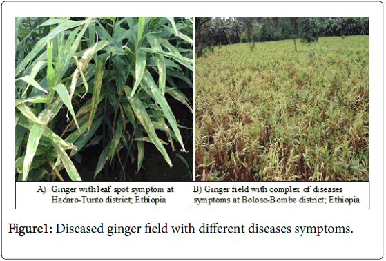
At Boloso-Bombe the incidence of leaf spot; characterized with its symptom of oval to elongated spots that later changed to whitish spot surrounded by dark brown margin with yellowish halo and caused by Phyllosticta zingiber was in the range of 0 to 55% in 2016 and 5 to 100% in 2017 [7-9]. At this district its maximum severity was not more than 60% in 2016 but, 2017 it is stretched to 85% (Table 1).
| Year of production | Diseases intensity | Range | Location | |||
|---|---|---|---|---|---|---|
| Boloso-Bombe District | Hadaro-Tunto District | |||||
| Ginger wilt | Ginger leaf spot | Ginger wilt | Ginger leaf spot | |||
| 2016 | Incidence (%) | minimum | 25 | 0 | 30 | 0 |
| maximum | 85 | 55 | 100 | 30 | ||
| Severity (%) | minimum | 30 | 0 | 35 | 5 | |
| maximum | 70 | 60 | 85 | 25 | ||
| 2017 | Incidence (%) | minimum | 10 | 5 | 90 | 95 |
| maximum | 80 | 100 | 100 | 100 | ||
| Severity (%) | minimum | 5 | 2 | 10 | 5 | |
| maximum | 35 | 85 | 40 | 95 | ||
Table 1: The range of ginger wilt and leaf spot incidence and severity at the two study districts in 2016 and 2017 production season.
At Hadaro-Tunto, the incidence of ginger leaf spot was within the range of 0 to 30% in 2016 and 95-100% in 2017. Also its maximum severity was 25% in 2016 and 95% in 2017 (Table 1). At both locations relative to 2016 and 2017, in 2015 the lowest incidence and severity of leaf spot was recorded. These showed that both incidence and severity of ginger leaf spot was becoming worse and worse over years. Le, et al. (2014) reported that it is one of series ginger disease that affect ginger yield considerably [10].
As mentioned in the introductory part, ginger wilt epidemic was occurred in 2015 in all of ginger production areas of the country. Because of that, not only ginger wilt incidence and severity but also its effect on yield loss was high than ever in ginger production history in the study areas. In 2015 the average ginger wilt severity was as high as 78 and 85% in the field at both locations (Tables 2 and 3).
| Year of production | Disease intensity | |||||
|---|---|---|---|---|---|---|
| Ginger wilt | Ginger leaf spot | |||||
| Prevalence (%) | Incidence (%) | Severity (%) | Prevalence (%) | Incidence (%) | Severity (%) | |
| 2015 | 100 | 100 | 85 | 15 | 10 | 5 |
| 2016 | 100 | 55 | 50 | 45 | 22.5 | 30 |
| 2017 | 100 | 43 | 11.5 | 100 | 66.5 | 13.5 |
Table 2: Mean incidence, severity and prevalence of ginger wilt and leaf spot in field, since 2015 to 2017 production season at Boloso-Bombe District; Ethiopia.
| Year of production | Disease intensity | |||||
|---|---|---|---|---|---|---|
| Ginger wilt | Ginger leaf spot | |||||
| Prevalence (%) | Incidence (%) | Severity (%) | Prevalence (%) | Incidence (%) | Severity (%) | |
| 2015 | 100 | 100 | 78 | 19 | 15 | 2 |
| 2016 | 100 | 65 | 60 | 65 | 15 | 15 |
| 2017 | 100 | 95.5 | 28.2 | 100 | 100 | 26.8 |
Table 3: Mean incidence, severity and prevalence of ginger wilt and leaf spot in field, since 2015 to 2017 production season at Hadaro-Tunto District; Ethiopia.
At Boloso-Bombe, the maximum incidence of ginger wilt was 85% and 80% in 2016 and 2017, respectively. In 2016, at this location its severity was higher (30%-70%) compared with severity (5-35%) in 2017. This is due to less fevered environmental condition for disease development in 2017 in an area. Again, at Hadaro-Tunto district the trend of wilt incidence and severity in both years was followed similar fashion as Boloso-Bombe (Table 1).
At Boloso-Bombe district, the average ginger wilt incidence was 100, 55 and 43 percent in 2015, 2016 and 2017 production seasons, respectively. The highest ginger wilt incidence (100%) and severity (85%) was recorded on 2015 production season. While the lowest incidence (43%) and severity (11.5%) of wilt was recorded on 2017 production season (Table 2). In production years of 2015, 2016 and 2017 the prevalence of ginger wilt was 100% (Table 2).
The year of 2015 was the first highest devastating ginger wilt epidemics occurred in Ethiopia. In this year, the weather condition was moist with prolonged rainy season. Therefore, it made conducive condition for pathogen development and the occurrence of ginger wilt epidemics in the all ginger growing areas of the country. In the year of 2016 and 2017 the weather condition was relatively with low moisture and rain fall. This condition provides low severity of ginger wilt in 2016 and 2017 compared with 2015 production season.
At Hadaro Tunto district, average ginger wilt severity of 78, 60 and 28.2% was recorded in 2015, 2016 and 2017 production season. The highest wilt severity of 78% was recorded in 2015 followed by 60% in 2016. Here also the lowest wilt severity of 28.2% was recorded in 2017 (Table 3).
In 2015 however the ginger wilt epidemics was high, the average prevalence, incidence and severity of ginger leaf spot was low. At the time only 15% of ginger field was with symptom of leaf spot disease. But, in 2017 the prevalence of ginger leaf spot was 100%. While its average incidence and severity were 66.5 and 13.5%, respectively (Table 2). Also at Hadaro-Tunto district, 100% incidence and 26.8% of severity of ginger leaf spot was recorded with 100% of its prevalence (Table 3). This showed the distribution and intensity of ginger leaf spot in ginger production areas is increasing over time and potentially threatens ginger production.
Pathogen identification
However R. solanacearum was reported by Bekele, et al. as the causative agent of ginger wilt epidemics in Ethiopia, there was shortage of information about other fungal pathogen largely confounded with this pathogen and form disease complex in the other world where ginger production is common (Figure 1). In 2017 laboratory diagnosis was done at Areka Agricultural Research Centre on 22 rhizome and leaf specimens collected from farmer’s field of ginger that showed ginger wilt symptom, to identify the causative agent. The laboratory result showed R. solanacearum and Phyllosticta zingiberi was detected on 72.7% and 100% of specimens respectively. The same specimens were diagnosed for Fusarium and Pythium spp which are potential ginger wilt fungal pathogens in the other world [5,11,12]. The result showed 90.9% of specimens were positive for Fusarium oxysporum spp, but all specimens were negative to Pythium spp (Table 4).
| No. | Pathogen identified | Number of samples tested | Samples with positive diagnosis | Percentage of samples with pathogen identified |
|---|---|---|---|---|
| 1 | Fusarium oxysporum spp | 22 | 20 | 90.9 |
| 2 | Ralistonia solanacearum | 22 | 16 | 72.7 |
| 3 | Pythium spp | 22 | 0 | 0 |
| 4 | Phyllosticta zingiberi | 22 | 22 | 100 |
Table 4: Pathogen identified from wilt symptom ginger rhizome during laboratory diagnosis at and their incidence in percent.
Conclusion and Recommendation
This study identified fusarium wilt was caused by F. oxysporum and ginger leaf spot was caused by Phyllostica zingiberi as potential ginger production challenges in the studied areas; in addition to R. solanacearum which was reported previously in different parts of the country. Therefore, besides studying their biology and dynamics, focusing on developing management option to these complexes diseases is vital to increase ginger production and productivity specifically in the studied area and Ethiopia in general.
References
- Dohroo NP, Kansal S, Mehta P, Ahluwalia N (2012) Evaluation of eco-friendly disease management practices against soft rot of ginger caused by Pythium aphanidermatum. Plant Dis Res 27: 1-5.
- Rahim MA (1992) Spice and plantation crops in national economy: In: Proceeding of 6th National Horticulture Convention and Symposium, Bangladesh.
- Geta E, Kifle A (2011) Production, processing and marketing of ginger in Southern Ethiopia. J Horticul Fores 3: 207-213.
- Kassa B, kifelew H, Hunduma T (2016) Status of Ginger Wilt and Identification of the Causal Organism in Southern Nations Nationality and People Sates of Ethiopia. Int J Res Stud Agri Sci (IJRSAS) 2: 1-11.
- Dake GN (1995) Diseases of Ginger (Zingiber officinale Rosc.) and their Management. J Spi Arom Crop 4: 70-73.
- Jackson G (2010) Ginger Fusarium yellows. Pacific Pests and Pathogens Fact Sheet, In: Formation from Diseases of vegetable crops in Australia.
- Brahma RN, Nambier KKN (1982) Survival of Phyllosticta zingiberi Ramakr., causal agents of leaf spot disease of ginger. Plant Crop Res Inst pp: 123-125.
- Brahma RN, Nambier KKN (1984) Spore release and disposal in ginger leaf spot pathogen (Phyllosticta zingiberi Ramakr). Ind Soci Plant Crop pp: 541-554.
- Singh AK (2015) Efficiency of fungicides for the control of leaf spot of disease of ginger under the field conditions of Chhattisgarh (India). Afric J Agri Res 10: 1301-1305.
- Le DP, Smith M, Hudler GW, Aitken E (2014) Pythium soft rot of ginger: Detection and identification of the causal pathogens, and their control. Crop Protec 65: 153-167.
- Rahman MM, Mamun ANM (2009) Control of Rhizome rot diseases of ginger (Zingiber officinale Rosc) by chemical, soil amendments and soil antagonists. Agricul 7: 57-61.
Citation: Seid A, Tomas Z, Tamirat A (2020) Identification and Characterization of Ginger Wilt Pathogens Collected from Ginger Growing Areas of Southern Region. Adv Crop Sci Tech 8: 434. DOI: 10.4172/2329-8863.1000434
Copyright: © 2020 Tomas Z, et al. This is an open-access article distributed under the terms of the Creative Commons Attribution License, which permits unrestricted use, distribution, and reproduction in any medium, provided the original author and source are credited.
Share This Article
Recommended Journals
Open Access Journals
Article Tools
Article Usage
- Total views: 3312
- [From(publication date): 0-2020 - Apr 03, 2025]
- Breakdown by view type
- HTML page views: 2509
- PDF downloads: 803

