Hyperhomocystinaemia Causing Complete Unilateral Renal Artery Stenosis with Hypertensive Retinopathyâan Unusual Case Report
Received: 13-Feb-2023 / Manuscript No. roa-23-89402 / Editor assigned: 15-Feb-2023 / PreQC No. roa-23-89402 (PQ) / Reviewed: 28-Feb-2023 / QC No. roa-23-89402 / Revised: 02-Mar-2023 / Manuscript No. roa-23-89402 (R) / Published Date: 09-Mar-2023 DOI: 10.4172/2167-7964.1000429
Abstract
This report describes an unusual case of hypertensive retinopathy and choroidopathy with unilateral renal artery stenosis secondary to hyperhomocystinaemia.
Keywords
Hypertensive retinopathy; Choroidopathy; Renal artery stenosis; Hyperhomocystinaemia
Background
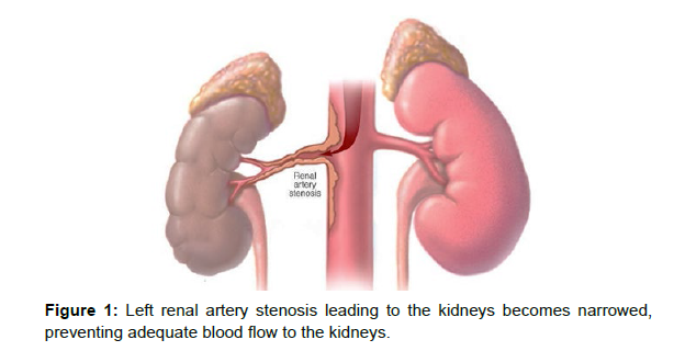
Renal artery stenosis is a narrowing of artery that carries blood to kidney (Figure 1) [1]. Renal artery stenosis often leads to secondary hypertension and kidney damage. Hypertensive retinopathy is a common clinical finding among hypertensive patients [2]. Hyperhomocystinaemina leads to renal artery stenosis can be considered as one of the rare cause [3]. However, numerous studies investigated the role of homocysteine as a predictor of vascular diseases most often seen in older people with atherosclerosis [4].
Case presentation
A 56 year old male came to our hospital in emergency department with history of blurring of vision in both eyes. He consulted ophthalmologist, where he was diagnosed with bilateral retinal haemorrhages. Blood pressure was found 250/140 millimetres of mercury (mmHg) at time of arrival as well as ST-T segment changes in echocardiogram. Therefore, the patient was admitted to our hospital for further management.
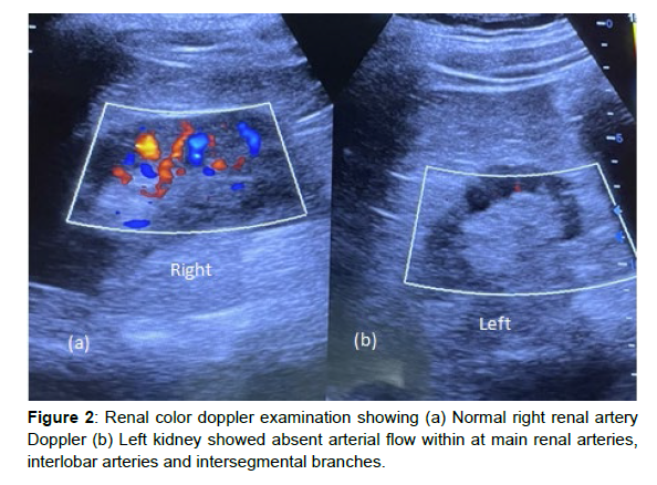
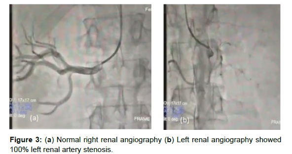
After admission he went through all the investigations. Blood investigations suggested altered lipid profile, significantly increased homocysteine levels, borderline HBA1C levels, significantly elevated urine micro albumin and altered creatinine. On ophthalmology examination patient was found to have retinopathy & choroidopathy. On sonography examination left kidney appeared small in size with grade I increase renal echo-pattern. Right kidney showed compensatory hypertrophy with grade I increase in echo-pattern. On renal color doppler (Figure 2) examination left kidney showed absent arterial flow within main renal arteries, interlobar arteries and intersegmental branches. However, the left renal vein appeared normal and showed normal colour filling. The findings suggested complete left renal artery stenosis. For further confirmation renal angiography was done (Figure 3) and it showed 100% left renal artery stenosis.
Conclusion
Hyperhomocystinaemia leads to renal artery stenosis can be considered as one of the rare cause [3]. However, numerous studies investigated the role of homocysteine as a predictor of vascular diseases. A positive association was demonstrated between plasma homocysteine levels and extent of atherosclerosis [3].However, Hypertensive retinopathy and choroidopathy is a common finding among hypertensive patients [2]. Renal artery stenosis (RAS) is the major cause of renovascular hypertension and may account for 1-10% of the 50 million cases of hypertension [4]. Ultrasound and doppler are useful in finding out renal artery disease progress early [5]. Intraarterial digital subtraction angiography is the standard diagnostic test for renal artery stenosis (RAS) [6].
References
- Safian RD, Textor SC (2001) Renal-artery stenosis. N Engl J Med 344: 431-442.
- Wong TY, Mitchell P (2004) Hypertensive retinopathy. N Engl J Med 351: 2310-2317.
- McCully KS (1969) Vascular pathology of homocysteinemia: implications for the pathogenesis of arteriosclerosis. Am J Pathol 56: 111-128.
- Hansen KJ, Edwards MS, Craven TE, Cherr GS, Jackson SA, et al. (2002) Prevalence of renovascular disease in the elderly: a population-based study. J Vasc Surg 36: 443-451.
- Hoffmann U, Edwards JM, Carter S, Goldman ML, Harley JD, et al. (1991) Role of duplex scanning for the detection of atherosclerotic renal artery disease. Kidney Int 39: 1232-1239.
- Erley CM, Bader BD, Berger ED, Tuncel N, Winkler S, et al. (2004) Gadolinium-based contrast media compared with iodinated media for digital subtraction angiography in azotaemic patients. 19: 2526-2531.
Indexed at, Google Scholar, CrossRef
Indexed at, Google Scholar, CrossRef
Indexed at, Google Scholar, CrossRef
Indexed at, Google Scholar, CrossRef
Citation: Dave K, Patel MP (2023) Hyperhomocystinaemia Causing CompleteUnilateral Renal Artery Stenosis with Hypertensive Retinopathy–an Unusual CaseReport. OMICS J Radiol 12: 429. DOI: 10.4172/2167-7964.1000429
Copyright: © 2023 Dave K. This is an open-access article distributed under theterms of the Creative Commons Attribution License, which permits unrestricteduse, distribution, and reproduction in any medium, provided the original author andsource are credited.
Share This Article
Open Access Journals
Article Tools
Article Usage
- Total views: 1647
- [From(publication date): 0-2023 - Mar 29, 2025]
- Breakdown by view type
- HTML page views: 1361
- PDF downloads: 286


