Hydatid Cyst of Limb Soft Tissues (11 Cases)
Received: 05-Nov-2018 / Accepted Date: 16-Feb-2019 / Published Date: 23-Feb-2019 DOI: 10.4172/2332-0877.1000391
Abstract
Purpose of the study: Hydatidosis is a parasitic disease caused by the larval form of tapeworm Echinococcus granulosis. Diagnosis is based on interview, clinic and imaging data. This study aims to identify the clinical, paraclinical, therapeutic and evolutionary features of hydatid cyst of the soft tissues in the limbs.
Materials and methods: Our retrospective study focused on a series of 11 cases of soft-tissue hydatid cyst, collected at the Orthopedic Traumatology Department of the IBN SINA Rabat Hospital, over a period of 13 years, between 2004 and 2017.
Results: Field of choice was that of female, sex ratio of 0.37 (3M/8W), of rural origin (72.7%), the average age is 43 years, which was consulted for swelling lasting for at least 3 months without alteration of the condition. The predilection was at the level of the thigh muscles 8 out of 11 (72.7%), of which 6 at the lodge adductors. Diagnosis is based on imaging and immunological reactions and has been confirmed by histology. All patients underwent surgical excision supplemented by antiparasitic chemotherapy.
Conclusion: Hydatid cysts of the soft tissues remain rare. Their often-asymptomatic nature and their slow evolution make their diagnosis late. The treatment is essentially surgical. The best way to fight against hydatid disease, whatever its location, is prevention.
Keywords: Echinococcosis; Primitive hydatid cyst; Soft tissues; Limbs; Surgery
Introduction
The hydatid cyst (KH), or hydatidosis, is an anthropozoonosis caused by the development in humans of the larval form of the dog taenia, Echinococcus granulosus (EG) [1]. This is a cosmopolitan affection raging particularly in traditional breeding countries, where dog-sheep promiscuity exists [2,3]. The muscular localization of hydatidosis is however much rarer and unusual even in endemic countries. The objective of this work was to approach the clinical, paraclinical, therapeutic and evolutionary peculiarities of this relatively rare affection, based on the study of our cases and the review of the literature.
Materials and Methods
We collected for this work 11 cases of soft-tissue hydatid cyst treated surgically at IBN SINA CHU in Rabat, Morocco over a period of 13 years, between 2004 and 2017. This is a retrospective study based on the record’s medical patients. We collected epidemiological, anamnestic, clinical, ultrasound and additional examinations. The analysis of these data allowed us to specify the characteristics of these hydatid locations. All the patients had a surgical treatment, the data of the operating report made it possible to note the different techniques applied and the operating difficulties.
Results
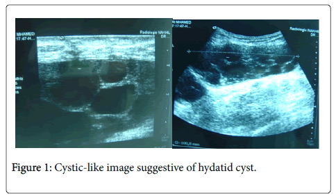
The average age of the patients was 43, with extremes of 24 to 61 years. Men were more affected than women: 3 men and 8 women, with a clear predominance among patients from rural areas: 8 patients. The notion of contact with the dog was found in 9 patients. 3 patients had a history of hydatid damage to the soft tissues already treated surgically. The reason for consultation was a swelling of the soft tissues gradually increasing in volume in 9 cases, and in 2 patients the diagnosis was made in a fortuitous way, following an assessment performed for another concomitant disease. The pain, spontaneous or on palpation, was found 5 times. Ultrasonography was performed 6 times; it showed signs in favor of hydatidosis in 5 cases (Figure 1) (Table 1).
| Location | Number of cases | Percentage |
|---|---|---|
| Thigh adductor | 6 cases | 54.5% |
| Posterior lodge of the thigh | 2 cases | 18.2% |
| Gluteal region | 1 case | 9.1% |
| Calf | 2 cases | 18.2% |
| Total | 11 cases | 100% |
Table 1: Localization was mainly of interest to the proximal muscles of the lower limbs.
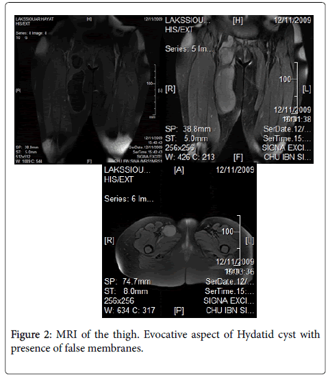
MRI is the exam of choice that allows to retain the diagnosis. In this series 9 patients have benefited from this review. She showed a hypointense aspect in T1 and hypersignal in T2, with the presence of multiloculated cystic formation and numerous vesicles girls at the level of the muscular compartments (Figure 2).
The extension assessment included chest x-ray and abdominopelvic ultrasound. They did not detect any other visceral localization, in particular hepatic or pulmonary. Hyper-eosonophilia was found in 2 patients and the hydatid serology was positive in 3 patients. The puncture of the cyst has not been performed; indeed, the diagnostic possibility of the hydatid cyst contra-indicates the puncture or biopsy given the risk of anaphylactic shock and local or remote dissemination.
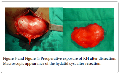
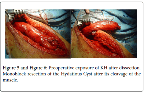
The treatment was surgical in all patients (Figures 3 and 4). A total perkystectomy was performed in all our patients, it is a monobloc resection of the cyst with protection of the operative field by compresses impregnated with oxygenated water (in 10 cases) (Figures 5 and 6). One case presented a second cyst located in the posterior tibial muscle and which had preoperative drainage because it adhered to the posterior tibial artery.
The opening of the cyst intraoperatively occurred once, without it followed by anaphylactic shock imposing an aspiration of the cyst with toilet cystic bed with hydrogen peroxide.
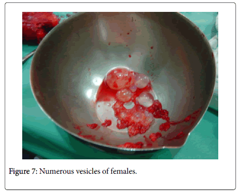
The evolution was favorable for all our patients with recovery and total healing, without recurrence. From an anatomopathological point of view: the macroscopic appearance of hydatidosis was typical at the opening of the operative specimen showing numerous vesicles bathing in the hydatid fluid, which was confirmed by histology (Figure 7).
Discussion
Muscle localization is rare and unusual, its frequency varies between 1% and 5% of all hydatid localizations [4]. This rarity can be explained by two reasons: the effectiveness of the first two filters (hepatic and pulmonary) for this parasitosis, making the lung and liver the two main locations of the hydatid cyst or the intramuscular development of cysts is hampered by muscular contractility and the lactic acid content of skeletal muscles. Thus, the skeletal muscle localization of the hydatid cyst is described as "unusual" [5]. The disease affects both sexes with a slight female predominance (sex ratio H/F=0.61) (because women are more caring than men in the herd and dogs, this female predominance is found for all anatomical locations of the hydatid cyst); in our series, 8 out of 11 patients are women.
Hydatidosis can be seen at any age, but reaches adults between 21 and 40 years of age. Ingestion may actually occur early in childhood, but is often expressed only in adulthood, given the slow course of the disease, but in our series, the preferred age was adults between 31 and 50 years (6 out of 11 cases), while young people aged 18-30 years were affected in only 2 cases and 3 cases in the over 50s. The muscular Echinococcus sits mainly in the proximal muscles of the lower limbs, as it has been observed in our patients. Indeed, the muscles of the thigh were reached 8 times out of 11 in our series, including 6 times in the adductor muscles. This selectivity for the proximal muscles would be related to the importance of blood flow.
The clinical symptomatology of Echinococcosis is not specific (Figure 7). The beginning of the affection is often insidious, it comes down to the appearance of a swelling gradually increasing in volume, of soft consistency, well limited and painless with conservation of the general state. This is the case of 8 of our patients. The pain may be present such is found in 5 of our patients with a functional gene in 3 cases. This hydatidosis may look noisy and simulate lipoma, hot abscess, or malignant or benign soft tissue tumors when the cyst is cracked or superinfected [6]. this is the case for one of our patients or we suspected a hot abscess of the calf. Affectation of the gluteal region can be revealed by cruralgia or paraesthesia due to compression of the sciatic nerve by the cyst, or most often by swelling increasing in volume [7]. During the interview, the rural origin and the contact with the dogs guide the diagnosis, which must be carried out pre-operatively. This makes it possible to avoid untimely gestures such as puncture or biopsy of the cyst. Radiologically Imaging is the most important component. Standard radiography eliminates calcification of cysts or bone involvement, but it is ultrasonography that is the key examination of the diagnostic orientation, it allows to specify the fluid nature of the swelling with sometimes visualization of vesicles girls. MRI is the most useful imaging medium for hydatid pathology of the soft tissues whenever the ultrasound appearance is atypical. In addition to the demonstration of vesicles and membranes, T2 hyposignal wall and periosteal enhancement allow the diagnosis of hydatidosis. The hydatid serology provides certainty in the diagnosis of echinococcosis when it is positive whereas its negativity does not exclude the diagnosis. The eosinophilia is inconstant and has only interest in the orientation of the diagnosis
However, the positive diagnosis is made by the histological study after surgical excision, in fact the macroscopic appearance of the hydatid cyst is typical with numerous vesicles, and membranes which bathes in the hydatid liquid.
The treatment is surgical. Surgery should be careful to avoid opening the cyst during dissection. The surgical field must be protected by a solution of hypertonic saline serum or hydrogen peroxide at the time of the surgical approach.
Monobloc resection with total perkystectomy is the ideal procedure, but not always feasible especially if the cyst is large, deep and making contact with the neighbouring vasculo-nervous elements: or aspiration of the cyst, followed by resection of the periyst is indicated for prevent accidental breakage and dissemination of the contents. Recent years have been marked by the development of percutaneous interventional radiology such as puncture-aspiration-injection and re-aspiration (PAIR), and percutaneous drainage without re-aspiration, which have improved mortality and morbidity hydatid cysts, but these conservative techniques have not been realized in our series.
A prophylactic drug treatment is recommended as a complementary treatment to surgery; preoperatively by 2 courses of 28 days spaced 14 days to sterilize the cyst and minimize the risk of dissemination and postoperatively for 6 months to 1 year with a dosage of transaminases before the start of medical treatment, then each trimester given its hepatic toxicity.
Conclusion
The muscular localization of the hydatid cyst is rare, even in highly endemic countries. Nevertheless, the diagnosis must be evoked in the epidemiological and clinical context. Ultrasound and MRI are the exams of choice to confirm this diagnosis thus avoiding the puncture.
The treatment is mainly surgical, taking the cyst and the periyst without breaking in, but a medical and surgical approach is now the rule to achieve a complete cure and avoid reinfections.
And as always in medicine, the best treatment is prevention, which remains essential for the fight against and reduction of the prevalence of human hydatidosis.
References
- Boussofara M, Sallem MR, Raucoules-Aimé M (2005) Anesthesia for surgery of the hydatid cyst of the liver. EMC-Anesthesia Resuscitation 2: 132-140.
- Khallouki M (2001) Hydatid cyst of the lung in children (about 124 cases).
- Klotz F, Nicolas X, Debonne JM, Garcia JF, Andreu JM (2000) Hydatid cysts of the liver. Encyclical. Med. Chir. (Editions Scientifiques et Médicales Elsevier SAS, Paris), Hepatology, 7-023-A-10, 16 p.
- Turki JE, Fredj, Ben F, Toumi S (2009) Pseudotumeur of the thigh: think of a hydatid cyst. J Internal Med 30: S428-S429.
- Salem B, Regaïeg N (2018) The hydatid cyst of soft tissues. Pan African Med J 7: 1-10.
- Bourée P, Thulliez P, Millat B (1982) Hydatidosis of the calf muscle. Bull Soc Path Exo 75: 201-204.
- Rafiqi k, Rafaoui A, Sirrajelhak M (2016) Primitive hydatid cyst of the thigh in a bodybuilder. J Sports Traumatol 33: 107-109.
Citation: Lazrek O, Sabri EM, Boufettal M, Bassir RA, Lamrani MO, et al. (2019) Hydatid Cyst of Limb Soft Tissues (11 Cases). J Infect Dis Ther 7:391. DOI: 10.4172/2332-0877.1000391
Copyright: © 2019 Lazrek O, et al. This is an open-access article distributed under the terms of the Creative Commons Attribution License, which permits unrestricted use, distribution, and reproduction in any medium, provided the original author and source are credited.
Share This Article
Recommended Journals
Open Access Journals
Article Tools
Article Usage
- Total views: 3799
- [From(publication date): 0-2019 - Apr 24, 2025]
- Breakdown by view type
- HTML page views: 3007
- PDF downloads: 792

