Hot Spot Sign
Received: 05-May-2022 / Manuscript No. roa-22-64121 / Editor assigned: 07-May-2022 / PreQC No. roa-22-64121 (PQ) / Reviewed: 20-May-2022 / QC No. roa-22-64121 / Revised: 23-May-2022 / Manuscript No. roa-22-64121 (R) / Published Date: 30-May-2022 DOI: 10.4172/2167-7964.1000381
Image Article
In Computed Tomography Imaging the focal hepatic hot spot sign shows as an area of intense focal wedge-shaped enlargement of the quadrate lobe of the liver in the arteria and venous phase [1]. Ishikawa originally described the focal hepatic hot spot symptom in 1983.
This abdominal enhancement is due to Porto systemic venous shunting between the SVC and portal vein via the internal mammary and Para umbilical veins along the ligamentum tears, secondary to superior vena cava obstruction.
Stanford and colleagues divided SVC syndrome into four types: Type I and Type II, which are defined as supra-azygous partial and near-complete obstruction of the SVC with ante grade flow in the azygos vein, Type III is defined as complete obstruction of SVC with reversal of azygous flow and Type IV as complete obstruction of SVC and azygous system with development of chest wall collaterals. When an enhanced CT of the abdomen is performed in a clinically unapparent obstruction this sign provides a clue to the diagnosis of thoracic SVC obstruction.
The characteristic location in the quadrate lobe of the liver as well as wedge shape enhancement in arterial and venous phase aid in distinguishing quadrate hot spot from focal hyper vascular liver lesion.
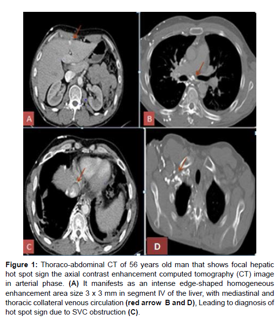
The hot spot sign is created by areas of focally increased blood flow that result from shunting this sign has been reported in Budd- Chiari syndrome the case of SVC syndrome (neoplasm of the thorax as kung carcinoma and lymphoma, Vasculo-Behcet’s disease) and mass of the liver (Abscess, Haemangioma, focal nodular hyperplasia and hepatocellular carcinoma) (Figure 1).
Figure 1: Thoraco-abdominal CT of 56 years old man that shows focal hepatic hot spot sign the axial contrast enhancement computed tomography (CT) image in arterial phase. (A) It manifests as an intense edge-shaped homogeneous enhancement area size 3 x 3 mm in segment IV of the liver, with mediastinal and thoracic collateral venous circulation (red arrow B and D), Leading to diagnosis of hot spot sign due to SVC obstruction (C).
Figure 1 explains Thoraco-abdominal CT of 56 years old man that shows focal hepatic hot spot sign the axial contrast enhancement computed tomography (CT) image in arterial phase. It manifests as an intense edge-shaped homogeneous enhancement area size mm in segment IV of the liver leading to diagnosis of hot spot sign due to SVC obstruction [2].
References
- Dickson AM (2005) The focal hepatic hot spot sign. Radiology 237: 647-648.
- Sureka B, Sullere A, Singh Khera P (2018) CT quadrate lobe hot spot sign. Middle East J Dig Dis 10: 192-193.
Indexed at, Google Scholar, Crossref
Select your language of interest to view the total content in your interested language
Share This Article
Open Access Journals
Article Tools
Article Usage
- Total views: 1902
- [From(publication date): 0-2022 - Dec 01, 2025]
- Breakdown by view type
- HTML page views: 1354
- PDF downloads: 548

