Histopathological Pattern of Nasopharyngeal Masses in Adults
Received: 09-Jun-2017 / Accepted Date: 23-Jun-2017 / Published Date: 30-Jun-2017 DOI: 10.4172/2161-119X.1000311
Abstract
Introduction: Nasopharyngeal mass resembles a confounding problem in adults because of the increased fear of malignancy with age advancement. Histopathological pattern of nasopharyngeal lesions varies from common benign lesions such as hyperplastic lymphoid tissue and benign nasopharyngeal cysts to malignant diseases such as lymphoma and nasopharyngeal carcinoma. Methods: This prospective study included all patients underwent a nasopharyngeal biopsy for a nasopharyngeal mass in 2 years period from October 2014 to September 2016. Blind biopsy was taken for cases clinically suspicious for having nasopharyngeal carcinoma. Patients histopathological findings were analyzed as regard demographic data and clinical presentation. Results: A total of 80 patients (45 males and 35 females) were included. Patient age ranged between 18 and 76 years (mean 38.7 years, SD 16.7). The most commonly detected pathological lesion was hyperplastic lymphoid tissue hyperplasia (48.75%). Malignant lesions constituted 48.75% of the study population (12.5% squamous cell carcinoma, 18.75% undifferentiated carcinoma and 17.5% malignant lymphoma). In addition, benign nasopharyngeal cysts were diagnosed in 2.5 % of cases. Conclusion: Benign lesions are more commonly detected in the nasopharynx in adult population particularly hyperplastic lymphoid tissue. Histopathological examination of nasopharyngeal mass is very essential to roll out malignancy which is not uncommon.
Keywords: Nasopharyngeal; Lymphoid hyperplasia; Carcinoma; Lymphoma; Histopathology
255136Introduction
Detection of mass in the nasopharynx of an adult is usually associated with suspicion of nasopharyngeal malignancy [1] Malignant diseases arising in the nasopharynx are asymptomatic in the early stage and may be confused with other benign lesions; therefore Patients with nasopharyngeal malignancy usually come with late presentation [2].
Adenoid tissue regression usually occurs at adolescence; however it may persist in adults causing significant nasal obstruction and histopathological examination is required to preclude the possibility of malignancy [1,3,4].
Benign lesions in the nasopharynx of adults also include cystic lesions such as branchial cleft cyst, Tornwaldt cyst and mucous retention cyst [5,6].
In this study we identify the various pathological entities detected in adult patients presented to our department with a nasopharyngeal swelling with analysis of the associated clinical and demographic data.
Methods
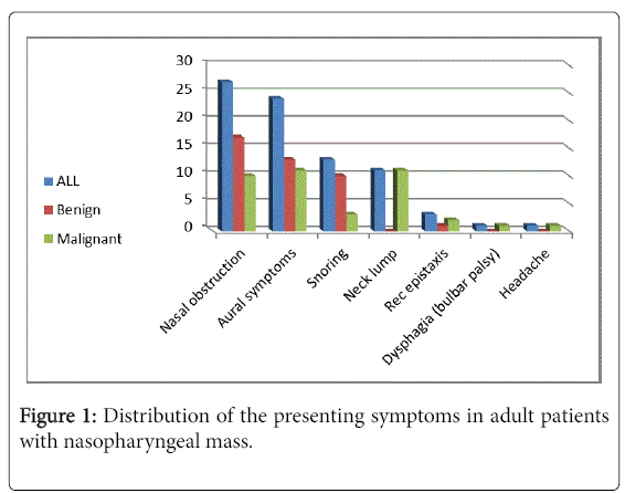
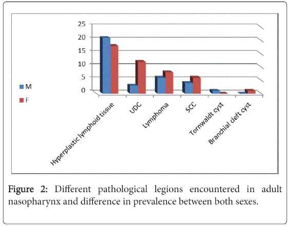
This is a prospective study of adult population (starting from 18years) undergoing endoscopic guided nasopharyngeal biopsy to exclude nasopharyngeal malignancy from October 2014 to September 2016 in our tertiary referral center. Institutional review board approval was obtained. All patients included in the current study presented to our outpatient clinic with nasopharyngeal masses. All cases of antrochoanal polyps and angiofibroma were excluded from the study. Demographic data (Age, gender, smoking status), clinical picture (presenting symptoms and signs) and clinical findings on nasopharyngeal endoscopy were recorded. Nasopharyngeal biopsies were obtained under general anesthesia. A 4 mm rigid endoscope was introduced through the inferior meatus till clearly visualizing the nasopharynx. A good view of the nasal cavity and the nasopharynx was possible in all cases and biopsy was performed using a punch forceps. Pathology reports were reviewed for biopsy results. Descriptive analysis was used for frequencies and means. Analysis was conducted using SPSS (SPSS Inc, Chicago, IL) and a p value <0.05 was considered significant (Figures 1 and 2).
Ethics, Consent and Permissions
Institutional review board approval was obtained from ethical committee at Sohag University, Egypt.
All study population were consented for participation in the study.
Results
A total of 80 patients (45 males- 56.25% and 35 females- 43.75%) were included in our study. Patients' age ranged between 18 and 76 years (mean ± SD=45.1 ± 13.6 years). Histopathological examination of nasopharyngeal biopsies revealed diversity of benign and malignant lesions. The detected benign pathological lesions included hyperplastic lymphoid tissue hyperplasia (48.75%), Tornwaldt cyst (1.25%) and Branchial cleft cyst (1.25%). Malignant lesions constituted 48.75% of the study population (12.5% squamous cell carcinoma, 18.75 % undifferentiated carcinoma and 17.5% malignant lymphoma) (Tables 1 and 2).
| Pathology | Number of cases | Percentage | Male | Female |
|---|---|---|---|---|
| Hyperplastic lymphoid tissue | 39 | 48.75% | 18 | 21 |
| Tornwaldt cyst | 1 | 1.25% | 0 | 1 |
| Branchial cleft cyst | 1 | 1.25% | 1 | 0 |
| Lymphoma | 14 | 17.5% | 8 | 6 |
| Undifferentiated carcinoma | 15 | 18.75% | 12 | 3 |
| Squamous cell carcinoma | 10 | 12. 5% | 6 | 4 |
| Total | 80 | 100% | 45 | 35 |
Table 1: Illustrates the numbers and percentages of different pathological nasopharyngeal lesions in the whole study population and numbers in each sex group.
| Presenting symptom | Number (percentage) | Number (percentage) of benign cases | Number (percentage) of malignant cases |
|---|---|---|---|
| Nasal obstruction | 27 (33.75%) | 17 (21.25%) | 10 (12.5%) |
| Aural symptoms | 24 (30%) | 13 (16.25%) | 11 (13.75%) |
| Snoring | 13 (16.25%) | 10 (12.5%) | 3 (3.75%) |
| Neck lump | 11 (13.75%) | 0 | 11 (13.75%) |
| Rec epistaxis | 3 (3.75%) | 1 (1.25%) | 2 (2.5%) |
| Dysphagia (bulbar palsy) | 1 (1.25%) | 0 | 1 (1.25%) |
| Headache | 1 (1.25%) | 0 | 1 (1.25%) |
| Total | 80 (100%) | 41 (51.25%) | 39 (48.75%) |
Table 2: Illustrates the distribution of patient main complaints in the whole study population and separately in malignant and benign lesions.
The duration of complaint ranged from six months to six years at time of presentation for benign lesions and from one month to one year in malignant lesions.
Out of 39 patients with malignant lesions 12 were smokers (all males); 6 patients had undifferentiated carcinoma, 4 had lymphoma and 2 had squamous cell carcinoma; however only six patients (all males) were smokers in the benign group. The mean age for patients having malignant lesions was 48.4 years with minimum 21 years and maximum 65 years.
There was no significant data regarding patient's jobs, or their residence.
Nine patients having malignant nasopharyngeal swellings presented with unilateral secretory otitis media while two patients presented with bilateral secretory otitis media. Only 2 cases with benign pathology had unilateral secretory otitis media, other cases had bilateral conditions including unsafe ear, central perforations and adhesive otitis media.
Discussion
The symptoms associated with nasopharyngeal malignancy are often vague and non specific which causes late diagnosis of the disease usually in an advanced stage [7]. Nasopharyngeal malignancy may be confused with enlarged nasopharyngeal lymphoid tissue in adults. The cause of adenoid hypertrophy in adults is not clearly understood; many factors are blamed including viral or bacterial infection with reactive lymphoid hyperplasia and external irritants such as dust, smoking and allergic disorders [1].
Routine nasopharyngeal endoscopic examination is not reliable to distinguish between malignant lesions and adenoid tissue in adults, the standard medical practice is to biopsy nasopharyngeal masses for histopathological examination [1,7].
Kamel and Ishak in 1990 reported exclusion of malignancy in many adult patients with nasopharyngeal masses and the diagnosis was made as chronic adenoiditis [1]. A study conducted in 2002 and included 30 cases reported a 15:1 ratio of benign: malignant nasopharyngeal masses; the study included both adults and children. Another study published in 2014 and included only adults starting from the age of 18 years reported an 18:1 ratio of benign: malignant nasopharyngeal masses [8,9].
In this study malignant nasopharyngeal diseases constituted 48.75 % of the group population which is markedly more than the percentage reported in other recent studies [8,9].
The hypertrophy of nasopharyngeal lymphoid tissue is reported in the first 4 years of life and its involution usually happens by the age of 6 to 16 years [10]. The reported aetiopathogenetic factors responsible for adenoid hypertrophy in adults included persistence of childhood adenoids due to chronic inflammation or re-growth of involuted adenoidal tissue as a result of irritants, infections or smoking. Studies also reported viral infection in immunocompromised patients particularly HIV infection or after organ transplantation [1,11].
The importance of age in assessing nasopharyngeal masses cannot be denied as malignant diseases usually develop later in life while benign masses are usually identified at a younger age. In our study, the mean age of adult patients with benign disease was 45.08 ± 15.6 years, while that of patients with malignant disease was 49.51 ± 12.06 years; this difference was significant (p<0.001). Meanwhile we should not forget that malignant nasopharyngeal disease was detected at young age of 21 years.
In this study we found no significant sex difference as regard adult adenoid hypertrophy, also any significant effect of smoking or immunodeficiency was not observed. We found important gender difference as regard development of malignant tumors where the male to female ratio was 2:1 however in patients with benign pathology the male to female ratio was 1:1.15.
The presence of a mass in the nasopharynx of an adult raises the suspicion for malignant disease; the coincidence of unilateral secretory otitis media is another warning sign for ruling out nasopharyngeal cancer [11].
The complaint of nasal obstruction and snoring was significantly higher in patients with benign lesions (21.25% and 16.25%, respectively) than in patients with malignant lesions (12.5% and 3.75%). The most common presentation in malignant group was neck lump and aural symptoms followed by nasal obstruction. Our results are in accordance to the literature evidence that unilateral OME increases suspicion for nasopharyngeal malignancy and the necessity of nasopharyngeal biopsy in those patients [12].
In 1997 Johannsson and his colleagues found that NPC was the most common malignant nasopharyngeal disease (82%), followed by plasmacytoma (4%), lymphoma (3%) and rhabdomyosarcoma (1%) [13]. Other studies in 1983 and in 2014 found that NPC was the most common malignancy, followed by lymphoma, which is similar to our results [2,9]
In this study found that reactive lymphoid hyperplasia is the most common pathology encountered in the nasopharynx of adult population followed respectively by nasopharyngeal carcinoma and nasopharyngeal lymphoma while nasopharyngeal cysts were the least detected lesions
Conclusion
Benign lesions are more commonly detected in the nasopharynx in adult population particularly hyperplastic lymphoid tissue. Histopathological examination of nasopharyngeal mass is very essential to rule out malignancy which is not uncommon.
References
- Kamel RH, Ishak EA (1990) Enlarged adenoid and adenoidectomy in adults: Endoscopic approach and histopathological study. J Laryngol Otol 104: 965-967.
- Hopping SB, Keller JD, Goodman ML, Montgomery WW (1983) Nasopharyngeal masses in adults. Ann Otol Rhinol Laryngol 92: 137-140.
- Wang WH, Lin YC, Weng HH, Lee KF (2011) Narrow-band imaging for diagnosing adenoid hypertrophy in adults: A simplified grading and histologic correlation. Laryngoscope  121: 965-970.
- Mattila PS, Tarkkanen J (1997) Age-associated changes in the cellular composition of the human adenoid. Scand J Immunol 45: 423-427.
- Chang ET, Adami HO (2006) The enigmatic epidemiology of nasopharyngeal carcinoma. Cancer Epidemiol Biomarkers Pre 15: 1765-1777.
- Sekiya K, Watanabe M, Nadgir RN, Buch K, Flower EN, et al. (2014) Nasopharyngeal cystic lesions: Tornwaldt and mucous retention cysts of the nasopharynx: Findings on MR imaging. J Comput Assist Tomogr 38: 9-13.
- Abu-Ghanem S, Carmel NN, Horowitz G, Yehuda M, Leshno M, et al. (2015) Nasopharyngeal biopsy in adults: A large-scale study in a non endemic area. Rhinology 53: 142-148.
- Biswas G, Ghosh SK, Mukhopadhyay S, Bora H (2002) A clinical study of nasopharyngeal masses. Indian J Otolaryngol Head Neck Surg 54: 193-195.
- Berkiten G, Kumral TL, Yildirim G, Uyar Y, Atar Y, et al. (2014) Eight years of clinical findings and biopsy results of nasopharyngeal pathologies in 1647 adult patients: A retrospective study. B-ENT 10: 279-284.
- Brodsky L, Koch RJ (1993) Bacteriology and immunology of normal and diseased adenoids in children. Arch Otolaryngol Head Neck Surg 119: 821-829.
- Yildirim N, Sahan M, KarslioÄŸlu Y (2008) Adenoid hypertrophy in adults: Clinical and morphological characteristics. J Int Med Res 36: 157-162.
- Glynn F, Keogh IJ, Ali TA, Timon CI, Donnelly M (2006) Routine nasopharyngeal biopsy in adults presenting with isolated serous otitis media: Is it justified? J Laryngol Otol 120: 439-441.
- Johannsson J, Sveinsson T, Agnarsson BA, Skaftason S (1997) Malignant nasopharyngeal tumours in Iceland. Acta Oncol 36: 291-294.
Citation: El-Taher M, Ali K, Aref Z (2017) Histopathological Pattern of Nasopharyngeal Masses in Adults. Otolaryngol (Sunnyvale) 7:311. DOI: 10.4172/2161-119X.1000311
Copyright: © 2017 El-Taher M, et al. This is an open-access article distributed under the terms of the Creative Commons Attribution License, which permits unrestricted use, distributionand reproduction in any medium, provided the original author and source are credited.
Share This Article
Recommended Journals
Open Access Journals
Article Tools
Article Usage
- Total views: 7699
- [From(publication date): 0-2017 - Dec 20, 2024]
- Breakdown by view type
- HTML page views: 6918
- PDF downloads: 781


