Research Article Open Access
Histopathological and Behavioral Changes in Oreochromis sp. after Exposure to Different Salinities
Marina Hassan1*, Zakariah MI2, Wahab W2, Muhammad SD1, Idris N1 and Jasmani S2
1Department of Aquaculture Science, Faculty of Fisheries and Aqua-Industry, University Malaysia Terengganu, Terengganu, Malaysia
2Institute of Tropical Aquaculture, University Malaysia Terengganu, Terengganu, Malaysia
- *Corresponding Author:
- Marina Hassan
Department of Aquaculture Science
Faculty of Fisheries and Aqua-Industry
University Malaysia Terengganu
Terengganu, Malaysia
Tel: +609 668 3205
E-mail: marina@umt.edu.my
Received Date: February 28, 2013; Accepted Date: March 02, 2013; Published Date: March 25, 2013
Citation: Hassan M, Zakariah MI, Wahab W, Muhammad SD, Idris N, et al. (2013) Histopathological and Behavioral Changes in Oreochromis sp. after Exposure to Different Salinities. J Fisheries Livest Prod 1:103. doi:10.4172/2332-2608.1000103
Copyright: © 2013 Hassan M, et al. This is an open-access article distributed under the terms of the Creative Commons Attribution License, which permits unrestricted use, distribution, and reproduction in any medium, provided the original author and source are credited.
Visit for more related articles at Journal of Fisheries & Livestock Production
Abstract
A study was carried out in the laboratory on the adaptability and tolerance of the Tilapia fingerlings, Oreochromis sp. to different salinities. This data has provided important information on the possibility of its culture in marine environment or brackish water. We investigated the histopathological changes and behavioral changes of the fish challenged with four different salinity treatments including a control (0, 5, 20 and 35 ppt) for 96 hours. The Tilapia fingerlings with the size 10-14 cm total length acclimated successfully to freshwater before introduced to hyper-saline environment. The results showed that all fish survive in 0 ppt and 5 ppt, while 75% death in 20 ppt and 100% death in 35 ppt. The mortality rate was increased with increased of salinity. Fish exposed to different salinities exhibited clinical signs agitated behavior, respiratory distress, abnormal nervous behavior and death were recorded. Degeneration, necrosis, hemorrhage and hyperplasia of kidney and gills were observed as major histopathological changes.
Keywords
Oreochromis sp.; Histopathology; Behavior; Salinity
Introduction
Tilapia, Oreochromis sp is an economically important fish cultured in Southeast Asia and is in high demand in Malaysia. To date, the consumption of saltwater tilapia fish has been increased because of their tasty flesh and not too strong fishy taste rather than freshwater tilapia. Tilapias are popular cultured species because of their high environmental tolerant characteristics. The rapid growth of tilapia, their resistance to poor quality, ability to grow under sub-optimal nutritional conditions, and high fecundity, all make them well suited for aquaculture Lawson [1]. Moreover, Tilapia is a good candidate for aquaculture especially in developing countries where there are high levels of animal protein deficiencies. The industry has a good potential to build up in Malaysia based on the local and Asia market. Besides, the saltwater Tilapia are capable to survive in the low salinity (<10 ppt) but the conditions may be stressful to the fish. The salinity stress responses are change behavior of the fish Claudia [2], growth rate of Tilapia Lawson [1] increases oxygen consumption and decreases specific growth rate El-Dahhar [3]. Baroiller et al. [4] suggested that the salinity 37 ppt to 40 ppt might not suitable for Tilapia culture. There are some species of Tilapia are potentially culture in brackish water farming such as Oreochromis niloticus, Oreochromis mossambicus, Oreochromis aureus, Oreochromis spilurus, Oreochromis hornorum, Sarothherodon melanotheron and hybrid red tilapia Dennis et al. [5].
The sudden salinity changes may impact the physiological condition of the fish and the tolerance limits of the fish will cause stress and lead to decrease the immune system level. Rearing Tilapia in saltwater could have no different between freshwater. The Tilapia appeared normal and healthy through the external observation in saltwater but the level of stress with the sudden introduced to different salinity level unknown. Hence, to get the better understanding of this situation, this present study was to elucidate effects of salinity on the behavior and histopathological changes of the fish. The severity of internal organs lesions were reflected with the behavior of the fish.
Materials and Methods
Fish
The Tilapia fish (Oreochromis sp.) were obtained from Terengganu and was examined live. They were transferred to the laboratory at University Malaysia Terengganu and kept in aerated aquaria containing freshwater. Sixteen fish of Tilapia with 10-14 cm total length (TL) were used for the experiment. The fish were acclimatized in laboratory conditions for a few weeks during which they were fed with commercial fish pellets.
Salinity tolerance experimental
For the salinity tolerance experiment, the tanks were set up by filling the tank (33.0 cm x 15.2 cm x 20.3 cm) with ½ of water. The different salinity of water were prepared include; 0 (control), 5 ppt, 20 ppt and 35 ppt. The observation was started by placing 4 tilapia fish randomly in each tank with the same range of size and 2 replicate in each aquarium containing different salinity as well as in the control. No water change and feeding throughout the experiment. There were four tilapia transferred from freshwater directly into salinities of 0 (control), 5 ppt, 10 ppt, 20 ppt and 35 ppt and hold for a period of seven days at room temperature. At the beginning, the test was checked hourly and the data were taken for the first 8 hours, then daily for the next 3 days. The death of fish were recorded when the opercula movement and the tail beat stopped and the fish no longer responded to mechanical stimulus. The observed dead fish were removed from the water in time to avoid the deterioration of the water quality.
Behavior observation
Tilapia fish were exposed to the different salinity. The abnormal stress behaviors are observed by visual assesmently as suggested by Aysel [6]. Behavioral responses of fish such as convulsions, equilibrium status or imbalance, fin movement, hyperactivity and swimming rate were observed as suggested [6,7]. The behavioral response of the tilapia was conducted at 1–8 hours, and every 12 hours during the salinity stress test.
Histopathological study
After examination of behavior, the organs such as gills and kidney from the dead fish were sampled and fixed in 10% formalin. After 96 hours, all the fish were killed and organs such as gills and kidney were taken and fixed in 10% formalin. The tissue samples were prepared by using the standard methods for histopathological techniques. After that, the tissue were stained with Haematoxylin and Eosin and observed under light microscope and histopathological was evaluated and described.
Results
Effects of salinity on the behavioral responses
The fish exhibited a normal response with no mortality when treated between 0 ppt and 5 ppt salinity. Various levels of response such as restlessness or hyper activeness or erratic behavior were displayed on salinities 20 ppt and 35 ppt. Oreochromis sp. exposed to high salinities exhibited signs of agitated behaviors, respiratory distress and abnormal nervous behaviors (Table 1-3). At first 3 hours throughout the test on 20 ppt and 35 ppt, the fish showed in initial frequent surface to bottom, aggression and sometime tried to jump out of the aquarium. They also expressed highly increased opercular movements which indicating respiratory distress and accompanied by excessive secretion of mucus Anur [8]. Abnormal nervous behaviors such as sudden darts, state of motionless and different postures were observed. The fish were become very weak, settle at bottom and died. After 96 hours, the behavior of the control fish were normal and no mortality was recorded (Figure 1). The exhibited signs and mortality rate increased with increased of salinity level. Fish exposed to the 20 ppt and 35 ppt showed 75% and 100% mortality respectively as shown in Table 4.
| Clinical signs | Salinity (ppt) | |||||
|---|---|---|---|---|---|---|
| 0 | 5 | 20 | 35 | |||
| Aggression | − | + | + | +++ | ||
| Jumping | − | − | ++ | +++ | ||
| Stunned posture | − | + | ++ | +++ | ||
| FSBM | − | − | ++ | +++ | ||
| Erratic swimming | − | + | ++ | +++ | ||
Frequent Surface to Bottom Movements (FSBM), None (-), Weak (+), Moderate (++) and Strong (+++)
Table 1: Agitated behaviors after exposure 96 hours.
| Clinical signs | Salinity (ppt) | ||||||
|---|---|---|---|---|---|---|---|
| 0 | 5 | 20 | 35 | ||||
| Opercula movement | − | − | + | +++ | |||
| Air gulping | − | − | ++ | +++ | |||
| VPES | − | − | ++ | +++ | |||
| EMS | − | + | +++ | +++ | |||
Vertical Posture with Exposed Snouts (VPES), Excessive Mucus Secretion (EMS), None (-), Weak (+), Moderate (++) and Strong (+++)
Table 2: Respiratory distress after exposure 96 hours.
| Clinical signs | Salinity (ppt) | |||||
|---|---|---|---|---|---|---|
| 0 | 5 | 20 | 35 | |||
| SSM | − | − | ++ | ++ | ||
| State of motionless | − | − | ++ | ++ | ||
| Sudden darts | − | − | + | +++ | ||
| DP | − | − | +++ | +++ | ||
| Death | − | − | ++ | +++ | ||
Sluggish and Swirling Movements (SSM), Different Postures (DP), None (-), Weak (+), Moderate (++) and Strong (+++)
Table 3: Abnormal nervous behavior after exposure 96 hours.
| Concentration (ppt) | Numbers | 12 h | 24 h | 48 h | 72 h | 96 h | Total of dead fish | Percentage % |
|---|---|---|---|---|---|---|---|---|
| 0 | 4 | 0 | 0 | 0 | 0 | 0 | 0 | 0 |
| 5 | 4 | 0 | 0 | 0 | 0 | 0 | 0 | 0 |
| 20 | 4 | 0 | 0 | 1 | 2 | 0 | 3 | 75 |
| 35 | 4 | 4 | - | - | - | - | 4 | 100 |
| Total of death fish per time | 4 | 0 | 1 | 2 | 1 | 7 |
Table 4: Mortality rate of Oreochromis sp. on exposure to salinity per treatment.
Effects of salinity on the histopathological responses
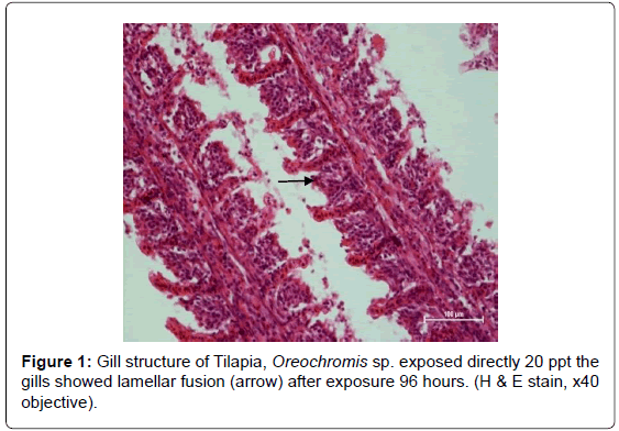
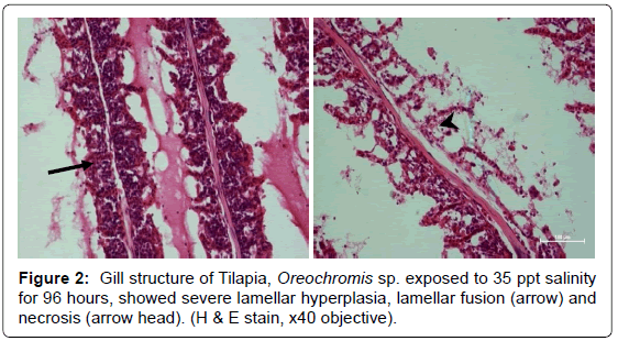
The gills sections from the tilapia, Oreochromis sp. in the control group presented a normal appearance and not reveal any histopathological lesions in the tissues. The histological lesions were occurred severely with increased of the salinity (Table 5). Histopathological lesions observed in the gills of the fish exposed to the salinity showed different lesions which include from hyperplasia of the epithelium, fusion of secondary lamellae and necrosis. At 5 ppt salinity, the gills were start to change but with slightly changes. For the 20 ppt salinity, the fish was started showed the severely affecting of the gills which appearance on hyperplasia and fusion of lamellae while at 35 ppt salinity it showed a very severe lesion which necrosis of the tissue was appear (Figure 2).
| Result/Organs | Gills | Kidney |
|---|---|---|
| 0ppt | Normal | Normal Slightly hydropic degeneration |
| 5ppt | Hyperplasia of the epithelium | Hydropic degeneration Narrowing tubular lumen |
| 20ppt | Hyperplasia Lamellar fusion | Tubular necrosis Edema in Bowman’s capsule Hydropic degeneration Haemorrhage |
| 35ppt | Hyperplasia Lamellar fusion Necrosis | Gross lesion-Kidney very fragile (sample not taken)-necrosis |
Table 5: The histopathological responses of Oreochromis sp. to different salinity level for 96 hours.
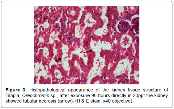
The histopathology of control fish kidney showed normal appearance of glomerulus but slightly degeneration on the renal tubules. There were mild lesions with low salinity at the 5 ppt where the hydropic degeneration and narrowing tubular lumen was appearing. The histopathological lesions were severely observed in high salinity between 20 ppt and 35 ppt such as hydropic degeneration, edema in Bowman’s capsule, hemorrhage and necrosis into the surrounding tissue (Figure 3). At 35 ppt salinity it showed very severe lesions where the necrosis was observed in the kidney. Some of the kidney samples exposed to 35 ppt were not taken because it was very fragile and difficult to observe by histopathologicaly. Gross appearance from the dead fish, it was swollen, hemorrhage and soft.
Discussion
From the investigations of this study, it is proved Tilapia is tolerated with 0 ppt and 5 ppt salinity. In both salinity of 0 ppt and 5 ppt, normal behavior and no mortality were recorded. However, in 5 ppt salinity there was only a weak of excessive mucus secretion was observed on the fish. This is an indication that the fish were perfectly able to regulate their body physiology within this salinity. There were 100% mortality recorded in 35 ppt salinity, indicates that the fish has developed osmoregulatory failure. According to Deacon and Hecht [9] the mortality observed after 24 hours the fish exposed to 20 ppt salinity could be consequence of the progressive deterioration in the osmotic and ionic regulatory mechanisms including the fish inability to control the excessive water loss, leading to osmoregulatory exhaustion, collapse and finally death. In another study Lawson [1], the mortality was due to stress, duress and less resistance of the fish to this salinity. There was no mortality found between 0 ppt and 5 ppt which indicated the fish were able to withstand a wide salinity range. According to [10,11] the survival of the fish depends on the ability of the body fluids to function at least for short time in an abnormal range of abnormal internal osmotic and ionic concentrations. The fish can regulate the body fluid to restore the level of osmotic pressure to near normal. The migration or abruptly transferred of fish from freshwater to seawater will normally lead to increase osmotic concentration of fish blood serum and change in ionic contents [12,13].
The restlessness or erratic behavior in high salinities indicates the fish were approaching their tolerance limits and loss of water at fast rate to external medium from the fish [1]. This also may be due to biochemical body derangement including hepatic compromise Fadina [14]. The fish exposed to different salinity exhibited changes in behavior such as convulsions, equilibrium status or imbalance, fin movement, hyperactivity and swimming rate were observed in this study. From the present study, respiratory distress such as increase opercular activities, gulping of air, vertical movement and excessive mucus was recorded in 20 ppt and 35 ppt. For increased opercular frequency activities has been reported as an adaptive mechanism to hyper saline environments [15-18]. This sign is may be due to excessive mucus secretions because the mucus on the gills reduces respiratory activity in fishes and unable for fish to actively carry out gaseous exchange [8,19].
The progressive decrease in the opercular frequency led to swimming close to the water surface in order to increase the oxygen intake in the water surface Soares [20]. The increase of the salinity concentration interrupted the respiratory system in the fish and caused lamellar hyperplasia Reid [21]. The severe lesions in gill were observed in fish exposed to 35 ppt are hyperplasia, lamellar fusion and necrosis. This is in accord with Mallat [22] who concluded that the most common gill lesions which induced by toxic substances and other chemicals are necrosis, hyperplasia and lamellar fusion. A higher salinity in the water produce lamellar lesions such as necrosis which lead to death after exposed the fish to 35 ppt salinity. The degeneration of gills also causes a dysfunction of fish gas exchange ability causing an anoxic internal behavior Ajani [23].
Direct transfer from freshwater to higher salinity conditions cause the fish a strong respiratory distress. This can be showed by histopathological observation when the fish were exposed at 35 ppt salinity exhibited a severe lesion on the gills such as hyperplasia, lamellar fusion and necrosis. This indicates that the fish were approaching their tolerate limits and developed osmoregulatory failure [1]. A higher salinity and toxic in the water produced severe lesions on the gills and kidney such as severe hydropic degeneration, edema and necrosis which lead to mortality Yahona [24]. Several abnormal nervous behaviors such as sluggish and swirling movements with different postures, state of motionless, sudden darts and death may cause by the failure of the kidney function where the histopathological lesions were observed on the fish kidney Oti [25]. The lesions of the kidney tissue are including hydropic degeneration, edema in Bowman’s capsule, hemorrhage and necrosis. The histopathology changes in kidney lead to mild of hydropic degeneration of renal tubules when the fish were exposed to 5 ppt salinity. In 20 ppt, the Oreochromis sp. started to show severe lesions of the kidney while in the 35 ppt salinity; gross necrosis was found on the fish. Histopathological changes in fish tissue and residue levels of test substances in fish are very important parameter for deriving the maximum acceptable concentration of chemicals in the context of fish culture requirements Svobodova [26].
Acknowledgement
We would like to thanks to Institute of Tropical Aquaculture/AKUATROP Laboratory management and Institute Marine Biotechnology/IMB for the support, Mr. Raja Zakaria Raja Abdullah from Perniagaan Ikan Jaya, Kelantan for the fish sponsor and Mr. Nor Azri Shah Norhan , Mr. Muhammad Zaim Md Desa, Mr. Muhammad Embong for their help.
References
- Lawson EO, Anetekhai MA (2011) Salinity tolerance and preference of hatchery reared Nile Tilapia, Oreochromis niloticus (Linnaeus1758). Asian J Agr Sci 3: 104-110.
- Claudia H, Jeffrey CW (2009) Morphologic Effects of the Stress Response in Fish. ILARJ 50: 387-396.
- El-Dahhar AA, Kamel EA, El-Wakil H, Sh. Grana Y (2011) Effect of salinity stress on growth of three strains of Egyptian Nile Tilapia (Oreochromis niloticus). J Arab Aquat Soc 6: 251-269.
- Baroiller JF, Clota F, Cotta HD, Derivaz M, Lazard J, et al. (2000) Seawater Adaptability to Two Tilapia Species (S. melanotheron and Oreochromis niloticus) and their reciprocal F1 Hybrids. Proceedings of the fifth International symposium on Tilapia in Aquaculture.
- Dennis M, Riza A, Wilfredo C, Ma Severa FK, Roman S, et al. (2004) https://ag.arizona.edu/oip/ista6/ista6/ista6web/pdf/426.pdf
- Aysel CKB, Ayhan O (2010) Acute Toxicity and Histopathological effects of sublethal fenitrothion on Nile tilapia, Oreochromis niloticus. J Pest Biochem Physiol 97: 32-35.
- Rand GM (1985) Behavior Fundamentals of Aquatic Toxicology Methods and Applications, Hemisphere Publishing Corporation, Washington, USA.
- Anur AN, Jasmani S, Ikhwanuddin M, Hassan M (2011) Effects of a methanolic extract of Melaleuca cajuputi on African catfish (Clarias gariepnus). Proceedings of the International Fisheries Symposium.
- Deacon N, Hecht T (1999) The effect of reduced salinity on growth, food conversion and protein efficiency ratio in juvenile spotted grunter, Pomadasys commersonnii (Lecepede) (Teleostei Haemulidae). Aquaculture Resource 30: 13-20.
- Kurata H (1959) Preliminary report on the rearing of herring larvae. Bull. Hokkaido Resource Fish Laboratory 20: 117-138.
- Holliday FGT, Jones MP (1967) Some effects of salinity on the development of eggs and larvae of the Plaice (Pleuronectes platessa). Marine Biology Association UK 47: 39-48.
- Gordon MS (1959) Ionic regulation in the Brown trout (Salmon trutta). Journal of Experimental Biology 36: 227-252.
- Miles HM, Smith LS (1968) Ionic regulation in migrating juvenile Coho Salmon, Oncorhynchus kisutch. Comp Biochem Physiol 26: 381-398.
- Fadina OO, Taiwo VO, Ogunsanmi AO (1991) The effects of single and repetitive oral administrative of common pesticide and alcohol on rabbits. Tropical Veterinary 17: 97-106.
- Morgan JD, Iwama GK (1991) Effects of salinity on growth, metabolism and ion regulation in juvenile rainbow and steelhead trout (Oncorhynchus mykiss) and fall chinook salmon (Oncorhynchus tshawytscha). Can J Fish Aquat Sci 48: 2083-2094.
- Iwama GK, Takemura A, Takano K (1997) Oxygen consumption rates of tilapia in freshwater, seawater, and hyper saline seawater. J Fish Biol 51: 886-894.
- Morgan JD, Sakamoto T, Grau EG, Iwama GK (1997) Physiological and respiratory responses of the Mozambique tilapia (Oreochromis mossambiqus) to salinity acclimation. Comp Biochem Physiol A: Physiology 117: 391-398.
- De Boeck G, Vlaemink A, Van der Linden A, Blust R (2000). Salt Stress and resistance to hypoxic challenges in the common carp (Cyprinus carpio L.). J Fish Biol 57: 761-776.
- Konar SK (1970) Nicotineas a fish poison. Progressive Fish-Culturist 32: 103-104.
- Soares MGM, Menezes NA, Junk WJ (2006) Adaptations of fish species to oxygen depletion in a central Amazonian floodplain lake. Hydrobiologia 568: 353-367.
- Ried SG, Sundin L, Milson WK (2006) The Physiology of Tropical Fishes, Elsevier, Academic Press, Italy.
- Mallat J (1985) Fish gill structural changes induced by toxicants and other irritants: a statistical review. Can J Fish Aquat Sci. 42: 630-648.
- Ajani F, Olukunie OA, Agbede SA (2007) Hormonal and haematological responses of Clarias gariepinus (Burchell, 1822) to nitrate toxicity. Journal of Fisheries 2: 48-53.
- Yahona M, Pablo E (2007) Behavioral and gill histopathological effects of acute exposure to sodium chloride in moneda (Metynnis orinocensis). J Environ Toxicol Pharmacol 25: 365-372.
- Oti EE, Ukpabi UH (2000) Acute toxicity of milk extract thevetia periviana (person) to African freshwater catfish, Clarias gariepinus (tengels). Africa J Sci Technol 1: 132-138.
- Svobodova Z, Vykusova B, Machova J (1994) The Effects of Pollutants on Selected Hematological and Biochemical Parameters in Fish. In: Sublethal and Chronic Effects of Pollutants on Freshwater Fish, Muller R, Lloyd R (Eds.). Fishing News Books 39-52, UN.
Relevant Topics
- Acoustic Survey
- Animal Husbandry
- Aquaculture Developement
- Bioacoustics
- Biological Diversity
- Dropline
- Fisheries
- Fisheries Management
- Fishing Vessel
- Gillnet
- Jigging
- Livestock Nutrition
- Livestock Production
- Marine
- Marine Fish
- Maritime Policy
- Pelagic Fish
- Poultry
- Sustainable fishery
- Sustainable Fishing
- Trawling
Recommended Journals
Article Tools
Article Usage
- Total views: 16263
- [From(publication date):
September-2013 - Jan 15, 2025] - Breakdown by view type
- HTML page views : 11588
- PDF downloads : 4675


