Histopathologic Patterns of Soft Tissue Tumors in Jimma University Medical Center, Southwest Ethiopia: A 5-Year Retrospective Study
Received: 07-Feb-2020 / Accepted Date: 21-Feb-2020 / Published Date: 28-Feb-2020 DOI: 10.4172/2161-0681.1000380
Abstract
Background: Soft tissue tumors are a complex group of neoplasms with differentiation towards mesenchymal tissue occurring in all age groups. Although pathologically diverse, they frequently exhibit similar clinical presentations and radiological features. Therefore, correct histopathologic diagnosis is crucial. Despite histopathologic diagnosis is widely practiced in study area very few studies were found on histopathologic pattern of soft tissue sarcoma. Therefore, this study was aimed to describe histopathology pattern of soft tissue tumors in Jimma University Medical Center, 2018.
Materials and methods: A cross sectional study design was employed on five years patient’s biopsy record of Jimma University Medical Center pathology department from September 2013 to August 2018 G.C. Structured check lists was used to collect data. Data were entered into Epi data v.3.1 and exported to SPSS V.20 for analyses. Data was described using frequency, proportions and median. Finally result was presented using tables, figures and narrative form.
Results: A total of 268 patients were diagnosed with soft tissue tumor over the last five years with malignant to benign ratio of 1:2.6 and sex ratio of Male to Female 1:1.01. The age group at which soft tissue tumors commonly occur was from 21-30 (26.9%) years of age. The most favored site of occurrence (59.5%) is the lower limb. From the different histologic subtypes of sarcoma, the top three are Malignant Fibrous Histiocytoma (27.0%), which is common in adults followed by Fibrosarcoma (18.9%), and Rhabdomyosarcoma (17.6%) which is common in youngsters.
Conclusion: Benign tumors are three times more common than malignant. The most common soft tissue sarcoma in children is Rhabdomyosarcoma and in adults Malignant Fibrous Histiocytoma and larger scale study needs to be planned to look at more aspects that could possibly contribute to the causes of soft tissue tumors.
Keywords: Histopathology; Patterns; Soft tissue tumor; Sarcoma; Ethiopia
Abbreviations
DFSP: Dermato Fibro Sarcoma Protuberance; FNAC: Fine Needle Aspiration Cytology; IRB: Institutional Review Board; IQR: Inter Quartile Range; JUMC: Jimma University Medical Center; MFH: Malignant Fibrous Histiocytoma; RMS: Rhabdomyosarcoma; STS: Soft Tissue Sarcoma; STT: Soft Tissue Tumors; SPSS: Statistical Package for Social Sciences; WHO: World Health Organization.
Introduction
Soft Tissue Tumors (STT) is a complex group of neoplasms with differentiation towards mesenchymal tissue occurring in all age groups. It differs by various inherent features which includes histology, molecular genetics profiles, site predilection, and outcomes of care. Although pathologically diverse, they frequently exhibit similar clinical presentations and radiological features. Therefore, correct histopathologic diagnosis is crucial [1,2].
Within the various histo genetic categories, soft tissue tumors STT are usually divided into benign and malignant forms. Benign tumors outnumber malignant tumors (sarcoma) by a wide margin and especially lipoma is 100 times more common [3-5].
Soft Tissue Sarcoma (STS) includes approximately 40 malignant histological subtypes, and around 50% is most prominent in the extremities followed by trunk and retroperitoneal and head and neck. It constitutes 1% of all adult malignancies and about 12% of pediatric cancers. Annually the incidence is about 6 per 100000 persons [6-8].
Despite its rarity, sarcomas are accountable for relatively high morbidity and mortality especially in children and adolescents. It is capable of invasive, locally destructive growth with a tendency to recur and metastasize. Sarcomas are more common in older patients, with 15% affecting patients younger than 15 years and 40% affecting persons older than 55 years. Accordingly, as the population ages, as it is doing at a rapid rate, the incidence of these tumors will increase [9-11].
Clinically STT case presents with painless mass. But benign tumors like schwannoma and glomus tumors are painful. Some benign tumors have the capacity to recur, invade locally or a potential to be converted to malignancy. Therefore, such tumors need thorough evaluation and close follow up. After surgical excision benign tumors have a very high cure rate [12,13].
Outcome of the tumor depends on age of the patient, size and site of the tumors, tumors' grade and histological subtype. But tumor size greater than 5 cm, infiltration into surrounding structures, neurovascular bundle involvement, enlargement of loco-regional lymph nodes are all signs of high risk and poor prognosis. Therefore, early recognition, correct histological diagnosis and early surgical intervention are the most important factors to determine the prognosis of patients with malignant STTs [14,15].
Without any age classification STT can occur at any age. But variation was observed to age groups it most commonly affects. Sometimes it is more common at the 2nd and 3rd decades of life. But in another study increment in incidence was observed as age advances and maximum numbers of cases are reported in 51-60 years of age [16-18].
There is a limited study in Africa about soft tissue tumors and only a single soft tissue cytopathology was accessed in Ethiopia. Therefore, in the absence of well-established cancer registry system and studies such record analysis provides useful information for understanding the changing trend and pattern. It can also help in undertaking further studies to identify the associated factors which are highly important for developing appropriate strategies and prioritizing control measures. Therefore, the objective of this study was to describe the histopathologic patterns of soft tissue tumors in Jimma University Medical Center, from September 2013 to August 2018 G.C.
Methodology
Study area and study period
A cross sectional study design was employed on five years patient’s biopsy record of Jimma University Medical Center pathology department from September 2013 to August 2018 G.C. Structured check lists was used to collect data. Data were entered into Epi data v. 3.1 and exported to SPSS V.20 for analyses. Data was described using frequency, proportions and median. Finally result was presented using tables, figures and narrative form.
The study was conducted in Jimma University Medical Center (JUMC), Pathology Department. The hospital is situated in Jimma town 345 KM to southwest of Addis Ababa: Ethiopia. The pathology department of JUMC is establishment in 1985 G.C and it is one of the high burden departments with two pathologists, 18 residents, and two histopathology technicians and 7 technical assistants. The services provided by the department include histopathology (biopsy), FNAC (fine needle aspiration cytology), hematopathology and fluid cytology. The average annual patient flows for cytopathologic and histopathology services are more than 5000 and 1600 per year respectively. The data was collected from August 1-30, 2018 G.C for the year between 2013 to 2018.
Study design
Facility based descriptive cross-sectional study design was used for all biopsies of patients sent to JUMC, Pathology Department during 2013 to 2018 G.C.
Sampling techniques
Primarily, all biopsy records with soft tissue tumors from Pathology department filled on biopsy request form and logbook during the last five years 2013 to 2018 G.C was identified. Secondly, among all identified soft tissue tumor biopsy records, records with benign and malignant soft tissue tumors diagnosis were also identified. Finally based on the data collection checklist important data was retrieved.
Data collection techniques
Data were collected using structured check lists adopted through reviewing of literatures included in this study and previous similar studies [18] that fulfill the objective of the study. Data were recorded on the prepared checklists by reviewing a histopathology record report of patients during the specified period. Two supervisors from junior pathology residents and two data collectors from histopathology technician were recruited for data collection.
Data quality assurance
To assure the quality of data, before the actual data collection, situational analysis was conducted to look at the records. Afterwards missed variables from the checklist were included into the extraction checklist. Two days training was provided for the data extractors on how to fill and refill data on the prepared check lists. As much as possible completeness of the data was assured by reviewing both logbook and registration.
Data processing and analysis
After checking completeness of the checklist, data was entered into Epi data v.3.1 and exported to SPSS V.20 for analysis. Before analysis data cleaning and grouping was done to make the data ready for analysis. Then descriptive statistics like frequency, percentage, mean was calculated to summarize the data. Finally, the obtained results were presented using tables, figures and narrative.
Ethical consideration
Ethical clearance was obtained from Institutional Review Board (IRB) of JUMC. Support letter for Permission to conduct study was also submitted to pathology department. During data extraction all records were retrieved with accuracy and patient name was not included to the data to keep confidentiality.
Results
A total of 284 biopsies of soft tissue were reviewed in the JUMC pathology department over the last five years and 16 were excluded from the study because of missed variables (diagnoses and size determination). From the included 268 soft tissue biopsies, 74 were malignant tumors and 194 were benign with malignant to benign ratio of 1:2.6.
The age at which soft tissue tumors commonly occur was between 21-30 years 72(26.9%) with a median age of 31.00 years and IQR (Interquartile Range) 50. Regarding sex distribution there was a slight female predilection with M: F ratio of 1:1.01. Benign soft tissue tumor is more common in female 107 (55.1%) but, the reverse is true in case of soft tissue malignancy/sarcoma and which is more common in male 48 (64.9%) and there was significant association between sex of the patient and soft tissue tumor (p value<0.05) (Table 1).
| Variable | Category | Type of tumor | Chi-square | |
|---|---|---|---|---|
| Malignant frequency (%) | Benign frequency (%) | |||
| Age of the patient | 10-Jan | 8 (10.8) | 26 (13.4%) | χ2=4.20 p value=0.53 |
| 20-Oct | 11(14.9%) | 34 (17.5%) | ||
| 21-30 | 17 (23.0% | 55(28. 4%) | ||
| 31-4 0 | 12 (16.2%) | 29 (14.5%) | ||
| 41-50 | 9 (12.2%) | 24 (12. 4%) | ||
| >50 | 17 (23.0%) | 26(13.4%) | ||
| Total | 74 (100.0%) | 194 (100.0%) | ||
| Sex of the patient | Male | 48 (64.9%) | 87 (44.9%) | χ2=8.59 p value=0.003 |
| Female | 26 (35.1%) | 107 (55.1%) | ||
| Total | 74 | 194 | ||
| Residence | Urban | 21 (28.4%) | 79 (40.7%) | χ2=3.49 p value=0.06 |
| Rural | 53(71.6%) | 115 (59.3%) | ||
| Total | 74 | 194 | ||
Table 1: Soft tissue tumor related socio demographic distribution of the patients from 2013 to 2018 G.C, JUMC. N=268.
The most common site of soft tissue tumor was in the lower extremity 106 (39.6%) followed by head and neck 75(27.9%). The anatomic site was found to be significantly associated with soft tissue tumor (p value<0.001). Similarly, the most common site for soft tissue sarcoma was lower limb 44 (59.5%) in which thigh accounts for 29 (39.2%) followed by trunk 11 (14.9%). Concerning the size of soft tissue tumor, 195 (72.8%) was Table 2).
| Variable | Category | Malignant Frequency (%) | Benign frequency (%0) | Chi-square |
|---|---|---|---|---|
| Site of tumor | Lower extremity | 44 (59.5%) | 62 (32.0%) | χ2=18.95, p value<0.001 |
| Head and neck | 10 (13.5%) | 65 (33.5%) | ||
| Trunk | 11 (14.9%) | 40 (20.6%) | ||
| Upper extremity | 9 (12.2%) | 27 (13.9.0%) | ||
| Total | 74 | 194 | ||
| Size of the tumor | 35 (47. 3%)160(82.5%)χ2=40.65, p value<0.001 |
Table 2: Soft tissue tumors characteristics in terms of site and size N=268.
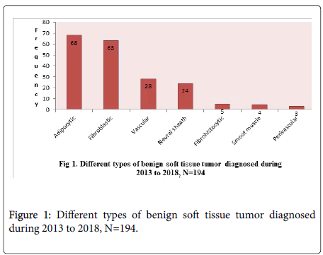
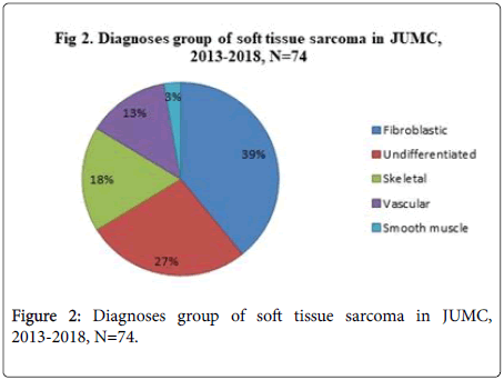
As it is explained above from the total biopsies reviewed benign tumor was diagnosed in 194 cases. From these total benign soft tissue tumors, the commonest histological type was Adipocytic which is Lipoma 68(35.1%) followed by fibroblastic in which Fibroma 61 (31.4%) was the commonest. Concerning soft tissue sarcoma/ malignancy from the total 74 cases diagnosed the commonest type was fibroblastic ground 29 (39.2%) followed by undifferentiated sarcoma 20 (27.0%) (Figures 1 and 2).
Regarding sex distribution of soft tissue sarcoma type, Fibrosarcoma 6(23.0%) was the commonest among females followed by Malignant Fibrous Histiocytoma 5(19.2%) and Rhabdomyosarcoma (RMS) 5(19.2%). But in males the commonest one is Malignant Fibrous Histiocytoma 15(31.2) followed by Fibrosarcoma 8(16.7%) and Rhabdomyosarcoma 8(16.7%) (Table 3).
| Diagnoses | Sex category | ||
|---|---|---|---|
| Male | Female | Total | |
| MFH* | 15(31.2) | 5(19.2%) | 20(27.0%) |
| Fibrosarcoma | 8(16.7%) | 6(23.0%) | 14(18.9%) |
| Rhabdomyosarcoma | 8(16.7%) | 5(19.2%) | 13(17.6%) |
| Myxofibrosarcoma | 7(14.6%) | 3(11.5%) | 10(13.5%) |
| DFSP** | 3 (6.3%) | 2(7.7%) | 5(6.7%) |
| Hemangioendothelioma | 2 (4.2%) | 3(11.5%) | 5(6.7%) |
| Angiosarcoma | 3 (6.3%) | 1(3.8%) | 4(5.4%) |
| Leiomyosarcoma | 1 (2.1%) | 1(3.8%) | 2(2.7%) |
| Hemangiopericytoma | 1 (2.1%) | - | 1(1.2%) |
| Total | 48(100%) | 26(100%) | 74(100%) |
MFH*- Malignant Fibrous Histocytoma, DFSP**- Dermato Fibro Sarcoma Protuberance.
Table 3: Soft tissue sarcoma patterns with respect to sex in JUMC from 2013 to 2018 G.C N=74.
From the total soft tissue sarcoma diagnosed (74), the most common top three types are MFH 20 (27.0%), Fibrosarcoma 14(18.9%), and RMS 13 (17.6%) and the least or uncommon type identified was Hemangiopericytoma. The occurrence of soft tissue sarcoma by age group was bimodal and it commonly affects people in the age of 21-30 years of age 17 (23.0%) and >50 years of age 17 (23.0%). In youngsters below the age of 10, the commonest type of sarcoma was found to RMS followed by Fibrosarcoma but, in adults MFH 20(27.0%) was the commonest. RMS accounts for 13(17.6%) of all sarcoma cases and all of them arise from thigh. Concerning their variant 3 of them were alveolar, one embryonal variant and 9 of the cases were not classified. The entire alveolar variant of RMS was diagnosed in adults between 3rd to 5th decades. Soft tissue sarcoma can affect different parts of the body. But the most common anatomic site was lower extremity for all the top three soft tissue sarcomas (Table 4).
| Age | Sarcomas type | ||
|---|---|---|---|
| MFH | Fibro sarcoma | RMS | |
| 1-10 | - | 3 | 4 |
| 10-20 | 8 | - | - |
| 21-30 | 4 | 3 | 2 |
| 31-40 | 1 | 5 | 3 |
| 41-50 | 2 | 1 | 1 |
| >50 | 5 | 1 | 3 |
| Total | 20 | 14 | 13 |
| Head and neck | 2 | 1 | 4 |
| Upper extremity | 4 | 1 | 2 |
| Trunk | 2 | 2 | - |
| Lower extremity | 12 | 10 | 7 |
| Total | 20 | 14 | 13 |
Table 4: Distribution of the commonest soft tissue sarcoma with respect to age and anatomic sites in JUMC from 2013 to 2018 G.C. N=74.
Discussion
In this study from the total patients diagnosed with soft tissue tumor over the last five years the majority was benign tumor which is predominantly Lipoma and Fibroma. Malignant accounts for one third of the soft tissue tumor and no sexual predilection seen for the soft tissue tumor. This finding was comparable to studies done in similar area in which benign tumor was much more common than malignant 82% and Lipoma accounts 70.5% [19] and study done in India and Pakistan [20,21]. However according to WHO 2013, Fletcher and Ackerman the ratio of benign to malignant was by far higher than this study finding [5] The reason for this variation can be explained that the clinicians at study area might fail to send the clear cut benign cases for histopathology and less health seeking behavior of the community for benign tumor which may be uncovered by future study.
In the present study Soft tissue sarcoma is more common in male and histologically MFH is most common soft tissue sarcoma type as whole and in male and Fibro sarcoma is most common sarcoma in females. However, in cytopathology study done in similar study area years back, DFSP was the most common and undifferentiated pleomorphic sarcoma (MFH) was the second commonest sarcoma [18]. This difference might be due to false positivity of the diagnosis in cytopathology and true negativity in histopathology. Therefore, there is a need of confirmation between cytopathology and histopathology diagnosis.
Distribution of sarcoma in terms of age showed bimodal occurrence with age of 50 years and between 11 to 20 years of age. Anatomically, two third of soft tissue sarcoma are found in the lower limb majorly on thigh followed by the trunk. This finding was in concordance with WHO 2013 soft tissue tumor and other literatures worldwide in which two third of soft tissue sarcomas were in the lower limb [1,20]. In addition, this finding was also comparable with American cancer society 2016 report and other studies conducted in different setting [9,20,21].
Rhabdomyosarcoma was the most common sarcoma in youngsters and the most sarcoma one in adult was MFH which is currently named as undifferentiated pleomorphic sarcoma.
Conclusion
Benign soft tissue tumors are more common than malignant. From benign soft tissue tumor groups Adipocytic (Lipoma) was the commonest. MFH was the predominant histological subtype commonly affecting adults and RMS affects youngsters. The most common anatomic site affected by sarcoma is lower extremity especially thigh. All patients operated for soft tissue tumor needs to be histological confirmed and Immunohistochemistry has been invaluable in categorization of some of these tumors and contributed to ensuring prompt and more accurate diagnosis in our center.
Author’s Contributions
MN contributes in the design of the study, analysis and manuscript write up. AK made the data analysis, co-write up and edition of the manuscript. Both authors critically revised the manuscript and have approved the final manuscript.
Funding
The study was fully funded by Jimma University and the funder had no role in study design, data collection and analysis, decision to publish or preparation of the manuscript.
Competing Interests
No competing interests exist
References
- Tuna M, Ju Z, Amos CI, Mills GB (2012) Soft tissue sarcoma subtypes exhibit distinct patterns of acquired uniparental disomy. BMC Med Genomics 5: 1-7.
- Thway K (2009) Pathology of soft tissue sarcomas. Clin Oncol 21: 695-705.
- Clark MA, Fisher C, Judson I, Thomas JM (2005) Medical progress: Soft- tissue Sarcomas in Adults. N Engl J Med 353: 701-711.
- Baig MA (2015) Histopathological study of soft tissue tumors (three years study ). Int J Sci Res 4: 1039-1049.
- Fletcher CDM (2012) Tumors of soft tissue. In: Diagnostic histopathology of tumors (4th edn). Boston, Massachusetts: Saunder, pp: 1796-1870.
- Zachary Burningham, Mia Hashibe, JDS (2012) The Epidemiology of Sarcoma. Clin Sarcoma Res 2: 383-398
- Malami Aliyu U, Okuofo EC, Oluchukwu Okwonna C, Sahabi SM (2018) Clinicopathological pattern of soft tissue sarcoma in a tertiary health institution in North Western Nigeria. Int J Res Med Sci 6: 1632–1538.
- Von Mehren M, Randall RL, Benjamin RS, Boles S, Bui MM et al. (2016) Soft Tissue Sarcoma, Version 2.2016, NCCN Clinical Practice Guidelines in Oncology. J Natl Compr Cancer Netw 14:758-786.
- Stiller CA, Trama A, Serraino D (2013) Descriptive epidemiology of sarcomas in Europe: Report from the RARECARE project. Eur J Cancer 49: 684-695.
- Shuchita Lakra P (2015) Histopathological spectrum of soft tissue tumors in a teaching hospital. iOSR J Dent Med Sci 14: 2279-2861.
- Jain P, Shrivastava A, Malik R (2014) Clinic morphological assessment of soft tissue tumors. Sch J Appl Med Sci 2:886-890.
- Abhivardhan D, Sivakumar DLR (2015) Clinical study of soft tissue sarcoma cases in a south-indian teaching hospital. JMSCR 3: 5631-5639.
- Hasan SI, Suliman YA, Hassawi AB (2010) Soft tissue tumors: Histopathological study of 93 cases. Ann Coll Med Mosul 36: 92-98.
- Jain S, Jadav K (2017) Histopathology of soft tissue tumors in association with immunohistochemistry. Int J Biomed Adv Res 8: 327-336.
- Harpal S, Richika R, Ramesh K (2016) Histopathological pattern of soft tissue tumors in 200 cases. Ann Int Med Dent Res 2: 6-11.
- BEZABIH M (2001) Cytological diagnosis of soft tissue sarcomas. Cytopathology 12: 177-813.
- B OM, I A, Misauno M A, Mancha D G, Onche I I et al. (2015) Soft Tissue Sarcoma. The Experience at JOS University Teaching Hospital. JOS Nigeria. IOSR J Dent Med Sci Ver 14: 47-49.
- Vinitha Samartha, Shreya Hegde, Zulfikar Ahmed UN (2015) Histopathological Study of Malignant Soft Tissue Tumors. J Evol Med Dent Sci 4: 3320-3328.
- Habib Reshadi, Alireza Rouhani, Saeid Mohajerzadeh, Marvan Moosa, Asghar Elmi M (2014) Prevalence of Malignant Soft Tissue Tumors in. Arch Bone Jt Surg 2: 106-110.
- Yusuf I, Iliyasu Y, Mohammed A (2013) Histopathological study of soft tissue sarcomas seen in a teaching hospital in Kano, Nigeria. Niger J Basic Clin Sci 10: 70-5.
- Toro JR, Travis LB, Hongyu JW, Zhu K, Fletcher CDM (2006) Incidence patterns of soft tissue sarcomas, regardless of primary site, in the Surveillance, Epidemiology and End Results program, 1978-2001: An analysis of 26,758 cases. Int J Cancer 119: 2922-2930.
Citation: Nigussie M, Kebede A (2020) Histopathologic Patterns of Soft Tissue Tumors in Jimma University Medical Center, Southwest Ethiopia: A 5-Year Retrospective Study. J Clin Exp Pathol 10: 380. DOI: 10.4172/2161-0681.1000380
Copyright: © 2020 Nigussie M, et al. This is an open-access article distributed under the terms of the Creative Commons Attribution License, which permits unrestricted use, distribution, and reproduction in any medium, provided the original author and source are credited.
Select your language of interest to view the total content in your interested language
Share This Article
Recommended Journals
Open Access Journals
Article Tools
Article Usage
- Total views: 3218
- [From(publication date): 0-2020 - Dec 13, 2025]
- Breakdown by view type
- HTML page views: 2339
- PDF downloads: 879


