Hip Pain Revealing Metastatic Prostate Cancer
Received: 25-Dec-2018 / Accepted Date: 24-Jan-2019 / Published Date: 31-Jan-2019 DOI: 10.4172/2167-0846.1000338
Abstract
Background: Prostate cancer is the most common malignant tumor and the second most common cause of cancer associated mortality in males. Bone metastasis is frequent, usually multiple and osteoplastic. Presentation of a pure osteolytic and solitary metastasis from a prostate carcinoma is extremely rare.
Methodology: We report a case of prostate cancer in a 54-year-old man who presented with progressive severe right hip pain and stiffness with no urinary symptom. An MRI of the pelvis revealed a metastasis to the right hip. A prostate biopsy revealed prostate adenocarcinoma.
Results: In the literature, there are few cases of a solitary osteolytic bone metastasis from carcinoma of the prostate, and especially in the pelvis. only one such case seems to be reporting.
Keywords: Hip pain; Prostate carcinoma; Osteolytic bone metastasis; Revealing metastasis
Case Report
A 54-year-old man was admitted to our service, have in his past medical history a gastric ulcer treated in 2006. He presents since 6 months a gradually progressive severe right hip pain and stiffness, restricting physical activities and walking. In a context of fever, thrill and weight loss quantified at 4 kg, without other joint signs or associated signs.
The clinical exam noted a patient in good general stat, weighing 60 kg for a height of 1 m 68 cm. Exam of the musculoskeletal system find a limp, with painful limitation of all mobility of the right hip. The contralateral hip and the other joints were intact. Somatic exam including pulmonary, cardiovascular, abdominal, do not find any anomaly. There was no sign of peripheral tumor syndrome.
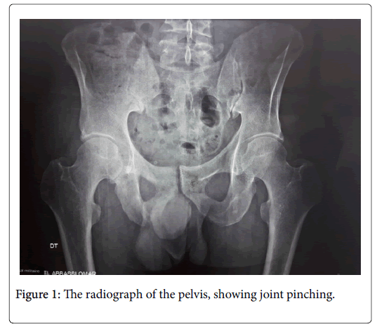
The radiograph of the proximal right femur shows a posterior and inferior coxofemoral narrowing with no objective bone lesions (Figure 1).
The articular echography of right hip was showing an articular effusion, who was preleved to exclude a risk of infection. The result shows mechanical liquid.
An inflammatory syndrome is founding with C reactive protein (CRP) at 28 mg/l and Erythrocyte Sedimentation Rate (ESR) at 50 mm in first hour. In addition, Tumor marker studies revealed an elevated level of Prostate-Specific Antigen (PSA) at 989 ng/l. the rest of biological blood exam was normal.
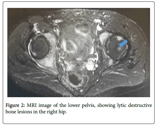
MRI imaging of the right hip show lytic destructive bone lesions suspect of metastatic lesions (Figure 2).
Cause of the high level of PSA, we have explored prostate. The rectal touch reveled nodular prostate increased volume, with high volume in echography showing prostate at 60 g with significant post-void residue at 100 ml.
The extension assessment, notably by bone scintigraphy and Wholebody bone scan, confirmed the solitary metastatic localization.
The diagnosis retained was that of osteolytic metastases unique to prostate adenocarcinoma.
Discussion
Hip pain has many origins; however, usually clinical exam associated to simple radiographic, biologic exam and advanced imaging allows you to specify the etiology.
However, pain of hip is not always reaching the joint hip. Pelvis and the upper end of the femur are often headquarters for primary or secondary tumor localization.
In the clinical case presented, given the young age of the patient and the absence of personal or family history of neoplasia, the possibility of a tumor cause for us was unlikely. Our etiological investigation was more concerned with the search for an infectious cause or chronic inflammatory rheumatism such as spondyloarthritis or rheumatoid arthritis.
Bone metastases are the most common bone tumoral pathology. Prostate cancer is involved in about 29% of cases. These bone metastases occur in approximately 70% of patients with advanced prostate cancer and about 30% are diagnosed at the time of diagnosis [1]. A number of bone metastases occur during the follow-up of a known cancer and pose few diagnostic problems. In contrast, the identification of the primary tumor is difficult in about half of cases when the metastasis is inaugural and moreover isolated: the origin is not found in 4 to 10% of cases.
Prostate cancer (PCa) is the most common malignant tumor in men after age 50. This increase in the incidence of PCa is explained by earlier detection cause of use the prostate specific antigen (PSA) in routine practice.
Bone represents one of the sites most frequently affected by metastases in this cancer; more than 70% of patients with castrationresistant cancer will develop bone metastases [2].
Cancer cells spread to the bone by direct extension, arterial embolization or venous spread. The data confirmed that bone metastases are more common in patients with high levels of PSA and poorly differentiated tumors at the biopsy, regardless of the age of the patient [3].
The incidence of revealing bone metastases varies according to the hospital structures that support them. It is uncommon in oncology centers where recruitment is based on the early diagnosis of the most common tumors, particularly breast and prostate cancer. On the other hand, they are most often involved in the follow-up of a known and treated primary cancer (Table 1).
| Site of the primary tumor | Main histological type | Frequency (%) |
|---|---|---|
| breast | adenocarcinoma | 32,6 |
| Bronchi | Squamous cell carcinoma | 16,8 |
| Unknown | Variable | 10,9 |
| Prostate | adenocarcinoma | 7,7 |
| ORL | Squamous cell carcinoma | 7,4 |
| Kidney | adenocarcinoma | 4,2 |
| Skin | Melanoma | 1,9 |
| Bladder | Transitional carcinoma | 1,6 |
Table 1: Origin of the main bone metastases and their main identified histological type during autopsy.
In our case, the cancer was unknown and it is the hip ache in connection with to bone metastasis that revealed it. Bone metastases are expressed by bone pain; those if they are described in literature as being the most often felt as "deep", permanent but of varying acuteness, crescent, increased by mobilization and efforts, rebels to simple analgesics. They often cause nocturnal awakenings giving it an "inflammatory" character. However, all types of pain can be observed, and the characteristics of the pain are often not linked to the seat, the volume of the metastasis or the nature of the primary tumor.
Bone metastases can also be expressed by a complication, spontaneous fractures or occurring after minimal trauma, hypercalcemia or neurological compressions, including bone marrow compression in 7% of cases leading to paraplegia in the absence of immediate surgery, the most appearing in cases over 60 years old. This explains the usual management in rheumatology or internal medicine, and more rarely, from the outset in orthopedic surgery or neurosurgery.
Bone metastases are generally multiple and osteoplastic, solitary in only 10% of cases [4]. Our observation has the particularity of the extremely rare mode of revelation by a unique osteolytic metastasis.
With the omission of pelvic lymph node involvement, which represents a locoregional development of the disease, the most common distant metastatic site of prostate cancer (CaP) is bone tissue; in particular, that of the axial skeleton and proximal long bones. In fact, metastases in the lungs, liver and adrenal glands have been found regularly but very infrequently compared to bone metastases, which accounted for more than 90% of remote metastases. Other locations are much rarer and are published in the form of clinical cases.
Only one case similar to the one reported by Agheli et al. [5] has been found in the literature. A 70-year-old male treated for type 2 diabetes, high cholesterol, and coronary artery disease. The father's family history includes his death from prostate cancer at the age of 45 [6].
Indeed, it has long been known that having a positive family history and/or a certain ethnic background such as Afro-Caribbean is a risk factor for PCa development. Evidence from twin studies where monozygotic twins were compared with dizygotic twins [7] as well as studies of familial PrCa highlights this. First-degree relatives of PCa patients have twice the risk of developing the disease compared with the general population [8]. In men diagnosed under the age of 60 years, the risk to their first-degree relatives is >4-fold that of those without a family history [9].
He had severe pain in his right hip and stiffness that hindered his physical activity and walking. Despite no urinary symptoms, imaging revealed a single lytic lesion of the upper 1/3 of the right femur related to bone metastases of prostate adenocarcinoma.
The rectal exam finds a prostate of normal, non-nodular texture, moderately enlarged in volume. The blood test result was normal except the Alkaline phosphates (ALP), which was raised at 210 U/L. Our patient had a disturbed inflammatory balance and normal PAL levels.
Brown et al. reported two cases of localisation in the mandible. In addition, the metastasis was osteolytic, and suspected to be of prostatic origin. However, the nature of the primitive could not be specified. Ansari et al. in turn, published single osteolytic metastases of prostate cancer in a 60-year-old radial-head man.
Conclusion
Bone marrow metastases are associated with major joint morbidity in advanced prostate cancer and require multidisciplinary management. They are very common during this malignant condition, usually multiple and thought-provoking, occurring on prostate cancer known at an advanced stage of its development. Our case is thus characterized by its sciatica of the primary tumor, and on the other hand by its individual colonization and its very rare bone type.
Disclosure of Interest
The authors declare that they have no conflicts of interest concerning this article.
References
- Jemal A, Siegel R, Ward E, Murray T, Xu J, et al. (2007) Cancer statistics. CA Cancer J Clin 57: 43-66.
- Lebret T, Méjean A (2008) Pathophysiology of bone metastases from prostate cancer and the role of bisphosphonates in treatment. Cancer Treat Rev 18: 349-356.
- Salonia A, Gallina A, Camerota TC, Picchio M, Da- Pozzo MFLF, et al. (2006) Bone metastases are infrequent in patients with newly diagnosed prostate cancer: Analysis of their clinical and pathologic features. Urology 68: 362-366.
- Ansari MS, Nabi G, Aron M (2003) Solitary radial head metastasis with wrist drop: a rare presentation of metastatic prostate carcinoma. Urol Int 70: 77-79.
- Agheli A, Patsiornik Y, Chen Y (2009) Prostate carcinoma, presenting with a solitary osteolytic bone lesion to the right hip. Radiol Case Rep 4: 288.
- Conroy T, Malissard L, Dartois D, Luporsi E, Stines J, et al. (1988) Histoire naturelle et eÌvolu- tion des metastases osseuses. AÌ€ propos de 429 observations. Bull Can 75: 845-857.
- Lichtenstein P, Holm NV, Verkasalo PK, Iliadou A, Kaprio J, et al. (2000) Environmental and heritable factors in the causation of cancer analyses of cohorts of twins from Sweden, Denmark, and Finland. N Engl J Med 343: 78-85.
- Goldgar DE, Easton DF, Cannon-Albright LA, Skolnick MH (1994) Systematic population-based assessment of cancer risk in first-degree relatives of cancer probands. J Natl Cancer Inst 86: 1600-1608.
- Lange EM (2010) Male reproductive cancers: epidemiology, pathology and genetics. Can Gen Spr 1: 449-467.
Citation: Youssoufi T, Haddani FZ, Guich A, Hassikou H (2019) Hip Pain Revealing Metastatic Prostate Cancer. J Pain Relief 8:338. DOI: 10.4172/2167-0846.1000338
Copyright: © 2019 Youssoufi T, et al. This is an open-access article distributed under the terms of the Creative Commons Attribution License, which permits unrestricted use, distribution, and reproduction in any medium, provided the original author and source are credited.
Select your language of interest to view the total content in your interested language
Share This Article
Recommended Journals
Open Access Journals
Article Tools
Article Usage
- Total views: 13863
- [From(publication date): 0-2019 - Nov 29, 2025]
- Breakdown by view type
- HTML page views: 12879
- PDF downloads: 984


