High-Resolution ENT Video Endoscope with Superior Image Quality Equivalent to that of Gastric Video Endoscopes
Received: 07-Feb-2014 / Accepted Date: 28-Mar-2014 / Published Date: 04-Mar-2014 DOI: 10.4172/2161-119X.1000166
Abstract
Background and study aims: To assess the usability of high resolution fiberscope which has equivalent image quality to that of the esophageal and gastric video endoscopes
Patients and methods: Image resolution of this endoscope was estimated by the United States Air Force (USAF) resolution test chart. Clinical application was done between January and December 2010 and transnasal observation of the larynx and hypopharynx were performed during this period. These examinations were done for screening and follow-up for patients with hypopharyngeal and laryngeal disorders.
Results: This endoscope could distinguish features on a scale of nearly 20 μm, and abnormal vascular patterns on the mucosal surface characteristic of carcinomas were clearly observed under a conventional light source. In addition, these changes on the mucosal surface became more apparent with use of the i-SCAN®. Nevertheless, the handling of this video endoscope was similar to that of popular ENT video endoscopes, and all patients tolerated its use well.
Conclusion: This new device may dramatically improve pharyngolaryngeal examination in ENT clinics.
Keywords: Early diagnosis, Intraepithelial papillary capillary loops, Narrow-band imaging, Video endoscope
246256Introduction
At present, esophageal cancer can be detected at an early stage by observing characteristic vascular abnormalities such as abnormal Intraepithelial Papillary Capillary Loops (IPCLs) [1-4]. Pharyngeal carcinoma has been detected in the same manner, and carcinomas of the head and neck can be detected early with the use of video endoscopes in the upper gastrointestinal tract [5-8]. However, anatomical restrictions limit the size of the Charge-Coupled Device (CCD) used in video endoscopes. ENT video endoscopes with Narrow Band Imaging (NBI) are also reported to be useful in the detection of such vascular abnormalities; however, their image quality is lower compared to that of gastric video endoscopes [5]. The transnasal gastric video endoscope is much smaller and is becoming popular with clinicians. However, this scope t is designed for gastric observation; its handling is not suitable for observations in the ENT field. In addition, the image quality is not comparable to that of commonly used oral gastric video endoscopes. In this article, we report our experience using a new video endoscope with image quality equivalent to that of the latest gastric video endoscopes.
Materials and Methods
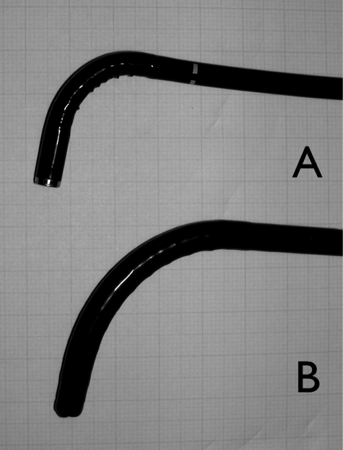
An ENT (ear nose throat) video endoscope (Pentax® VNL-1590 STi; Hoya Co., Ltd.; Tokyo, Japan) was used for this study. Its diameter was relatively large; the tip and middle diameters are φ 5.6 and 5.1, respectively. However, its radius of curvature was nearly the same as that of a general ENT fiberscope (Figure 1a and b). Both the diameter and radius of curvature were much smaller than those of gastric video endoscopes. The size of the CCD was nearly equivalent to that of the latest gastric video endoscopes. The focal length was slightly closer to the lens—the minimum distance was 3 mm-thus allowing for precise examination of the mucosa. Image resolution was estimated by the United States Air Force (USAF) resolution test chart.
The development of this endoscopic system began from 2007. We started using this video endoscope in our office clinic in August 2009 after minor modifications and adjustments were made to it. Final specifications were decided in late 2009. Between January and December 2010, we observed the larynx and hypopharynx using this scope. Prior to examination, all patients underwent application of nasal spray with adrenalin and lidocaine. All examinations were recorded as digital video throughout the procedure. The study protocol complied with the Declaration of Helsinki and the Institutional Review Board (Tokyo Medical and Dental University No 401). No financial supports were given to this study.
Results
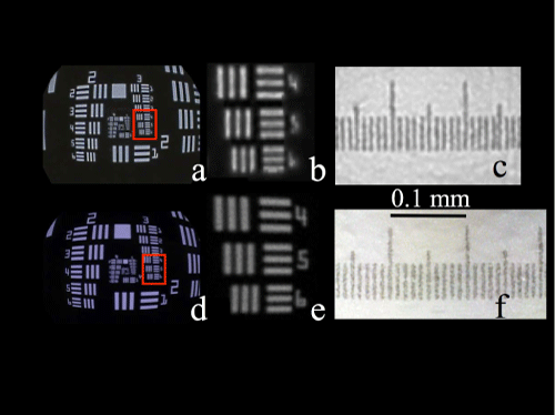
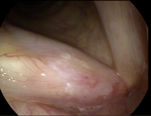
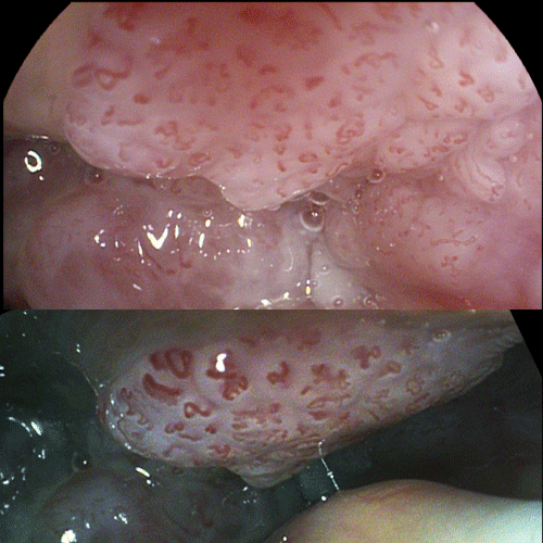
This endoscope could distinguish at least 32 line pairs/mm on a USAF chart; hence, it could distinguish a line of at least 16 μm in width (Figure 2). Between January and December 2010, transnasal observation of the larynx and hypopharynx using this scope were performed 356 times during this period. Due to severe nasal deviation, one patient required the application of gauze soaked with adrenalin and lidocaine to the nasal cavity. Other patients safely underwent examination after nasal pretreatment, that is, no major side effects such as epistaxis were noted. Image quality was much better than that of general ENT video endoscopes (Figures 3 and 4). This video endoscope has an image modification function (i-Scan®), and all lesions, including vascular structures, were more clearly observed using this function (Figure 4). Abnormal IPCLs that appeared on the surface of carcinomas were directly and easily observed with the VNL-1590 STi.
Figure 2: Estimation of resolution by USAF chart. Various sized white bars are spaced in a line at regular intervals equivalent to the width of a bar. The chart is consisted from 6 “Groups” and each group has 6 “Elements” which have thee vertical and horizontal bars. Size of bars gradually shrinks from Group 2, Element 2 to Group 5 Element 6 in this figure. Figure 2a shows an image obtained using a general ENT video endoscope, Figure 2c shows and image obtained using theVNL-1590 STi, Figure 2b and 2d show observation inside the frame: Group 3, Element 4 to 6. Compared to the conventional video endoscope, the VNL-1590 STi more clearly depicts these bars. Figure 2e (dotted frame) and 2f (dotted oval frame) are images of Group 4 and 5, respectively, on the USAF chart. The VNL-1590 STi can distinguish between 32 (line width = 16 μm) and 36 (line width = 14 μm) vertical line within 1 mm (Group 5, Element 1 and 2) and horizontal line pars of 22.62 (22 μm; Group 4, Element 4).
Discussion
Several diagnostic techniques related to vascular abnormalities seen in cases of carcinoma have been reported. In particular, abnormal IPCLs have been reported as an early finding in esophageal and pharyngeal carcinomas [1-4]. Magnifying endoscopy coupled with NBI has been reported to be useful for detecting IPCLs [3,4]. With the use of these superior video endoscopes, hypopharyngeal as well as mesopharyngeal carcinomas were detected during diagnostic examinations for esophageal and gastric diseases by gastroenterologists and surgeons [7]. ENT video endoscopes with NBI were also reported to be useful in the detection of such vascular abnormalities. These abnormalities were usually detected with NBI as demarcated brownish areas or scattered brown spots [5].
In the ENT clinic, transnasal examination of patients in a sitting position has several advantages compared to transoral examination with patients in the lateral position [8,9]. Examination in a sitting position is more natural and comfortable for patients. It also enables examination while the patient is performing various tasks or under certain conditions. For example, examinations during swallowing, the Valsalva maneuver, head torsion, and neck flexion enable clear visualization of the fossa of Rosenmuller, the pyriform sinus, and the posterior wall of the hypopharynx, subglottis, and glottic ventricle [10]. Therefore, these techniques are helpful for superior endoscopic examinations. The radius of curvature of the VNL-1590 STi is nearly the same as that of a general ENT video endoscope; therefore, the clinician can easily observe anatomical structures under various conditions that facilitate examination. Nevertheless, the VNL-1590 has higher resolution and can distinguish features on a scale of less than 20 μm. According to the reports of the size of abnormal vasculature in early esophageal carcinoma, the average caliber of an IPCL was 12.9 ± 3.9 μm in m1, 14.5 ± 3.9 μm in m2, and 18.1 ± 5.2 μm in m3 cancer [2]. For cancers that invade the submucosa, the average caliber of the tumor vessels was 26.1 ± 11.6 μm. The resolution of the VNL-1590 STi was nearly equivalent to these abnormal vessels themselves, and these vessels could be directly observed, rather than merely as brownish areas or scattered brown spots.
Some techniques and devices, including the use of dyes (such as iodine) and NBI, have facilitated detection of carcinoma at an early stage [3-7]. However, these procedures or devices require drug administration or a specialized light source. The VNL-1590 STi has an image modification function called i-Scan® that facilitates detection of not only the blood vessels but also other structures such as cartilage and ligaments [11-16]. Image modification is different from NBI and is done instantaneously, and modified images appear natural (Figure 4). However, the image resolution of the VNL-1590 STi is high enough to detect subtle changes on the mucosal surface and very tiny keratotic changes or abnormal IPCLs can be detected with this video endoscope with conventional examination procedures.
Conclusion
Despite its relatively large size, the VNL-1590 STi is easy to handle and can be used for daily clinical examinations. The image quality is excellent and nearly equivalent to that of the latest gastric video endoscopes. It is valuable for pharyngeal and laryngeal examinations in ENT clinics.
References
- René Lambert (2013) Role of Endoscopy in Screening and Treatment of Gastrointestinal Cancer. J Gastrointest Dig Syst S2: 006.
- Goda K, Dobashi A, Yoshimura N, Chiba M, Fukuda A, et al. (2013) Clinicopathological features of narrow-band imaging endoscopy and immunohistochemistry in ultraminute esophageal squamous neoplasms. Dis Esophagus.
- Goda K, Tajiri H, Ikegami M, Yoshida Y, Yoshimura N, et al. (2009) Magnifying endoscopy with narrow band imaging for predicting the invasion depth of superficial esophageal squamous cell carcinoma. Dis Esophagus 22: 453-460.
- Watanabe A, Tsujie H, Taniguchi M, Hosokawa M, Fujita M, et al. (2006) Laryngoscopic detection of pharyngeal carcinoma in situ with narrowband imaging. Laryngoscope 116: 650-654.
- Katada C, Nakayama M, Tanabe S, Koizumi W, Masaki T, et al. (2008) Narrow band imaging for detecting metachronous superficial oropharyngeal and hypopharyngeal squamous cell carcinomas after chemoradiotherapy for head and neck cancers. Laryngoscope 118:1787-1790.
- Nonaka S, Saito Y (2008) Endoscopic diagnosis of pharyngeal carcinoma by NBI. Endoscopy 40: 347-351.
- Katada C, Tanabe S, Koizumi W, Higuchi K, Sasaki T, et al. (2010) Narrow band imaging for detecting superficial squamous cell carcinoma of the head and neck in patients with esophageal squamous cell carcinoma. Endoscopy 42: 185-190.
- Sato K, Nakashima T (2002) Office-based videoendoscopy for the hypopharynx and cervical esophagus. Am J Otolaryngol 23: 341-344.
- Tsunoda A, Ishihara A, Kishimoto S, Tsunoda R, Tsunoda K (2007) Head torsion technique for detailed observation of larynx and hypopharynx. J LaryngolOtol 121: 489-490.
- Hoffman A, Basting N, Goetz M, Tresch A, Mudter J, et al. (2009) High-definition endoscopy with i-Scan and Lugol's solution for more precise detection of mucosal breaks in patients with reflux symptoms. Endoscopy 41: 107-112.
- Atkinson M, Chak A (2010) I-Scan: chromoendoscopy without the hassle? Dig Liver Dis 42: 18-19.
- Hoffman A, Kagel C, Goetz M, Tresch A, Mudter J, et al. (2010) Recognition and characterization of small colonic neoplasia with high-definition colonoscopy using i-Scan is as precise as chromoendoscopy. Dig Liver Dis 42: 45-50.
- Hoffman A, Kiesslich R, Goetz M, Tresch A, Mudter J, et al. (2010) High definition colonoscopy combined with i-Scan is superior in the detection of colorectal neoplasias compared with standard video colonoscopy: a prospective randomized controlled trial. Endoscopy 42: 827-833.
- Dekker E, East JE (2010) Does advanced endoscopic imaging increase the efficacy of surveillance colonoscopy? Endoscopy 42: 866-869.
- Kodashima S, Fujishiro M (2010) Novel image-enhanced endoscopy with i-scan technology. World J Gastroenterol 16: 1043-1049.
- Lee CK, Lee SH, Hwangbo Y (2011) Narrow-band imaging versus I-Scan for the real-time histological prediction of diminutive colonic polyps: a prospective c omparative study by using the simple unified endoscopic classification. Endoscopy 74-3: 603-609.
Citation: Tsunoda A, Tsunoda K, Sumi T, Kishimoto S, Kitamura K (2014) High-Resolution ENT Video Endoscope with Superior Image Quality Equivalent to that of Gastric Video Endoscopes. Otolaryngology 4:166. DOI: 10.4172/2161-119X.1000166
Copyright: © 2014 Tsunoda A, et al. This is an open-access article distributed under the terms of the Creative Commons Attribution License, which permits unrestricted use, distribution, and reproduction in any medium, provided the original author and source are credited.


