Make the best use of Scientific Research and information from our 700+ peer reviewed, Open Access Journals that operates with the help of 50,000+ Editorial Board Members and esteemed reviewers and 1000+ Scientific associations in Medical, Clinical, Pharmaceutical, Engineering, Technology and Management Fields.
Meet Inspiring Speakers and Experts at our 3000+ Global Conferenceseries Events with over 600+ Conferences, 1200+ Symposiums and 1200+ Workshops on Medical, Pharma, Engineering, Science, Technology and Business
Editorial Open Access
High-Resolution MR Neurography: Application in Peripheral Nerve Disease
| Theodoros Soldatos* | |
| Department of Research and Medical Imaging, National and Capodestrian University of Athens, Athens, Greece | |
| Corresponding Author : | Theodoros Soldatos Department of Research and Medical Imaging National and Capodestrian University of Athens Athens, Greece E-mail: tsoldat1@jhmi.edu |
| Received January 28, 2012; Accepted February 01, 2012; Published February 06, 2012 | |
| Citation: Soldatos T (2012) High-Resolution MR Neurography: Application in Peripheral Nerve Disease. Radiol Open Access. 1:e102. doi: 10.4172/2167-7964.1000e102 | |
| Copyright: © 2012 Soldatos T. This is an open-access article distributed under the terms of the Creative Commons Attribution License, which permits unrestricted use, distribution, and reproduction in any medium, provided the original author and source are credited. | |
Visit for more related articles at Journal of Radiology
Abstract
| Editorial |
| Although the evaluation of peripheral neuropathies has traditionally relied on clinical examination and electrodiagnostic studies, there has been an increasing demand from treating physicians for information about the injury type, location of injured nerve stumps, presence or absence of neuroma and underlying etiology of neuropathy, particularly in cases where surgical intervention is contemplated. In this setting, magnetic resonance (MR) imaging of peripheral nerves, also referred to as MR Neurography, has been gaining increasing popularity, because of advances in MR hardware and the development of new imaging techniques. The implementation of 3 Tesla MR Scanners, in particular, has offered higher resolution and contrast imaging of peripheral nerves, which has in turn provided exquisite anatomic and lesion conspicuity [1]. |
| In current clinical practice, MR Neurography may be applied to: a) confirm clinical suspicion of peripheral neuropathy, by directly showing the nerve abnormality or regional muscle denervation changes, b) assess the extent of the abnormality in nerve injuries, or the disease load in diffuse peripheral nerve lesions, such as hereditary neuropathies and neurofibromatosis, c) depict lesions causing nerve entrapment or impingement [2,3], d) exclude peripheral neuropathy by showing normal nerves and regional muscles, e) detect incidental lesions in the region of interest that mimic neuropathy symptoms [4] and f) provide imaging guidance for perineural medication injections. |
| Typically, MR Neurography techniques utilize a combination of fat-saturated T2-weighted (T2W), short inversion time recovery (STIR), or T2 spectral adiabatic inversion recovery turbo spin-echo (T2 SPAIR TSE) images for the detection of the nerve signal, contour and size changes, as well as T1-weighted (T1W) spin-echo or fluidattenuated long inversion recovery (FLAIR) images for the anatomic assessment of the involved areas, based on the abundant intra- and perineural fat. In cases of suspected tumor or infection, pre- and postcontrast fat-suppressed T1W images are additionally acquired. |
| Axial T1W and fat-suppressed T2W images serve as the mainstay in MRN interpretation for prudent assessment of peripheral nerve imaging characteristics, such as signal intensity, course, caliber, fascicular pattern, size, and perineural fibrosis or mass lesions [5]. Normal peripheral nerves demonstrate intermediate signal intensity, similar to skeletal muscle on T1W images and intermediate to minimally increase on T2W images, and exhibit smooth course without focal deviations, uniformly sized fascicles, which are also isointense to skeletal muscles, clean surrounding perineural fat planes, and caliber similar to the adjacent arteries, which gradually decrease proximally to distally. In cases of neuropathy, the signal intensity of the nerve increases abnormally, approaching the fluid-like signal intensity of the adjacent vessels on T2W images. Additionally, the abnormal nerve may demonstrate a) focal or diffuse enlargement, featuring size greater than the adjacent artery, b) enlargement or disruption of single or multiple fascicles, c) focal or diffuse deviations or discontinuity, d) enhancement, which is present in tumors and infections, e) enhancement of perineural fat planes. Apart from the aforementioned direct nerve-related features, secondary skeletal muscle signal intensity changes can indicate or further confirm peripheral neuropathy. Skeletal muscles may show acute denervation changes (edema-like signal) as early as 24 hours from the onset of neuropathy, sub-acute changes (edema-like SI and minimal fatty replacement) weeks to months after injury, as well as chronic changes (fatty replacement and atrophy), months to years after nerve injury. When direct nerve-related findings are subtle or absent, secondary muscle signal intensity changes could be considered highly indicative of neuropathy when they demonstrate regional nerve distribution, they are diffuse rather than focal, and there is no fascial and subcutaneous edema to suggest infectious or traumatic myopathy. |
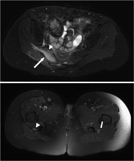
| In our institution, MR Neurography has been established as anintegral component of the diagnostic algorithm in both neurology and nerve injury patients [6] (Figure 1). We expect that the method will gain even more clinical utility in the following years, as clinicians discover its usefulness and MR hardware and imaging techniques evolve. |
References |
|
Figures at a glance
 |
| Figure 1 |
Post your comment
Relevant Topics
- Abdominal Radiology
- AI in Radiology
- Breast Imaging
- Cardiovascular Radiology
- Chest Radiology
- Clinical Radiology
- CT Imaging
- Diagnostic Radiology
- Emergency Radiology
- Fluoroscopy Radiology
- General Radiology
- Genitourinary Radiology
- Interventional Radiology Techniques
- Mammography
- Minimal Invasive surgery
- Musculoskeletal Radiology
- Neuroradiology
- Neuroradiology Advances
- Oral and Maxillofacial Radiology
- Radiography
- Radiology Imaging
- Surgical Radiology
- Tele Radiology
- Therapeutic Radiology
Recommended Journals
Article Tools
Article Usage
- Total views: 14255
- [From(publication date):
March-2012 - Mar 08, 2025] - Breakdown by view type
- HTML page views : 9832
- PDF downloads : 4423
