Herlyn-Werner-Wunderlich Syndrome on Imaging: Case Report
Received: 04-Sep-2023 / Manuscript No. roa-23-113209 / Editor assigned: 06-Sep-2023 / PreQC No. roa-23-113209 / Reviewed: 18-Sep-2023 / QC No. roa-23-113209 / Revised: 25-Sep-2023 / Manuscript No. roa-23-113209 / Published Date: 30-Sep-2023 DOI: 10.4172/2167-7964.1000486
Abstract
Herlyn-Werner-Wunderlich Syndrome is a rare congenital anomaly affecting the urogenital tract, specifically involving the Mullerian ducts and mesophrenic ducts. It is characterized by a triad of uterine didelphys with a blocked hemivagina and the absence of a kidney on the same side.
Symptoms typically manifest following menarche, presenting as pelvic pain, dysmenorrhea, and the development of a pelvic mass. In rare cases, it may lead to primary infertility in later years.
Diagnosis of this condition is typically accomplished through ultrasound and MRI imaging techniques.
In this case report, we present the clinical scenario of a 13-year-old patient who had experienced her third menstrual cycle and presented with pelvic pain lasting for a week.
Keywords
Didelphys uterus; Hemivagina; Hematocolpos; Imaging
Introduction
Herlyn-Werner-Wunderlich Syndrome (HWWS), also known as OHVIRA, is a rare congenital malformation syndrome characterized by a triad of uterine didelphys with an obstructed hemivagina and missing kidney on the same side. This condition results from an uncommon combination of müllerian duct anomalies (MDAs) and mesonephric duct malformation within the female urogenital tract [1].
While the exact prevalence of this syndrome is unknown, it is estimated to affect between 0.1% to 3.8% of the population. The syndrome was initially described by Herlyn and Werner in 1971, and in 1976, Wunderlich further characterized the association of right renal aplasia with a bicornuate uterus and a simple vagina with isolated accumulation of menstrual blood in the vagina [2].
Patients with HWWS typically remain asymptomatic until puberty and often develop symptoms shortly after their first menstruation. These symptoms commonly include pelvic pain, painful menstruation, or the presence of a palpable vaginal or pelvic mass.
In this case report, we present a case of uterus didelphys with an obstructed hemivagina diagnosed at the age of 14 years. Through this case, we will discuss the clinical, diagnostic, and therapeutic aspects of this uterine malformation [3].
Case Report
A 13-year-old unmarried girl with no significant medical or surgical history presented with acute pain in her lower right abdomen, which had been persistent for the past 7 days. She had her first period at the age of 13, and her menstrual cycles were irregular.
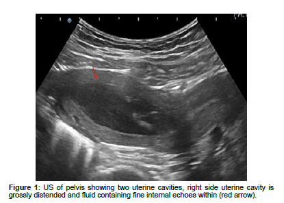
The patient did not report any fever, abnormal vaginal bleeding, nausea, vomiting, or diarrhea. During the physical examination, mild abdominal distension was observed along with tenderness in the lower right pelvic area. An abdominal ultrasound revealed the absence of the right kidney and a bicornuate uterus [4]. The right uterine horn appeared dilated and contained a cystic mass, with internal echoes indicating a hematoma. The left uterine horn and cervical canal appeared normal. Both ovaries were also found to be normal (Figure 1).
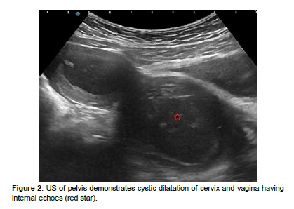
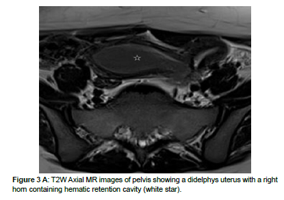
Following these findings, the medical staff recommended an MRI for further evaluation. The MRI images revealed a didelphys uterus with the left uterine horn pushed backward by a distended right uterine horn. Additionally, the right hemi-vagina was obstructed and contained a cavity filled with menstrual blood (Figure 2). On axial T2 views, it was evident that the right kidney was absent, compensated by hypertrophy of the left kidney [5].
Based on these investigations, the patient received a diagnosis of right renal agenesis, uterus didelphys with an obstructed right hemivagina, and hematometrocolpos.
Treatment commenced with hymenotomy, followed by the excision of the vaginal septum and drainage of a substantial amount of accumulated blood. Subsequently, reconstruction of the vaginal canal was performed, and a catheter was placed over the surgical resection site to prevent stenosis and facilitate drainage.
The primary goals of this treatment were to alleviate the patient’s pain and prevent potential complications, such as endometriosis and infertility (Figure 3A).
Discussion
Herlyn-Werner-Wunderlich Syndrome (HWWS) is a rare congenital malformation affecting the urogenital tract, specifically involving the Müllerian and mesophrenic ducts [6]. Overall, congenital anomalies of the Müllerian tract are estimated to affect approximately 2 to 3% of women [7].
The precise pathogenesis of the HWWS pattern is not yet completely understood, but it is believed to have multiple contributing factors [8]. It may result from factors such as non-development (agenesia or hypoplasia), defective vertical or lateral fusion, or resorption failure of the Müllerian ducts [6]. HWWS is characterized by the presence of uterine didelphys, obstructed hemivagina, and ipsilateral renal agenesis (Figure 3B) [6].
However, HWWS syndrome has been associated with various other malformations, including bicornuate and septate uterus, unilateral cervical obstruction, and incomplete vaginal septum [14]. Additionally, urinary tract anomalies may also occur, such as dysplastic kidney, ureteral anomalies like blind ectopic ureter with vaginal or cervical insertion, and paravaginal cervical cysts [14].
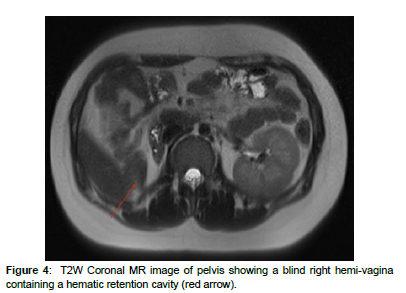
Uterus didelphys is classified as Class 3 according to the American Fertility Society classification [10]. According to the European Society of Human Reproduction and Embryology (ESHRE) classification, our patient can be categorized as the main class U3 (bicorporeal uterus), sub-class U3b (complete), with concurrent classes C2 (double ‘normal’ cervix) and V2 (longitudinal obstructing vaginal septum) (Figure 4) [11].
Patients with HWWS syndrome are typically asymptomatic until they reach puberty, at which point they may experience symptoms such as pelvic pain, dysmenorrhea, or the presence of a palpable mass in the vagina or pelvis [6]. The palpable mass often results from hematometrocolpos, which occurs due to the obstructed hemivagina.
Ultrasound is the initial imaging modality used to evaluate suspected Müllerian duct anomalies. It can aid in establishing the diagnosis by revealing a didelphys uterus and the presence of blood retention in the hemivagina and/or the uterine cavity [8, 12]. MRI is an excellent imaging modality for assessing complex Müllerian duct anomalies and associated pelvic findings [6]. It typically reveals complete duplication of the uterine and endocervical cavities, along with the presence of a hematoma collection connected to one of the endocervical canals due to the obstructed hemivagina [13]. Laparoscopy is considered the gold standard for diagnosing female genital tract abnormalities, but it is usually reserved for cases where MRI is unavailable or fails to provide a diagnosis [14].
Late diagnosis of this syndrome can lead to various complications, including endometriosis, pelvic adhesions, and infertility, all of which are related to chronic obstructed hematometrocolpos (Figure 5). Therefore, early diagnosis and treatment are essential to prevent these complications [9]. The treatment of choice is vaginal septum excision, typically performed using laparoscopic or endoscopic techniques, as it ensures adequate drainage for hematometrocolpos [9].
Conclusion
Herlyn-Werner-Wunderlich Syndrome (HWW), a rare congenital urogenital anomaly, presents with pelvic pain and dysmenorrhea from the first menarche.
Due to its rarity, diagnosing HWW syndrome can be challenging. It should be considered as one of the potential differential diagnoses in young patients experiencing pelvic symptoms, particularly when there is an associated renal abnormality or agenesis.
The diagnosis is typically confirmed through a combination of pelvic ultrasound and magnetic resonance imaging (MRI), which remains the preferred diagnostic approach.
The primary treatment for HWW syndrome is surgical, involving the complete removal of the vaginal septum to enable continuous drainage of menstrual retention.
References
- Aveiro AC, Miranda V, CabralAJ, Nunes S, Paulo F, et al. (2011) Herlyn-Werner-Wunderlichsyndrome: A rare cause of pelvic pain in adolescent girls. BMJ Case Rep 2011:bcr0420114147.
- Bhoil R, Ahluwalia A, Chauhan N (2016) Herlyn Werner WunderlichSyndrome with Hematocolpos: An Unusual Case Report of Full DiagnosticApproach and Treatment. Int J Fertil Steril 10 :136-140.
- Burgis J (2001) Obstructive Mülleriananomalies: case report, diagnosis, and management. Am J Obstet Gynecol 185: 338-344.
- Khaladkar SM, Kamal V, Kamal A, Kondapavuluri SK (2016) The Herlyn-Werner-WunderlichSyndrome -A Case Report with Radiological Review. Pol J Radiol 24: 395-400.
- Wunderlich M (1976) Unusual form of genitalmalformation with aplasia of the right kidney. Zentralblatt fur Gynakologie98: 559-562.
- Del Vescovo R, Battisti S, Di Paola VL, Piccolo CL,Cazzato RL,et al. (2012) Herlyn-Werner-Wunderlichsyndrome: MRI findings, radiological guide (two cases and literaturereview), and differential diagnosis. BMC Med Imaging 12: 1-10.
- Shavell VI, Montgomery SE, Johnson SC (2009) complete septate uterus,obstructed hemivagina, and ipsilateral renal anomaly: pregnancy coursecomplicated by a rare urogenital anomaly. Arch Gynecol Obstet 280: 449-452.
- Han B, Herndon CN, Rosen MP, Wang ZJ, Daldrup-Link H(2010) Uterine didelphys associatedwith obstructed hemivagina and ipsilateral renal anomaly (OHVIRA)syndrome. Radiol Case Rep 5: 327.
- Negrão E, Flor-de-Lima B, Dias SC, Guimarães L,Madureira AJ (2022) Herlyn-Werner-Wunderlichsyndrome: A case report in a young woman, with literature review. Radiol Case Rep 17: 1991-1995.
- Ghasemi M, Esmailzadeh A (2019) An unusual appearance of thepost-pubertal Herlyn-Werner-Wunderlich syndrome with acute abdominal pain:A case report. Int J Reprod Biomed 17: 851.
- Grimbizis GF, Gordts S, Di Spiezio Sardo A (2013) The ESHRE/ESGE consensus on theclassification of female genital tract congenital anomalies. Hum Reprod 28: 2032-2044.
- Kozłowski M, Nowak K, Boboryko D, Kwiatkowski S,Cymbaluk-Płoska A (2020) Herlyn-Werner-Wunderlich syndrome: Comparison of twocases. Int J Environ Res Public Health 17: 7173.
- Salastekar N, Coelho M, Majmudar A, Gupta S (2019) Herlyn-Werner-Wunderlichsyndrome: A rare cause of abdominal pain and dyspareunia. Radiol Case Rep 14: 1297-1300.
- Park NH, Park HJ, Park CS, Park SI (2008) Herlyn-Werner-WunderlichSyndrome with unilateral hemivaginal obstruction, ipsilateral renalagenesis, and contralateral renal thin GBM disease: a case report withradiological follow-up. J Korean Soc Radiol 62: 105-109
Indexed at, Google Scholar, Crossref
Indexed at, Google Scholar, Crossref
Indexed at, Google Scholar, Crossref
Indexed at, Google Scholar, Crossref
Indexed at, Google Scholar, Crossref
Indexed at, Google Scholar, Crossref
Indexed at, Google Scholar, Crossref
Indexed at, Google Scholar, Crossref
Indexed at, Google Scholar, Crossref
Indexed at, Google Scholar, Crossref
Indexed at, Google Scholar, Crossref
Indexed at, Google Scholar, Crossref
Citation: Rostoum S, Houssaini AS, Zhim M, Diallo ID, Chorfa SH, et al. (2023) Herlyn-Werner-Wunderlich Syndrome on Imaging: Case Report. OMICS J Radiol 12: 486. DOI: 10.4172/2167-7964.1000486
Copyright: © 2023 Rostoum S, et al. This is an open-access article distributed under the terms of the Creative Commons Attribution License, which permits unrestricted use, distribution, and reproduction in any medium, provided the original author and source are credited.
Select your language of interest to view the total content in your interested language
Share This Article
Open Access Journals
Article Tools
Article Usage
- Total views: 1845
- [From(publication date): 0-2023 - Oct 19, 2025]
- Breakdown by view type
- HTML page views: 1531
- PDF downloads: 314


