Guben Xiezhuo Recipe Alleviated Colon Dialysis on Urotoxin-Induced Intestinal Barrier Injury and Systemic Inflammation
Received: 11-Oct-2021 / Manuscript No. CMB-21-44357 / Editor assigned: 13-Oct-2021 / PreQC No. CMB-21-44357 (PQ) / Reviewed: 08-Jan-2022 / QC No. CMB-21-44357 / Revised: 13-Jan-2022 / Manuscript No. CMB-21-44357 (R) / Accepted Date: 13-Jan-2022 / Published Date: 20-Jan-2022
Abstract
Background: Chronic kidney disease (CKD) is a major disease that endangers public health around the world. The urinary toxins produced in the disease can induce intestinal barrier damage and systemic inflammation, which further aggravates the disease and creates a vicious circle. Therefore, this work carried out cell-level research to determine the ability of Guben Xiezhuo Recipe for colon dialysis to repair the intestinal barrier and to interfere with the entire process.
Methods: T84 cells were divided into six groups, one of which was a control group, and the experimental components were divided into five groups according to the level of urinary toxin content and the different drugs added. Perform a series of tests on six groups. Immunofluorescence technique was used to analyze the expression characteristics of intestinal epithelial tight junction protein Occludin in intestinal epithelial cells in the intestinal barrier injury model. Western Blot technology was used to analyze the expression of ZO-1 and Cluadin-1 in intestinal epithelial cells in the intestinal barrier injury model. RT-PCR technology was used to analyze the expression changes of Keap1, Nrf2, ROS, and NF-κB mRNA in intestinal epithelial cells. ELISA was used to analyze the content of inflammatory signal hypersensitive C-reactive protein and interleukin-6 (IL-6) in cell culture fluid.
Results: The results of immunofluorescence analysis and Western Blot analysis demonstrated the ability of urinary toxins produced by CKD to destroy the intestinal barrier. The results of RT-PCR analysis explained the mechanism of urinary toxins destroying the intestinal barrier from the molecular biology level. The results of ELISA analysis indicate that the intestinal barrier damage will further induce systemic inflammation. Through the comparison of the above analysis results in the six groups, it can be shown that Guben Xiezhuo Colon Dialysis Recipe has an excellent therapeutic effect on intestinal epithelial injury.
Conclusion: Through the detection and comparison of the contents of intestinal barrier-related proteins and inflammatory signal factors in each group, it is proved that the Guben Xiezhuo Recipe colon dialysis has an excellent therapeutic effect on the repair of intestinal barrier damage and further inhibits the deterioration of CKD, which also provides an optional auxiliary strategy
Keywords
Chronic kidney disease; Micro ecosystem; Probiotics; Centrifuge tube
Introduction
The incidence of chronic kidney disease (CKD) has been increasing year by year, and it has become a serious disease that threatens the health of the public health throughout the world [1]. End stage renal disease (ESRD) is the final stage of the development of various chronic kidney diseases, during which the multi-system functions of the body will undergo undesirable changes. Although with the development of medical technology, the survival time and quality of life of ESRD patients have been greatly improved, there is still no specific method to delay the progression of renal failure before renal replacement therapy, especially for patients with CKD [2].
In patients with CKD chronic kidney disease, the intestinal micro ecosystem will be disturbed during the progression of the disease, resulting in imbalance of the intestinal flora, including a decrease in probiotics and an increase in pathogenic bacteria that can produce urinary toxins [3]. The result is aggravation of the intestines urinederived toxins accumulate in the blood and cannot be cleared in time by the damaged kidneys, and renal function further declines, eventually forming a vicious circle between the intestines and kidneys. Disordered intestinal flora can also destroy the intestinal epithelial barrier function, causing intestinal urinary toxins and conditional pathogenic bacteria to translocate and enter the blood circulation, activate the intestinal mucosal immune system, induce systemic micro-inflammatory reactions, and aggravate the kidneys damage [4].
Nuclear transcription factor E2-related factor (Nrf2) belongs to the Cap-n-colar (CNC) leucine zipper transcription activator family and is the main regulator of many genes encoding antioxidants, antidote and cytoprotective molecules [5]. Nrf2 induces a series of antioxidant protective protein codes, promotes the regulation of the redox environment in cells, and becomes an important pathway for cells to resist oxidative stress. Nrf2 stimulants such as lubusulfurin and watercress have been used as Chinese herbal medicine for the treatment of inflammatory and tumor diseases for hundreds of years. Research in recent years has shown that activation of the Nrf2 signaling pathway can reduce organ ischemia and repeated infusion injuries. CDDO- 9,11-dihydrotrifluoroamide (Dh404) is a synthetic triterpenoid. Studies have shown that, dh404 produces anti-inflammatory and anti oxidative stress effects by stimulating the Nrf2 signaling pathway. It has a protective effect on chronic kidney disease in rats [6].
Pyrrolidine dithiocarbamate(PDTC), is a specific inhibitor of NF- κB, which can inhibit the activation of NF-κB and reduce inflammation. It is also an antioxidant, which can directly eliminate free radicals and inhibit the generation of free radicals [7].
Claudin is mainly located on the side of epithelial cells near the lumen. Its function is to close the intercellular space and prevent the free entry and exit of materials inside and outside the epithelial layer of the lumen. It is the material basis for the selection of permeabilization of epithelial cells [8]. Tight junctions are a composite structure formed under the interaction of multiple proteins, which are mainly composed of two components, which are transmembrane protein and cytoplasmic protein. The main transmembrane proteins are occludin and claudin-1. Both occludin and claudin-1 are four-pass transmembrane proteins, and the amino and carboxyl ends are located in the cytoplasm [9]. Tight junctions can effectively prevent the paracellular transport of bacteria, toxins and other substances in the intestinal cavity through regulation under physiological conditions, and maintain the integrity of the intestinal mucosal epithelial barrier function.
Zonulaocludens-1(ZO-1) is a protein that is related to tightly connect. In recent years, it has been discovered that it is related to the maintenance and regulation of epithelial fence and barrier function, and also participates in the regulation of cell material transport, maintenance of epithelial polarity, cell proliferation and differentiation, tumor cell metastasis and other important processes [10]. Since the structure and function of ZO-1 are closely related to other members of tight junctions, in most cases, as long as ZO-1 is damaged, the functions of tight junctions will change accordingly, so ZO-1 is often used to observe the tight junctions of various tissues. Indicators of barrier function and permeability function, such as blood-brain barrier, intestinal epithelial cells, retinal pigment epithelial cells, etc [11].
Nrf2 is a key factor in the cellular oxidative stress response. It is regulated by kelch-like ECH-associated protein 1(Keap-1) and interacts with the antioxidant response element to regulate antioxidant proteins and phase II detoxification enzymes expression [12].
In this work, immunofluorescence was used to analyze the expression characteristics of intestinal epithelial tight junction protein occludin in the intestinal barrier injury model. This work uses Western Blot technology to analyze the expression of proteins ZO-1 and Cluadin-1 in intestinal epithelial cells of the intestinal barrier injury model, and analyzes the mRNA of Keap-1, Nrf2, ROS, and NF-κB in intestinal epithelial cells by RT-PCR technology. The above test is used to evaluate the destruction and repair effect of the intestinal epithelial barrier function of CKD patients. In this work, Elisa was used to analyze the inflammatory signal hypersensitive C-reactive protein and interleukin-6 (IL-6) content in the cell culture medium to evaluate the inflammatory response caused by CKD and the reduction of inflammatory response by colon dialysis with Guben Dialysis effect.
Materials and Methods
Experiment design
Cultivate T84 cells at 37°C and 5% CO2 with DMEM/F12 medium containing 10% fetal calf serum (FCS) and 50 U/L penicillinstreptomycin in cell culture flasks, and replace the medium every 1-2 days. Then induce and establish an intestinal epithelial injury model with low and high doses of urea. We divided the cells into six groups in the experiment. Only DMEM/F12 and 10% FCS were added to the control group. Compared with the control group, the low-dose urea model group (low-urea group) added an extra 42 mg/dl of urea, while the high-dose urea model group (high-urea group) increased the added urea concentration to 72 mg/dl. On the basis of the highurea group, Nrf2 agonist dh404, NF-κB receptor blocker PDTC and FSKL medicated serum were added to the Nrf2 agonist group (Nrf2 activator group), NF-κB receptor blocker group (NF-κB inhibitor group) and Guben Xiezhuo Colon Dialysis Recipe-contained serum group (TCM+FSKL group).
Immunofluorescence technique was used to analyze the expression characteristics of intestinal epithelial tight junction protein Occludin in intestinal epithelial cells in the intestinal barrier injury model. Western Blot technology was used to analyze the expression of ZO-1 and Cluadin-1 in intestinal epithelial cells in the intestinal barrier injury model. RT-PCR technology was used to analyze the expression changes of Keap-1, Nrf2, ROS, and NF-κB mRNA in intestinal epithelial cells. ELISA was used to analyze the content of inflammatory signal hypersensitive C-reactive protein and interleukin-6 (IL-6) in cell culture fluid.
Immunofluorescence analysis
In each group of cells, 1% BSA was applied for blocking at room temperature for 30 min to block non-specific epitopes. Incubate the specific primary antibody according to the recommended instructions for the antibody and let it stand overnight in a humidified box at 4. Take out the slices the next day and rewarm at room temperature for 30 min. Select the corresponding immunofluorescence secondary antibody and then incubate at 37℃ for 30 min in the dark. Under dark conditions, stain the nucleus with DAPI. Add anti-fluorescence quencher for mounting. Finally, use a fluorescence microscope to observe and take pictures.
Western blot analysis
Collect cells from each group, and add 200 μl of cell lysate to each six-well plate. After sonication, the cells were lysed on ice for 1 hour. The lysed cell sample was centrifuged at 12,500 rpm for 15 minutes at 4. Then, transfer the supernatant in the centrifuge tube to a clean centrifuge tube. GAPDH protein quantification kit was used to quantify protein concentration. The measured protein samples were stored at -80. In Western blot electrophoresis, the protein loading concentration was 50 μg per well. After SDS-PAGE electrophoresis, the membrane was transferred and blocked. ZO-1 and Cluadin-1 (1: 500, anti-human, Abcam, USA) primary antibody were diluted to use concentration. The samples were incubated overnight on a shaker at 4°C. After washing with PBS, the samples were incubated with the secondary antibody (1: 1000, anti-human, Abcam, USA) for 30 minutes at room temperature in the dark. Finally, the developer was used for development and photography.
RT-PCR analysis
The intestinal epithelial cells were treated with TRIzol reagent to extract the total RNA in the cells. Revert Aid TW first Strand cDNA Synthesis Kit was used to synthesize the first chain of DNA. QuantiNova SyBr Green PCR Kit was used to perform PCR analysis. Reaction conditions: pre-denaturation at 95 for 1 minute, denaturation at 95°C for 30 seconds, annealing at 60 for 30 seconds, and elongation at 72 for 30 seconds, running 40 cycles.
ELISA analysis
Five standard wells were set on the ELISA-coated plate. The standard samples were added to the wells according to the concentration requirements and serially diluted. The sample volume in each well was 50μl. Then a blank control well and the sample well to be tested were set. The blank control well would not add sample and enzyme-labeled reagent; the other steps were the same with the sample well. Add 40μl of sample diluent to the sample well, and then add 10μl of the sample. The final dilution of the sample is 5 times. Seal the plate with a sealing film and incubate at 37 for 30 minutes. After incubation, wash the plate with washing solution and dry it. After drying, add 50μl of enzyme-labeled reagent to each well except the blank well. Repeat the incubation and washing steps. Then add 50μl of developer A and 50μl of developer B in each well sequentially, and develop color at 37 for 15 minutes in the dark, and lastly add 50μl of stop solution to stop the reaction (the blue turns to yellow immediately). Set the blank control well as zero, and measure the absorbance (OD value) of each well in sequence at 450nm wavelength.
Statistical analysis
The experimental results are expressed as mean ± standard deviation. Statistical analysis was performed using SPSS 22.0 software. The figures were produced with Origin 2020 software.
Results
Immunofluorescence analysis
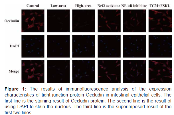
Tight junction protein Occludin can effectively prevent the paracellular transport of bacteria, toxins and other substances in the intestinal cavity through regulation under physiological conditions, and maintain the integrity of the intestinal mucosal epithelial barrier function. Figure 1 shows the results of immunofluorescence analysis. From the results, as the dose of urea increases, the expression of Occludin also decreases. Nrf2 agonists can inhibit the damage of intestinal epithelial cells, and NF-κB receptor blockers can inhibit the inflammatory response. Therefore, the expression of Occludin in these two groups increased. Moreover, the expression level of Occludin in the experimental group was higher than that in the Nrf2 activator group and NF-κB inhibitor group, which was close to the control group. It showed that the therapeutic effect of the prescription is better.
Western blot analysis
Figure 1: The results of immunofluorescence analysis of the expression
characteristics of tight junction protein Occludin in intestinal epithelial cells. The
first line is the staining result of Occludin protein. The second line is the result of
using DAPI to stain the nucleus. The third line is the superimposed result of the
first two lines.
The functions of tight junction protein Claadin-1 is similar to Occludin. The structure and function of tight junction protein ZO-1 are closely related to other similar proteins. In most cases, as long as ZO-1 is damaged, the function of tight junctions will change accordingly. Therefore, ZO-1 is often used as a reference of tightly connect barrier function and permeability function of difference tissues. Figure 2 shows the results of Western blot analysis. Figure 2 shows the results of Western blot analysis. From the results, the relative expression trend of Claadin-1 and ZO-1 is consistent with that of Occludin in immunofluorescence analysis, which is closely related to the similar functions of this three. The results also proved the excellent effect of the prescription treatment.
The symbol * means P<0.05, ** means P<0.01, *** means P
RT-PCR analysis
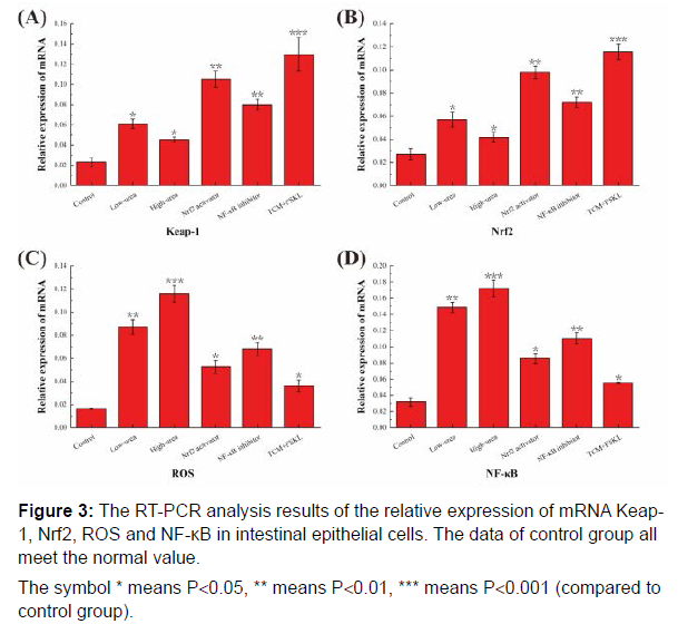
Nrf2 is a key factor in the cellular oxidative stress response. It is regulated by Keap1 and regulates the expression of antioxidant proteins and phase II detoxification enzymes by interacting with the antioxidant response element (ARE). As is shown in Figure 3A and 3B, Since the intestinal epithelial cells of the control group were not damaged, the mRNA expression of Keap-1 and Nrf2 related to damage inhibition was very low. The expression of two mRNAs in the other five groups was positively correlated with the degree of intestinal epithelial damage determined in previous experiments.
The more the expression of ROS and NF-κB mRNA, the more severe the degree of inflammation and the damage of intestinal epithelial cells. Therefore, as shown in Figure 3C and 3D, there is an obvious positive correlation between the expression of these two mRNAs and the degree of intestinal epithelial damage. The expression levels of each of the above mRNAs can all reflect the inhibitory effect of the prescription on the intestinal barrier injury induced by urinary toxin.
The symbol * means P<0.05, ** means P<0.01, *** means P
ELISA analysis
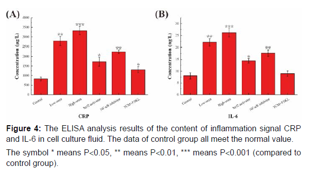
Both high-sensitivity C-reactive protein (CRP) and interleukin-6 (IL-6) are non-specific markers of the acute phase of systemic inflammation synthesized by the liver. Figure 4 shows the results of ELISA analysis. It can be seen from the results that the two inflammatory signal factors have a significant positive correlation with the degree of inflammation in each group, which is consistent with the protein expression trend related to intestinal epithelial injury in the aforementioned experiment, and jointly proves the therapeutic effect of the prescription.
The symbol * means P<0.05, ** means P<0.01, *** means P
Discussion
In this experiment, T84 cells were divided into six groups. In addition to the control group, the low-urea group and the high-urea group were used to compare the effect of the concentration of urinary toxin on the ability of intestinal epithelial cell barrier destruction. The result of the comparison between the two groups is that the higher the urinary toxin concentration, the greater the damage. In addition, the other three groups were added with different drugs on the basis of the high-urea group to further judge the therapeutic effect of Guben Xiezhuo Colon Dialysis Recipe.
Tight junction proteins are mainly located on the side of epithelial cells near the lumen, whose functions are to close the intercellular space and prevent the free entry and exit of materials inside and outside the epithelial layer of the lumen. They are the material basis for the selection of permeabilization of epithelial cells [13]. Tight junctions are a composite structure formed by the interaction of multiple proteins, which are mainly composed of two components: transmembrane proteins and cytoplasmic proteins. The main transmembrane proteins are occludin and claudin-1. Both occludin and claudin-1 are fourpass transmembrane proteins, and the amino and carboxyl ends are located in the cytoplasm. Tight junctions can effectively prevent the paracellular transport of bacteria and toxins in the intestinal cavity through regulation under physiological conditions, and maintain the integrity of the intestinal mucosal epithelial barrier function [14]. ZO-1 is also an important tight junction protein. In addition to maintaining the intestinal barrier function, ZO-1 is also involved in important processes such as regulating cell material transport, maintaining epithelial polarity, cell proliferation and differentiation, and tumor cell metastasis [15]. This project is to detect the content of occluding, claudin-1 and ZO-1 through immunofluorescence analysis and Western Blot analysis to determine the degree of damage to the intestinal epithelial cell barrier.
The reactive oxygen species (ROS) produced by oxidative stress directly or indirectly damages the physiological functions of intracellular proteins, lipids, nucleic acids and other macromolecular substances, which are the pathophysiological basis of many diseases [16]. The body has formed a complex oxidative stress response system. When exposed to electrophilic reagents or active oxygen stimulation, it can induce a series of protective proteins to alleviate cell damage. Nrf2 is a key factor in the cellular oxidative stress response. It is regulated by Keap1 and interacts with the antioxidant response element (ARE) to regulate the expression of antioxidant proteins and phase II detoxification enzymes [17]. This project is to detect the mRNA content of Keap-1, Nrf2 and ROS through RT-PCR analysis to determine the severity of the systemic inflammatory response caused by intestinal epithelial cell barrier damage induced by urinary toxin.
Hypersensitivity C-reactive protein is a non-specific marker synthesized by the liver in the acute phase of systemic inflammation. IL-6 plays a central role in the acute inflammatory response. It can mediate the acute phase response of the liver and stimulate the production of CRP and fibrinogen. A variety of infectious diseases can lead to increased serum IL-6 levels, and IL-6 levels are also closely related to patient prognosis [18]. NF-κB participates in the response of cells to external stimuli, such as cytokines, radiation, heavy metals, viruses and so on. NF-κB plays a key role in the process of cellular inflammation and immune response. Misregulation of NF-κB can cause autoimmune diseases, chronic inflammation, and many cancers [19]. This project is to detect the content of NF-κB mRNA, CRP and IL-6 through RT-PCR analysis and ELISA analysis to further determine the overall situation of systemic inflammation.
Through a series of tests and comparisons on the contents of intestinal barrier-related proteins and inflammatory signal factors in each group, it is proved that the Guben Xiezhuo Recipe colon dialysis has an excellent therapeutic effect on the repair of intestinal barrier damage and further inhibits the deterioration of CKD, which also provides an optional auxiliary strategy. Nevertheless, whether Guben Xiezhuo Colon Dialysis Recipe can further treat the chronic kidney disease as the root cause of intestinal epithelial injury and systemic inflammation has yet to be concluded by further research.
Acknowledgments
We would like to acknowledge everyone for their helpful contributions on this paper.
Authors’ Contributions
HDH, YYT, HRW conceived and designed experiments; HDH, YYT, HRW performed experiments and data analysis; WQS, XDX provided technical support, data collection and analysis; and HDH, YYT, HRW wrote the manuscript. All authors provided their final approval of the submitted and published version.
Ethics Approval and Consent to Participate
The research protocol was reviewed and approved by the Ethical Committee and Institutional Review Board of the Minhang District Central Hospital.
Competing Interests
The authors declare that they have no competing interests.
Funding
The Science and Technology Commission of Shanghai Municipality (14DZ2260200, the project of Shanghai Key Laboratory of kidney and Blood Purification), National Natural Science Foundation of China (No.81774060).
References
- Lee JW, Lee SM, Chun J, Im JP, Seo SK, et al. (2020) Novel Histone Deacetylase 6 Inhibitor CKD-506 Inhibits NF-kappa B Signaling in Intestinal Epithelial Cells and Macrophages and Ameliorates Acute and Chronic Murine Colitis. Inflamm Bowel Dis 26(6): 852-862.
- Vaziri ND, Zhao YY, Pahl MV (2016) Altered intestinal microbial flora and impaired epithelial barrier structure and function in CKD: the nature, mechanisms, consequences and potential treatment. Nephrol Dial Transplant 31: 737-746.
- Meijers BK, Evenepoel P (2011) The gut-kidney axis: indoxyl sulfate, p-cresyl sulfate and CKD progression. Nephrol Dial Transplant 26: 759-761.
- Pahl MV, Vaziri ND (2015) The chronic kidney disease-colonic axis. Semin Dial 28: 459-463.
- Panieri E, Buha A, Telkoparan-Akillilar P, Cevik D, Kouretas D, et al. (2020) Potential Applications of NRF2 Modulators in Cancer Therapy. Antioxidants (Basel) 9(3): 193.
- Chen H, Fang Y, Li W, Orlando RC, Shaheen N, et al. (2013) NFkB and Nrf2 in esophageal epithelial barrier function. Tissue Barriers 1: e27463.
- Rao NAS, McCalman MT, Moulos P, Francoijs KJ, Chatziioannou A, et al. (2011) Coactivation of GR and NFKB alters the repertoire of their binding sites and target genes. Genome Res 21: 1404-1416.
- Marchiando AM, Shen L, Graham WV, Weber CR, Schwarz BT, et al. (2010) Caveolin-1-dependent occludin endocytosis is required for TNF-induced tight junction regulation in vivo. J Cell Biol 189: 111-126.
- Rom S, Heldt NA, Gajghate S, Seliga A, Reichenbach NL, et al. (2020) Hyperglycemia and advanced glycation end products disrupt BBB and promote occludin and claudin-5 protein secretion on extracellular microvesicles. Sci Rep 10(1): 18828.
- Li XF, Zhang XJ, Zhang C, Wang LN, Li YR, et al. (2018) Ulinastatin protects brain against cerebral ischemia/reperfusion injury through inhibiting MMP-9 and alleviating loss of ZO-1 and occludin proteins in mice. Exp Neurol 302: 68-74.
- Umeda K, Ikenouchi J, Katahira-Tayama S, Furuse K, Sasaki H, et al. (2006) ZO-1 and ZO-2 independently determine where claudins are polymerized in tight-junction strand formation. Cell 126: 741-754.
- Mitsuishi Y, Motohashi H, Yamamoto M (2012) The Keap1-Nrf2 system in cancers: stress response and anabolic metabolism. Front Oncol 2: 200.
- Vaziri ND, Wong J, Pahl M, Piceno YM, Yuan J, et al. (2013) Chronic kidney disease alters intestinal microbial flora. Kidney Int 83(2): 308-315.
- Glorieux G, Gryp T, Perna A (2020) Gut-Derived Metabolites and Their Role in Immune Dysfunction in Chronic Kidney Disease. Toxins (Basel) 12(4): 245.
- Chen LY, Xue YX, Zheng J, Liu XB, Liu J, et al. (2018) MiR-429 Regulated by Endothelial Monocyte Activating Polypeptide-II (EMAP-II) Influences Blood-Tumor Barrier Permeability by Inhibiting the Expressions of ZO-1, Occludin and Claudin-5. Front Mol Neurosci 11: 35.
- Huang QC, Zhan L, Cao HY, Li JB, Lyu YH, et al. (2016) Increased mitochondrial fission promotes autophagy and hepatocellular carcinoma cell survival through the ROS-modulated coordinated regulation of the NFKB and TP53 pathways. Autophagy 12(6): 999-1014.
- Cuadrado A, Rojo AI, Wells G, Hayes JD, Cousins SP, et al. (2019) Therapeutic targeting of the NRF2 and KEAP 1 partnership in chronic diseases. Nat Rev Drug Discov 18(4): 295-317.
- Vaziri D (2012) CKD impairs barrier function and alters microbial flora of the intestine: a major link to inflammation and uremic toxicity. Curr Opin Nephrol Hypertens 21: 587-592.
- Yang HL, Thiyagarajan V, Shen PC, Mathew DC, Lin KY, et al. (2019) Anti-EMT properties of CoQ0 attributed to PI3K/AKT/NFKB/MMP-9 signaling pathway through ROS-mediated apoptosis. J Exp Clin Cancer Res 38(1): 186.
Indexed at Google Scholar Crossref
Indexed at Google Scholar Crossref
Indexed at Google Scholar Crossref
Indexed at Google Scholar Crossref
Indexed at Google Scholar Crossref
Indexed at Google Scholar Crossref
Indexed at Google Scholar Crossref
Indexed at Google Scholar Crossref
Indexed at Google Scholar Crossref
Indexed at Google Scholar Crossref
Indexed at Google Scholar Crossref
Indexed at Google Scholar Crossref
Indexed at Google Scholar Crossref
Indexed at Google Scholar Crossref
Indexed at Google Scholar Crossref
Indexed at Google Scholar Crossref
Indexed at Google Scholar Crossref
Indexed at Google Scholar Crossref
Citation: He H, Huan H, Tang Y, Wang H, Sun W, et al. (2022) Guben Xiezhuo Recipe alleviated Colon Dialysis on Urotoxin-induced Intestinal Barrier Injury and Systemic Inflammation. Cell Mol Biol, 68: 224.
Copyright: © 2021 He H, et al. This is an open-access article distributed under the terms of the Creative Commons Attribution License, which permits unrestricted use, distribution, and reproduction in any medium, provided the original author and source are credited.
Share This Article
Recommended Journals
Open Access Journals
Article Usage
- Total views: 2468
- [From(publication date): 0-2022 - Apr 18, 2025]
- Breakdown by view type
- HTML page views: 1938
- PDF downloads: 530


