Grynfeltt's Hernia: Think it in Case of a Lumbar Mass
Received: 05-Sep-2022 / Manuscript No. roa-22- roa-22-74432 / Editor assigned: 07-Sep-2022 / PreQC No. roa-22-74432 (PQ) / Reviewed: 21-Sep-2022 / QC No. roa-22- roa-22-74432 / Revised: 23-Sep-2022 / Manuscript No. roa-22-74432 (R) / Published Date: 30-Sep-2022
Abstract
Lumbar hernia is a rare defect of the posterior abdominal wall, first described in 1672 by Barrett. Grynfelt and Lesshaft described herniation of the upper lumbar triangle in 1886 and 1870, respectively. It occurs due to weakening of the transversalis fascia and transversus abdominis fascia. As a consequence, this weakening allows the abdominal contents to protrude through the upper lumbar triangle.
Keywords
Grynfelt; Lesshaft Hernia; Lumbar mass; Imagery
Clinical Image
Grynfeltt -Lesshaft hernia is a rare defect of the posterior abdominal wall at the level of the upper lumbar triangle, described by Grynfelt and Lesshaft in 1886 and 1870, respectively [1]. It Occurs due to weakening of the transversalis fascia and transversus abdominis fascia allowing abdominal contents to protrude through the upper lumbar triangle, a region defined medially by the quadratus lumborum muscle, inferiorly by the internal oblique muscle and superiorly by the 12th rib. The contents may include retroperitoneal organs, intraperitoneal organs and retroperitoneal or epiploic adipose tissue [2].
They represent less than 2% of abdominal wall hernias and only a little over 300 cases reported in the literature [1]. They can be congenital (20%) or acquired in the majority of cases (82%) [1]. Advanced age, obesity, muscle atrophy and chronic obstructive pulmonary disease or any other condition that produces a persistent increase in intraabdominal pressure are the main risk factors [2].
Clinically, the patient may present with an asymptomatic lumbar mass, expansive on coughing, physical exertion or trunk anteflexion; a lumbar mass with back pain; or a lumbar mass with vague abdominal symptoms [2]. The main differential diagnosis is lipoma [1].
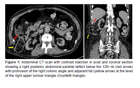
Ultrasound shows a subcutaneous mass, well circumscribed with fat content [2]. The CT scan is the examination of choice to study the anatomy of the lumbar region, the extension of the defect, the presence or absence of viscera in the hernia (Figure 1) and possibly the search for any complications [1,2] (Figure 1).
Figure 1: Abdominal CT scan with contrast injection in axial and coronal section showing a right posterior abdominal parietal defect below the 12th rib (red arrow) with protrusion of the right colonic angle and adjacent fat (yellow arrow) at the level of the right upper lumbar triangle (Grynfeltt triangle).
Described repair techniques include anatomic closure, covering the fascia with musculofascial flaps, and placement of mesh prostheses via the retroperitoneal or transabdominal laparoscopic approach [1,2].
References
- Ploneda-Valencia CF, Cordero-Estrada E, Castañeda-González LG, Sainz-Escarrega VH, Varela-Muñoz O, et al. (2016) Grynfelt-Lesshaft hernia a case report and review of the literature. Ann Med Surg (Lond) 7: 104-106.
- Zadeh JR, Buicko JL, Patel C, Kozol R, Lopez-Viego MA (2015) Grynfeltt Hernia: A Deceptive Lumbar Mass with a Lipoma-Like Presentation. Case Rep Surg 2015: 1-4.
Indexed at, Google Scholar, Crossref
Citation: Cherraqi A, Mandour J, Abide Z, Andour H, Fenni J, et al. (2022) Grynfeltt’s Hernia: Think it in Case of a Lumbar Mass. OMICS J Radiol 11: 401.
Copyright: © 2022 Cherraqi A, et al. This is an open-access article distributed under the terms of the Creative Commons Attribution License, which permits unrestricted use, distribution, and reproduction in any medium, provided the original author and source are credited.
Share This Article
Open Access Journals
Article Usage
- Total views: 1430
- [From(publication date): 0-2022 - Mar 29, 2025]
- Breakdown by view type
- HTML page views: 1071
- PDF downloads: 359

