Gingival Retiform Hemangioendothelioma A Case Report and Review of the Literature
Received: 17-Jan-2023 / Manuscript No. jdpm-23-87263 / Editor assigned: 19-Jan-2023 / PreQC No. jdpm-23-87263 (PQ) / Reviewed: 02-Feb-2023 / QC No. jdpm-23-87263 / Revised: 06-Feb-2023 / Manuscript No. jdpm-23-87263 (R) / Published Date: 13-Feb-2023 DOI: 10.4172/jdpm.1000139
Abstract
Hemangioendothelioma is a vascular tumor with microscopic features intermediate between those of hemangiomas and angiosarcomas. Hemangioendothelioma is a rare tumor, it can occur anywhere in the body. Its occurrence has been reported in lower extremities, liver, lungs and head and neck region.
Here we are reporting a gingival case of Hemangioendothelioma with review of literature. In March 2022, a 12-year-old female patient was referred to Pacific Northwest Kaiser oral pathology clinic for a gingival growth on buccal of tooth #22. The patient and her mother mentioned the lesion had been present for months. Complete removal of the lesion was done and sent for pathology examination. Microscopic examination was done by Kaiser Pathologist. The case was sent to bone / soft tissue pathologist, pediatric pathologist and dermatopathologist for a consult, and the final pathology result was retiform hemangioendothelioma.
The microscopic result was retiform hemangioendothelioma with a comment indicating this tumor typically would affect the distal extremities of young individuals. The term hobnail Hemangioendothelioma is used for Retiform and Dabska type tumors which are closely related.
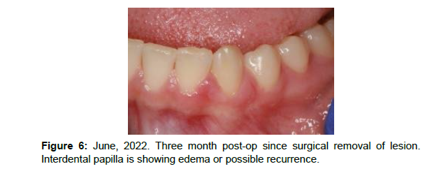
The biopsy site was checked in June 2022. Distal and facial gingiva of #22 was healed and was within normal limits. The interdental papilla between #22 and #23 showed edema or possible recurrent lesion. The biopsy site has been under surveillance. Recent follow up showed slight changes on the interdental papilla between #22 and #23 and a second biopsy has been recommended
Due to the nature of this lesion showing intermediate features and behaviors between a benign vascular lesion and an angiosarcoma, close observation is crucial.
Introduction
Gingival growths are mostly inflammatory or reactive in nature. Here we mention three of them which are the most common ones. Pyogenic granuloma is a tumor like lesion, red in color and very commonly seen on gingiva. It is considered a reactive lesion and nonneoplastic occurring due to local irritation, trauma, calculus or poor oral hygiene.
Another reactive gingival mass is peripheral giant cell granuloma. Unlike pyogenic granuloma which can occur on other sites of oral cavity, peripheral giant cell granuloma exclusively occurs on gingiva, or edentulous alveolar ridge and it has a mixed red and blue color.
Peripheral ossifying fibroma is a gingival growth which is red or pink in color. Some investigators believe it is a mature form of pyogenic granuloma, with fibrous maturation and calcification. A gingival mass or growth in children usually falls into one of the above categories mentioned. Juvenile spongiotic gingivitis can be localized and can be on the differential list, but its occurrence is less common. Hemangiomas, vascular malformations and hemangioendotheliomas can occur in oral cavity but their occurrence is not common, especially on gingiva.
Case Report
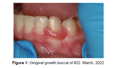
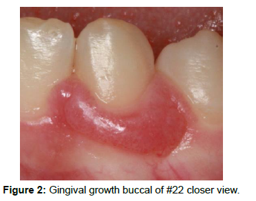
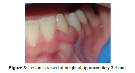
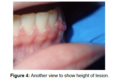
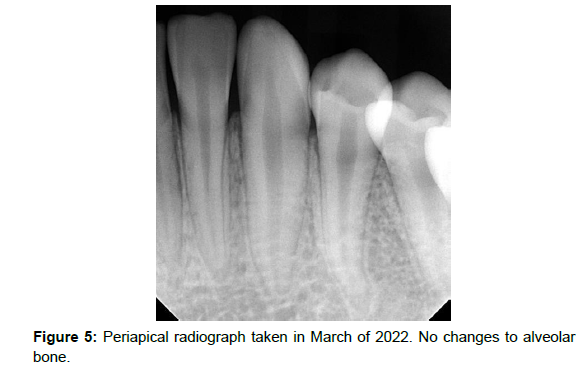
A 12-year-old female patient was referred to Pacific Northwest Kaiser oral pathology clinic by her dentist in March 2022. Medical history review showed mild Arnold Chiari type 1 and atopic dermatitis. A red gingival mass was noticed on buccal of #22 with extension to the interdental papillae on both sides measuring 1.4x0.4x0.3 cm. (Figures 1-4) The lesion was asymptomatic, the patient and her mother both believed it had been present for a long time. They mentioned it could be present for a year. PA was within normal limit and did not show any bone pathology. (Figure 5) After a consultation and explanation of the differential diagnosis, complete removal of the lesion was done and submitted to pathology lab. Differential diagnosis was pyogenic granuloma, peripheral giant cell granuloma and juvenile spongiotic gingivitis.
The surgery site was checked in June 2022. The buccal and distal gingiva of #22 healed but the interdental papilla between #22 and #23 showed edema and mild erythema. (Figure 6)
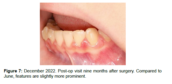
The patient and her mother were happy with the healing of the surgery site. Still chance of recurrence, features of the interdental papilla, along with details of the pathology result all were explained to them. Options were discussed and close monitoring of the biopsy site was decided. Biopsy site has been under surveillance since June 2022. Recent follow up in December 2022 showed buccal gingiva of #22 flat and within normal limit. The interdental papilla between #22 and #23 was slightly more prominent but it did not have the erythematous color of the original lesion and appeared to be more fibrotic. Lesion still remains asymptomatic and no pain upon palpation (See Figure 7) options was discussed with the patient and parents. Due to the nature of this neoplasm, a second biopsy has been recommended (Figure 7).
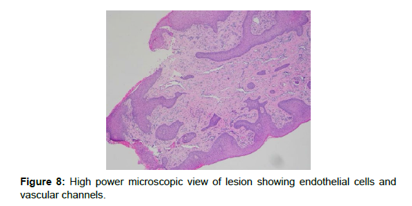
The case was sent for a consult to bone and soft tissue pathologist, pediatric pathologist, and dermatopathologist. The result was retiform hemangioendothelioma with a comment: This is a vascular proliferation arranged in a retiform pattern formed by plump CD31 positive endothelial cells that protrude into the lumina. There is significantly less CD34 than CD31 expression therefore a retiform hemangioendothelioma is the diagnosis. RHE is a low-grade variant of angiosarcoma typically affecting the distal extremities of young individuals. The term hobnail hemangioendothelioma is used for retiform and Dabska type tumors which are closely related. Rarely there can be multiple lesions with about two-thirds of lesions reported to recur particularly without wide local excision. The pathologist included two articles to support the information on the pathology report, Am J surg path January 1999 and Am j surg path February 1994.
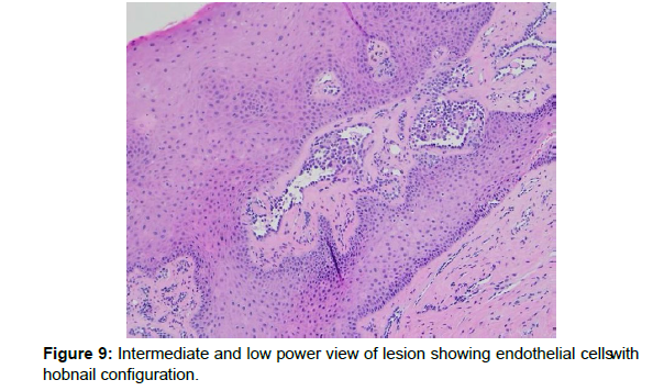
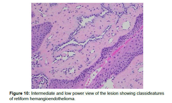
Microscopic description included a fragment of squamous mucosa with unremarkable epithelium. The stroma showed variably sized small to ectatic vascular spaces lined by a single layer of endothelial cells. The endothelial cells had a hobnail configuration with focal intravascular micropapillary growth. No mitotic figures were identified. (Figures 8-10) The cells were strongly positive with CD31 (Figure 11). They were negative with SOX10, S100, Desmin and Pankeratin. SMA highlighted the vessel walls.
Discussion
The term hemangioendothelioma describes vascular tumors with features intermediate between hemangiomas and angiosarcomas. Several types of hemangioendotheliomas have been reported including papillary intralymphatic angioendothelioma (also known as the Dabska tumor), kaposiform hemangioendothelioma, epithelioid hemangioendothelioma, pseudomyogenic hemangioendothelioma, composite hemangioendothelioma, and retiform hemangioendothelioma [1]. Given their rarity, statistics on prevalence of the hemangioendothelioma tumor family is limited. As of 2021, less than 40 cases each of the Dabska tumor, and its closest relative, the retiform hemangioendothelioma, have been reported [2,3].
Retiform hemangioendothelioma has been described as a low-grade angiosarcoma first introduced in 1994 by Calonje E [4]. Presentations of retiform hemangioendothelioma have most commonly involved the lower extremities, however current literature have reported sites including the liver, lungs, and head and neck regions [5] such as in the case described here. Retiform hemangioendothelioma affects mainly the younger population with a slight female predilection, although many cases report a wide range of ages. In a 2016 review, approximately 30% of retiform hemangioendothelioma cases occurred in patients under 20 years and 20% occurred in patients over 50 years [6]. Additionally, there is currently one reported case of congenital retiform hemangioendothelioma [7].
Clinical appearance has been described as slow growing indurated plaques and dermal or subcutaneous nodules [3]. Singular lesions as well as multiple and rapid growing lesions have been reported [8]. Dermal lesions have presented as well-defined erythematous patches reaching 4 x 3 cm in diameter [9], however, note the plaques have varied in size from 1-30 cm. Case reports involving the lower extremities have described ill-defined subcutaneous masses averaging 2.5-11cm in size with and without out overlying skin changes [7,10]. Lesions can range from asymptomatic to local discomfort, sharp-shooting pain [8] or systemic symptoms such as coughing and shortness of breath accompanying mediastinal involvement [5].
Intraoral reports of hemangioendothelioma include the gingiva, tongue, maxilla, buccal mucosa, and palate, with the epithelioid variant being the most common [11]. Presentations may show pink color, sessile or pedunculated, firm, painless masses [12]. Some of the literature has reported the tumors with ulcerations. The slow growth rate of these tumors may account for only 25% of oral cases presenting with resorption of underlying bone radiographically [13]. When present, radiographs have been described to have “saucer like bony erosion” underlying the lesions [13,14]. As mentioned above, less than 40 cases total of both the retiform hemangioendothelioma and the Dabska tumor have been reported. Approximately 14.3% of cases involve the head and neck [8], with the submandibular area the most common [15]. Other head and neck specific cases have described lesions as large spherical shaped and firm swellings extending from the masseteric area through the ear, ulcerating dermal lesions on the scalp, palatal and gingival swelling resembling gingival hyperplasia, and whitish/bluish nodules on posterior lateral tongue [3,13,14].
Classic histology of retiform hemangioendothelioma demonstrates proliferation of elongated arborizing vessels, arranged in a retiform pattern resembling that of the rete testis [1,8,9]. Both Dabska and retiform hemangioendothelioma show the blood vessels lined by hobnail endothelial cells protruding within the lumina [1, 16]. Given the higher incidence of metastasis and aggressive malignant potential, it is important to differentiate both entities from other vascular tumors such as the angiosarcoma, which shows mitosis and atypia within endothelial cells [3,5].
Reports in the literature have shown the closely related Dabska tumor presenting on the posterior border of the tongue as a multinodular painless exophytic mass with bluish overlying mucosa and irregular borders [14]. Differential diagnoses included the hemangioendothelioma as well as cystic tumors of salivary glands, angiosarcoma, and Kaposi sarcoma. Histological examination revealed dilated, thin-walled vascular spaces with hobnail endothelial cells and peripheral anastomosing blood vessels. Comparably, Sunil M, et al. [16] reported a case in which a 3.5 x 5 cm growth was detected intraorally. The mass was solitary, sessile, and erythematous. The mass extended from marginal gingiva to the interdental region extending over alveolar crest to palatal mucosa. The mass was soft in consistency and nontender, Slight bleeding upon palpation. Differential diagnosis included peripheral giant cell granuloma, inflammatory gingival hyperplasia, and peripheral ossifying fibroma. A periapical radiograph showed alveolar bone loss and pathological migration of maxillary teeth. Given the higher potential for metastasis and aggressive nature, these cases as well as further literature studies demonstrate the inability to diagnose retiform hemangioendothelioma clinically compared to a Dabska tumor and other vascular lesions, highlighting the importance of microscopic investigation to respond with appropriate treatment.
A similar case from Heera R, et al. [13] describes a patient with an ulcerated palatal swelling. Like our case of retiform hemangioendothelioma, this patient presented asymptomatically with a sessile, nonindurated mass without other relevant clinical findings. Common clinical differentials included those listed by the other cases above [12, 13]. Given the unpredictability and high recurrence rate of some hemangioendothelioma types, patients should be kept on regular follow-ups.
The retiform hemangioendothelioma specifically tends to have a high recurrence rate. It has been shown approximately 50% of retiform hemangioendotheliomas recur locally within 12 months of surgery [8]. Half of those recurring were detected within six months. A small number of cases report regional lymph node metastasis [17] with only one reported case of retiform hemangioendothelioma related death [3]. While the lesions themselves are not presumed to be fatal, cases of death involving hypovolemic shock post-surgery have been reported [8,18].
In our case described here, a healthy 12-year-old female patient presented with a gingival growth on buccal of tooth #22. The patient and her mother stated the lesion had been present for months. A periapical radiograph revealed underlying alveolar bone without signs of resorption. Like the cases described by Sunil M, et al. and Heera R, et al. [13,16], this lesion presented asymptomatically and appeared hyperplastic or pyogenic granuloma-like in nature. Given the small size and presumably relatively early detection of this lesion, it is unclear whether this lesion would have progressed to involve surface ulceration or underlying bone resorption as described by the previous cases.
Treatment of retiform hemangioendothelioma is traditionally performed with complete surgical resection with negative margins. In the case of cutaneous lesions, Mohs micrographic surgery is used. In patients with a large tumor size, high local recurrence, or lymph node metastasis, adjuvant radiation or chemotherapy treatments have been indicated [9,19]. Conservative amputations have also been used with success in large tumors of the extremities [20]. Given the location and the relatively small size of the lesion described in the case presented here, treatment involved complete surgical resection.
Conclusion
Gingival growths are commonly reactive such as pyogenic granulomas, peripheral giant cell granulomas and peripheral ossifying fibromas. The occurrence of this neoplasm intraorally and on the gingiva was interesting and rare. Since RHE has a high recurrence rate and it is a variant of angiosarcoma, close observation and monitoring is recommended after complete removal of the lesion.
References
- Requena L, Kutzner H (2013) Hemangioendothelioma. Semin Diagn Pathol 30: 29-44.
- Bhatia J, Tadi P (2022) Endovascular papillary angioendothelioma. StatPearls [Internet] Treasure Island (FL): StatPearls Publishing.
- Jothi EA, Sundaram M, Namasivayam J, Jothi M (2014) A rare presentation of retiform hemangioendothelioma in the external auditory canal. Case Rep Otolaryngol 2014: 715035.
- Calonje E, Fletcher CD, Wilson-Jones E, Rosai J (1994) Retiform hemangioendothelioma: A distinctive form of low-grade angiosarcoma delineated in a series of 15 cases. Am J Surg Pathol 18: 115-125.
- Chundriger Q, Tariq MU, Rahim S, Abdul-Ghafar J, Nasir Ud Din (2021) Retiform hemangioendothelioma: a case series and review of the literature. J Med Case Rep 15: 69.
- Zhang G, Lu Q, Yin H, Wen H, Su Y, et al. (2010) A case of retiform- hemangioendothelioma with unusual presentation and aggressive clinical features. Int J Clin Exp Pathol 3: 528-533.
- Manjunatha BS, Kumar GS, Vandana R (2009) Intraoral epithelioid hemangioendothelioma: an intermediate vascular tumor- a case report. Dent Res J (Isfahan) 6: 99-102.
- Bhutoria B, Konar A, Chakrabarti S, Das S (2009) Retiform hemangioendothelioma with lymph node metastasis: a rare entity. Indian J Dermatol Venereol Leprol 75: 60-62.
- Ranga SM, Kuchangi NC, Shankar VS, Amita K, Haleuoor BB, et al. (2014) Retiform hemangioendothelioma: an uncommon pediatric vascular neoplasm. Indian J Dermatol 59: 633.
- Liu Q, Ouyang R, Chen P, Zhou R (2015) A case report of retiform hemangioendothelioma as pleural nodules with literature review. Diagn Pathol 10: 194.
- Heera R, Cherian LM, Lav R, Ravikumar V (2017) Hemangioendothelioma of palate: a case report with review of literature. J Oral MAxillofac Pathol 21: 415-420.
- Hirsh AZ, Yan W, Wei L, Wernicke AG, Parashar B (2010) Unresectable retiform hemangioendothelioma treated with external beam radiation therapy and chemotherapy: a case report and review of the literature. Sarcoma 2010: 756246.
- Gordón-Núñez MA, Silva eM, Lopes MF, de Oliveira-Neto SF, Maia AP, et al. (2010) Intraoral epithelioid hemangioendothelioma: a case report and review of the literature. Med Oral Patol Oral Cir Bucal 15: e340-e346.
- Favia G, Limongelli L, Tempesta A, Maiorana E (2015) Dabska tumor of the tongue: a clinicopathological study with confocal laster scanning microscopy. Int J Case Rep Images 6: 46-50.
- Kim II-K, Cho HY, Jung BS, Pae SP, Cho HW, et al. (2016) Retiform hemangioendothelioma in the infratemporal fossa and buccal area: a case report and literature review. J Korean Assoc Oral Maxillofac Surg 42: 307-314.
- Sunil MK, Trivedi A, Trakroo A, Arora S (2016) Retiform hemangioendothelioma: a rare case report. J Indian Acad Oral Med Radiol 28: 292-295.
- Serel S, Serel BI, Uluc A, Heper AO, Gultan MS (2007) Congenital retiform hemangioendothelioma. Indian J Dermatol 52: 160-162.
- Albertini AF, Brousse N, Bodemer C, Calonje E, Fraitag S (2011) Retiform hemangioendothelioma developed on the site of an earlier cystic lymphangioma in a six- year-old girl. Am J Dermatopathol 33: e84-e87.
- Tan D, Kraybill W, Cheny RT, Khoury T (2005) Retiform hemangioendothelioma: a case report and review of the literature. J Cutan Pathol 32: 634-637.
- Reis-Filhho J, Paiva ME, Lopes JM (2002) Congenital composite hemangioendothelioma: a case report and reappraisal of the hemangioendothelioma spectrum. J Cutan Pathol 29: 226-231.
Indexed at, Google Scholar, Crossref
Indexed at, Google Scholar, Crossref
Indexed at, Google Scholar, Crossref
Indexed at, Google Scholar, Crossref
Indexed at, Google Scholar, Crossref
Indexed at, Google Scholar, Crossref
Indexed at, Google Scholar, Crossref
Indexed at, Google Scholar, Crossref
Indexed at, Google Scholar, Crossref
Indexed at, Google Scholar, Crossref
Indexed at, Google Scholar, Crossref
Indexed at, Google Scholar, Crossref
Citation: Warner L, Ghanee N, Oyama KA, Saraf S (2023) Gingival RetiformHemangioendothelioma A Case Report and Review of the Literature. J Dent PatholMed 7: 139. DOI: 10.4172/jdpm.1000139
Copyright: © 2023 Warner L, et al. This is an open-access article distributed underthe terms of the Creative Commons Attribution License, which permits unrestricteduse, distribution, and reproduction in any medium, provided the original author andsource are credited.
Share This Article
Recommended Journals
Open Access Journals
Article Tools
Article Usage
- Total views: 2230
- [From(publication date): 0-2023 - Apr 03, 2025]
- Breakdown by view type
- HTML page views: 1934
- PDF downloads: 296
