Image Open Access
Giant Plexiform Neurofibroma of the Urinary Bladder
Siddiqui MA1*, Sartaj S2, Rizvi SWA2 and Khan IA21Department of Radiology, Saint Louis University Hospital, MO, USA
2Jawaharlal Nehru Medical College, Aligarh, India
- *Corresponding Author:
- Mohammed Azfar Siddiqui
Department of Radiology, Saint Louis University Hospital
MO 63110, USA
Tel: 001-717-649-8167
E-mail: drazfarsiddiqui@gmail.com
Received date: July 03, 2017; Accepted date: July 05, 2017; Published date: July 11, 2017
Citation: Siddiqui MA, Sartaj S, Rizvi SWA, Khan IA (2017) Giant Plexiform Neurofibroma of the Urinary Bladder. OMICS J Radiol 6:266. doi: 10.4172/2167-7964.1000266
Copyright: © 2017 Siddiqui MA, et al. This is an open-access article distributed under the terms of the Creative Commons Attribution License, which permits unrestricted use, distribution, and reproduction in any medium, provided the original author and source are credited.
Visit for more related articles at Journal of Radiology
Keywords
Plexiform; Neurofibroma; Urinary bladder; Magnetic Resonance Imaging (MRI)
Clinical Description
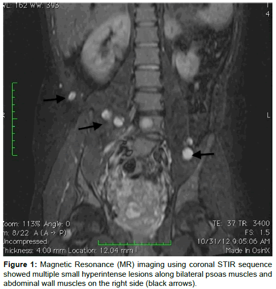
A 16-year-old boy without any established history of neurofibromatosis presented with progressive lumbar spinal deformity. There were no urinary or gastrointestinal symptoms. An initial radiograph of lumbar spine demonstrated grade 4 anterolisthesis of L5 vertebra over S1 vertebra along with posterior scalloping of L4 and L5 vertebral bodies. Further imaging with Magnetic Resonance (MR) Imaging using coronal STIR (Panel A) and sagittal post contrast fat-suppressed T1W (Panel B) sequences showed a large enhancing mass involving the postero-inferior wall of the urinary bladder. MR examination also demonstrated multiple small similar lesions along bilateral psoas muscles and abdominal wall muscles on the right side (Black Arrows). Surgical biopsy was done that confirmed the diagnosis of neurofibroma with malignant degeneration (Figures 1 and 2).
Figure 2: Magnetic Resonance (MR) imaging using sagittal post contrast fatsuppressed T1W sequence demonstrated a large enhancing mass involving the postero-inferior wall of the urinary bladder. It also demonstrated grade 4 anterolisthesis of L5 vertebra over S1 vertebra along with posterior scalloping of L4 and L5 vertebral bodies.
Relevant Topics
- Abdominal Radiology
- AI in Radiology
- Breast Imaging
- Cardiovascular Radiology
- Chest Radiology
- Clinical Radiology
- CT Imaging
- Diagnostic Radiology
- Emergency Radiology
- Fluoroscopy Radiology
- General Radiology
- Genitourinary Radiology
- Interventional Radiology Techniques
- Mammography
- Minimal Invasive surgery
- Musculoskeletal Radiology
- Neuroradiology
- Neuroradiology Advances
- Oral and Maxillofacial Radiology
- Radiography
- Radiology Imaging
- Surgical Radiology
- Tele Radiology
- Therapeutic Radiology
Recommended Journals
Article Tools
Article Usage
- Total views: 2995
- [From(publication date):
August-2017 - Mar 30, 2025] - Breakdown by view type
- HTML page views : 2193
- PDF downloads : 802


