Geniculate Ganglion Tumor Manifesting as Insidious Peripheral Facial Nerve Palsy
Received: 07-May-2018 / Accepted Date: 14-May-2018 / Published Date: 21-May-2018
Clinical Description
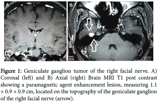
A 20-year-old woman presented with a 9-month history of slowly progressive right peripheral facial nerve palsy. Clinical assessment showed Grade II House-Brackmann right facial nerve palsy [1]. Otoscopy and laboratory examination were normal. History of traumatic brain injury was negative. Magnetic resonance imaging of the brain showed a T1 hypointense, T2 hyperintense and paramagnetic agent enhancement lesion, measuring 1.1 x 0.9 x 0.9 cm, located on the geniculate ganglion topography of the of the right facial nerve (Figure 1). Therapeutic options were discussed with the patient and the wait and- scan strategy was instituted, since she had a near-normal facial nerve function [2]. Attention to this clinical–radiologic correlation may help physicians make correct diagnoses.
References
- House JW, Brackmann DE (1983) Facial nerve grading system. Otolaryngol Head Neck Surg 93: 184-193.
- Lahlou G, Nguyen Y, Russo FY, Ferrary E, Sterkers O, et al. (2016) Geniculate ganglion tumors: clinical presentation and surgical results. Otolaryngol Head Neck Surg 155: 850-855.
Citation: Fernandes EP, Brooks JBB, Prosdócimi FC, Oliveira RA, Silveira GL, et al. (2018) Geniculate Ganglion Tumor Manifesting as Insidious Peripheral Facial Nerve Palsy. Neurol Clin Therapeut J 2: 107.
Copyright: © 2018 Fernandes EP, et al. This is an open-access article distributed under the erms of the Creative Commons Attribution License, which permits unrestricted use, distribution, and reproduction in any medium, provided the original author and source are credited.
Select your language of interest to view the total content in your interested language
Share This Article
Open Access Journals
Article Usage
- Total views: 4071
- [From(publication date): 0-2018 - Nov 17, 2025]
- Breakdown by view type
- HTML page views: 3153
- PDF downloads: 918

